. 2002 Feb 1;21(3):334-43.
doi: 10.1093/emboj/21.3.334.

NF1/X represses PDGF A-chain transcription by interacting with Sp1 and antagonizing Sp1 occupancy of the promoter

Affiliations

NF1/X represses PDGF A-chain transcription by interacting with Sp1 and antagonizing Sp1 occupancy of the promoter

Louise A Rafty et al. EMBO J. .

Abstract

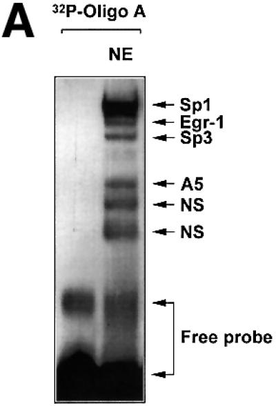
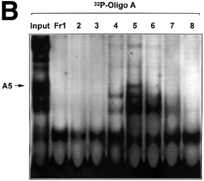
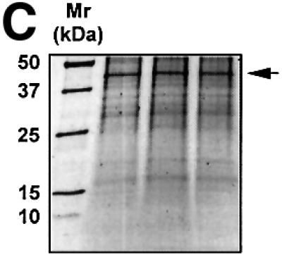
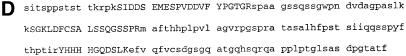
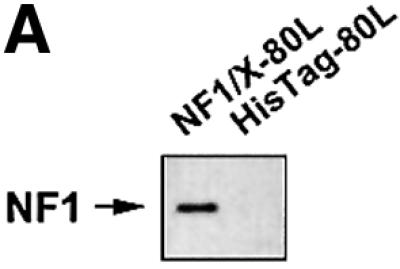
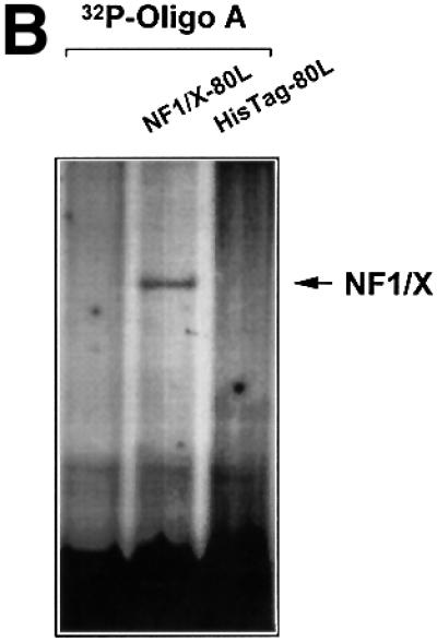
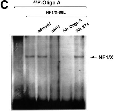
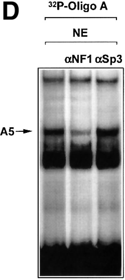
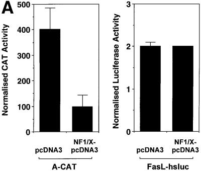
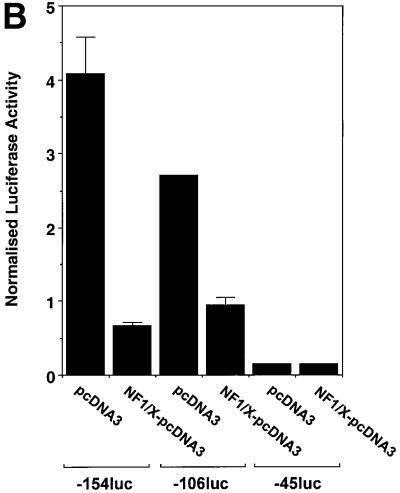
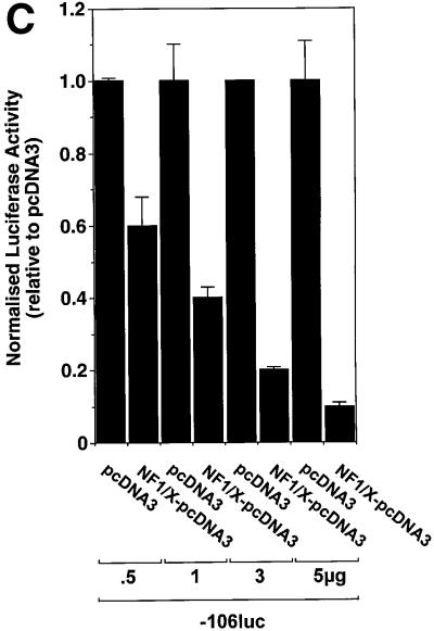
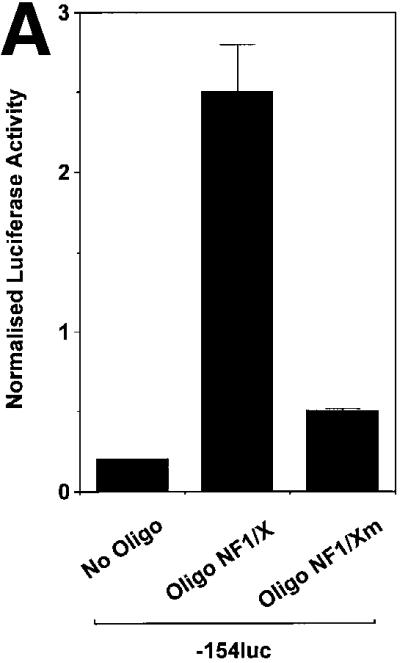
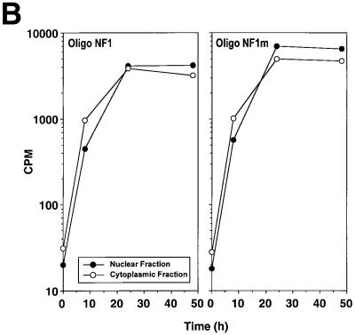
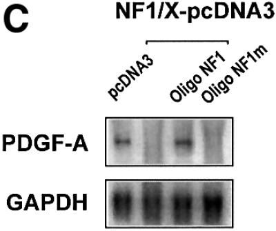
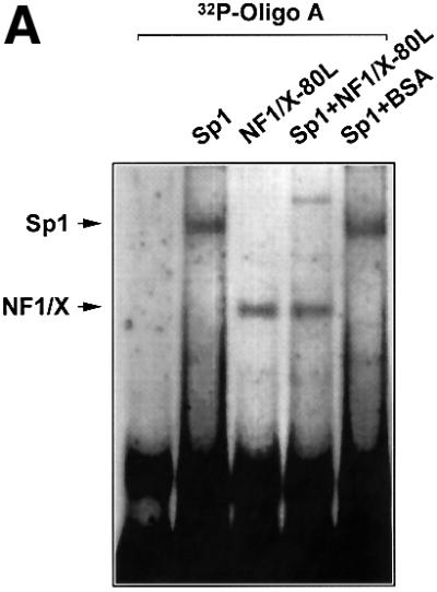
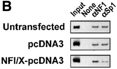
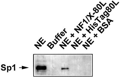
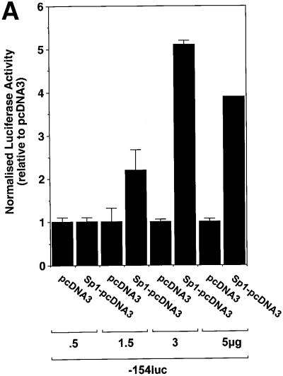
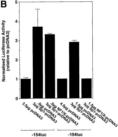
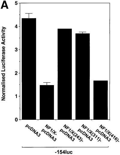
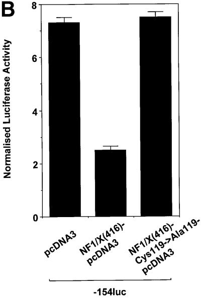
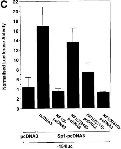
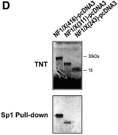
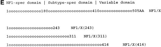
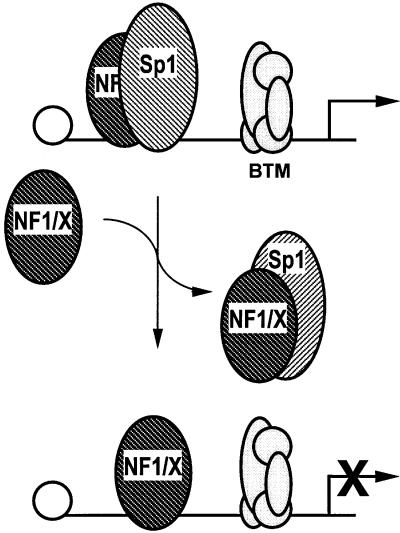
The regulatory mechanisms mediating basal and inducible platelet-derived growth factor (PDGF)-A expression have been the focus of intense recent investigation, but repression of PDGF-A expression is largely unexplored. Here we isolated a nuclear factor that interacts with the proximal region of the PDGF-A promoter using bulk binding assays and chromatography techniques. Peptide mass fingerprint and supershift analysis revealed this DNA-binding protein to be NF1/X. NF1/X repressed PDGF-A promoter-dependent transcription and endogenous mRNA expression, which was reversible by oligonucleotide decoys bearing an NF1/X-binding site. Mutation in the DNA-binding domain of NF1/X abolished its repression of PDGF-A promoter. NF1/X antagonized the activity of a known activator of the PDGF-A chain, Sp1, by inhibiting its occupancy of the proximal PDGF-A promoter. NF1/X physically and specifically interacts with Sp1 via its subtype-specific domain and blocks Sp1 induction of the promoter. NF1/X residues 311-416 mediated NF1/X suppression of basal PDGF-A transcription, whereas residues 243-416 were required for NF1/X repression of Sp1-inducible promoter activity. These findings demonstrate that repression of PDGF-A gene transcription is governed by interplay between NF1/X and Sp1.


Figures

None
Fig. 1. The proximal PDGF-A promoter interacts with nuclear protein. (A) EMSA was performed using nuclear extracts from WKY12-22 cells and [32P]Oligo A (5′-GGGGGGGGCGGGGGCGGGGGCGGGGGAGG-3′, sense strand; Khachigian et al., 1995). The first lane contains probe alone. The identities of the protein components of the complexes have been delineated by supershift analysis in Day et al. (1999). NE, nuclear extract; NS, non-specific complexes based on oligonucleotide competition analysis. (B) EMSA was performed using [32P]Oligo A and fractions from size-exclusion chromatography. Unfractionated nuclear extract in EMSA is shown in the ‘Input’ lane. Fractions were collected in 1:1 mixture of buffers C and D prior to EMSA. Gel electrophoresis was performed as described in Materials and methods. The arrow indicates complex A5. (C) SYPRO-stained 1D SDS–PAGE of proteins eluted from EMSA gel fragments precipitated with methanol. The protein species indicated by the arrow (triplicate samples) were excised from each lane (three bands pooled) for MS/MS analysis [(see D)]. (D) MALDI-TOF analysis of complex A5. Peptides (in capitals) corresponding to Rattus norvegicus NF1/X.
None
Fig. 2. NF1 interacts with the proximal PDGF-A promoter. (A) Recombinant NF1/X (1 µg) produced from construct NF1/X-80L and the control backbone HisTag vector (HisTag-80L) were analysed for NF1 immunoreactivity by western blot analysis using polyclonal antibodies to NF1. (B) Recombinant NF1/X protein (5 µg) and eluate from control backbone HisTag-80L vector were incubated with [32P]Oligo A prior to EMSA. The arrow represents Oligo A-bound recombinant NF1/X. (C) Competition and antibody elimination analysis. The indicated antibody (1 µg) was incubated with the binding mixture for 10 min before the addition of the [32P]Oligo A. For competition studies, a 50-fold molar excess of unlabelled oligonucleotide was incubated with the binding mixture for 10 min prior to the addition of the probe. The sequence of E74 is 5′-AGCTTCTCTAGCTGAATAACCGGAAGTAACTCATCGTCG-3′ (sense strand). (D) Interaction of endogenous NF1 protein with proximal PDGF-A promoter. Nuclear extracts from WKY12-22 cells were incubated with [32P]Oligo A with or without pre-incubation with 1 µg of polyclonal NF1 or Sp3 antibodies for 10 min prior to the addition of the 32P-labelled probe. Bound species were resolved by non-denaturing electrophoresis and visualized by autoradiography. The arrow denotes complex A5 containing NF1.
None
Fig. 3. Repression of PDGF-A promoter activity by NF1/X. (A) WKY12-22 cells were transiently co-transfected with 2 µg of A-CAT or 2 µg of a luciferase construct driven by 1.2 kb of the Fas ligand promoter, along with either 3 µg of NF1/X–pcDNA3 or pcDNA3. Twenty-four hours after transfection, the cells were lysed and assessed for CAT or luciferase activity. CAT activity was normalized to the concentration of protein in the lysate. (B) Serial 5′ deletion and transient transfection analysis using PDGF-A promoter constructs and NF1/X. COS-7 cells were transfected with the indicated PDGF-A promoter–luciferase constructs together with 3 µg of either NF1/X-pcDNA3 or pCDNA3. Twenty-four hours after transfection, the cells were lysed and assessed for luciferase activity. (C) Overexpression of NF1/X represses PDGF-A chain transcription in a dose-dependent manner. COS-7 cells were transiently transfected with 2 µg of –106luc and the indicated amounts of NF1/X or pcDNA3. Twenty-four hours post-transfection, the cells were lysed and assessed for luciferase activity. In experiments involving luciferase reporters, the cells were also transfected with 2 µg of pRL–TK to normalize for transfection efficiency. Firefly luciferase activity was normalized to Renilla. Identical results were obtained in WKY12-22 cells.
None
Fig. 4. NF1/X oligonucleotide decoys rescue the PDGF-A promoter from repression. (A) Cells were transfected with 8 µg of –106luc either alone or together with 1 µM Oligo NF1/X or Oligo NF1/Xm, as indicated in the figure. The cells were also transfected with 2 µg of pRL–TK to normalize for transfection efficiency. Firefly luciferase activity was normalized to Renilla. Luciferase activity in the cell lysates was determined after 24 h. (B) Oligo NF1/X and Oligo NF1/Xm localize in the nucleus upon transfection with similar kinetics. Nuclear and cytoplasmic extracts of cells that had previously been transfected with 32P-labelled Oligo NF1/X or Oligo NF1/Xm (500 fmol, 1 × 10⁶ c.p.m.) were prepared 8, 24 and 48 h post-transfection. Radioactivity associated with each fraction was determined in a β-scintillation counter. (C) NF1/X represses endogenous PDGF-A expression and this can be reversed by Oligo NF1/X but not NF1/Xm. Northern blotting analysis was performed using total RNA of WKY12-22 cells 8 h after transfection with 20 µg of either NF1/X– pcDNA3 or pcDNA3 and 1 µM Oligo NF1/X or Oligo NF1/Xm. Hybridization was performed with 32P-labelled PDGF-A and GAPDH cDNA prior to washing and autoradiography. Sequence of Oligo NF1/X (5′-TATTTTGGATTGAAGCCAATATGATAATGA-3′) and Oligo NF1/Xm (5′-TATTTGTTATTGAAGCCAATATGATAATGA-3′).
None
Fig. 5. NF1/X inhibits Sp1 occupancy of the proximal PDGF-A promoter. (A) EMSA was performed with [32P]Oligo A and 1 µg of recombinant Sp1 or NF1/X-80L, either alone or together. BSA was added in lieu of NF1/X where indicated. Sp1 and NF1/X complexes are indicated by arrows. (B) ChIP analysis in WKY12-22 cells transfected with pcDNA3, NF1/X–pcDNA3 or untransfected. Chromatin cross-linked protein–DNA complexes were immunoprecipitated using antibodies to NF1, Sp1 or no antibody and the PDGF-A promoter amplified by PCR.
None
Fig. 6. NF1/X physically interacts with Sp1. Recombinant NF1/X-80L (or eluate from the HisTag backbone control or BSA) was incubated with COS-7 cell nuclear extract (NE) prior to precipitation with nickel–resin suspension and endogenous Sp1 detection by western blotting analysis and chemiluminescence. Lane 1 represents unfractionated NE containing Sp1 immunoreactivity; lane 2, NE buffer C/D; lane 3, Sp1 (out of NE) precipitated with NF1/X-80L; lane 4, no Sp1 detected using the HisTag-80L backbone; lane 5, no Sp1 detected when BSA used. The arrow indicates immunoreactive Sp1.
None
Fig. 7. NF1/X represses Sp1 induction of the PDGF-A promoter. (A) Sp1 transactivates the PDGF-A promoter. Cells were transiently transfected with 2 µg of –154luc and the indicated amounts Sp1–pcDNA3 or pcDNA3. (B) NF1/X inhibits Sp1 transactivation of the PDGF-A promoter. The cells were transiently transfected with 3 µg of Sp1–pcDNA3 and the indicated amount of NF1/X or pCDNA3. Twenty-four hours after transfection, the cells were lysed and luciferase activity determined. The cells were also transfected with 2 µg of pRL–TK to normalize for transfection efficiency. Firefly luciferase activity was normalized to Renilla.
None
Fig. 8. Repression domain of NF1/X is mediated by the subtype-specific domain. (A) Residues 311–416 in NF1/X mediate inhibition of basal PDGF-A expression. Cells were transiently transfected with 2 µg of –154luc and 1.5 µg of NF1/X, NF1/X(243), NF1/X(311) or NF1/X( 416) in pcDNA3. (B) Mutation of Cys119 (to Ala) in NF1/X abrogrates its repression of the PDGF-A promoter. Transfections were performed with 2 µg of –154luc and 3 µg of NF1/X(416)–pcDNA3 or NF1/X(416)-Cys119 (to Ala)–pcDNA3. (C) Amino acids 243–416 in NF1/X mediate inhibition of Sp1-inducible PDGF-A expression. Transfections were performed with 2 µg of –154luc, 3 µg of Sp1–pcDNA3 and 1.5 µg of either NF1/X, NF1/X(243), NF1/X(311) or NF1/X(416) in pcDNA3. Twenty-four hours post-transfection, the cells were lysed and luciferase activity determined. The cells were also transfected with 2 µg of pRL–TK to normalize for transfection efficiency. Firefly luciferase activity was normalized to Renilla. (D) Immunoprecipitation experiments using in vitro transcribed/translated [35S]Met-labelled NF1/X(416), NF1/X(311) and NF1/X(243) in rabbit reticulocyte lysates and Sp1 antibodies. The top blot demonstrates the electrophoretic mobility of all three proteins on denaturing SDS–PAGE; the bottom blot is the product of immunoprecipitation analysis with Sp1 antibodies and electrophoretic resolution. (E) Schematic representation of NF1/X truncations used in this study. The various domains within NF1/X and their lengths (not to scale) are indicated in the schematic. The numbered residues indicate the last NF1/X amino acid in each construct.
None
Fig. 9. Model of NF1/X repression of PDGF-A promoter activity. NF1/X physically interacts with Sp1 (which regulates basal and inducible PDGF-A transcription) and prevents Sp1 occupancy of the PDGF-A promoter, shutting down gene expression as a consequence. The influence of NF1/X on other promoter-bound nuclear factors is presently unclear. BTM, basal transcriptional machinery.

Similar articles

Cited by

References

    1. Adams A.D., Choate,D.M. and Thompson,M. (1995) NF1-L is the DNA-binding component of the protein complex at the peripherin negative regulatory element. J. Biol. Chem., 270, 6975–6983. - PubMed
    1. Apt D., Chong,T., Liu,Y. and Bernard,H.-U. (1993) Nuclear factor 1 and epithelial cell-specific transcription of human papillomavirus type 16. J. Virol., 67, 4455–4463. - PMC - PubMed
    1. Bergsten E., Uutela,M., Li,X., Pietras,K., Ostman,A., Heldin,C.-H., Alitalo,K. and Eriksson,U. (2001) PDGF-D is a specific, protease-activated ligand for the PDGF β-receptor. Nature Cell Biol., 3, 512–516. - PubMed
    1. Blomquist P., Li,Q. and Wrange,O. (1996) The affinity of nuclear factor 1 for its DNA site is drastically reduced by nucleosome organization irrespective of its rotational or translational position. J. Biol. Chem., 271, 153–159. - PubMed
    1. Bonthron D.T., Morton,C.C., Orkin,S.H. and Collins,T. (1988) Platelet-derived growth factor A chain: gene structure, chromosomal location and basis for alternative mRNA splicing. Proc. Natl Acad. Sci. USA, 85, 1492–1496. - PMC - PubMed

Publication types

Substances