Comparative Study
. 2002 Jan;13(1):262-75.
doi: 10.1091/mbc.01-07-0320.

Actin dependence of polarized receptor recycling in Madin-Darby canine kidney cell endosomes

Affiliations
Comparative Study

Actin dependence of polarized receptor recycling in Madin-Darby canine kidney cell endosomes

David R Sheff et al. Mol Biol Cell. 2002 Jan.

Abstract

Mammalian epithelial cell plasma membrane domains are separated by junctional complexes supported by actin. The extent to which actin acts elsewhere to maintain cell polarity remains poorly understood. Using latrunculin B (Lat B) to depolymerize actin filaments, several basolateral plasma membrane proteins were found to lose their polarized distribution. This loss of polarity did not reflect lateral diffusion through junctional complexes because a low-density lipoprotein receptor mutant lacking a functional endocytosis signal remained basolateral after Lat B treatment. Furthermore, Lat B treatment did not facilitate membrane diffusion across the tight junction as observed with ethylenediaminetetraacetic acid or dimethyl sulfoxide treatment. Detailed analysis of transferrin recycling confirmed Lat B depolarized recycling of transferrin from endosomes to the basolateral surface. Kinetic analysis suggested sorting was compromised at both basolateral early endosomes and perinuclear recycling endosomes. Despite loss of function, these two endosome populations remained distinct from each other and from early endosomes labeled by apically internalized ligand. Furthermore, apical and basolateral early endosomes were functionally distinct populations that directed traffic to a single common recycling endosomal compartment even after Lat B treatment. Thus, filamentous actin may help to guide receptor traffic from endosomes to the basolateral plasma membrane.


Figures

Figure 1
Figure 1
Effects of cytochalasin D and latrunculin of MDCK actin cytoskeleton. Fixed MDCK cells were labeled with Alexa-568 phalloidin after treatment with cytochalisn D (25 μg/ml) or Lat B (0.4 μg/ml). Confocal sections from apical (top row) and basal (bottom row) regions of the cell are shown. Stress fibers are visible in the basal section of untreated cells.
Figure 2
Figure 2
Latrunculin B redistributes polarized receptors in MDCK cells. MDCK cells expressing both the FcR-LDLR chimera [FcLR(5-22)] and human TfnR were grown on Transwell filters. Live cells were labeled with Texas Red-2.4G2 (anti-FcR) and Alexa-488-Tfn from both sides. In control cells, FcLR(5-22) was expressed almost entirely on the apical surface (A and B, left), whereas the TfnR was found at the basolateral surface (A and C, left) After treatment with Lat B (0.4 μg/ml for 30 min), the TfnR redistributed into both the apical and basolateral domains, as does the FcLR (right). Both receptors colocalized along the margins of the basolateral domain as well as in patches in the apical domain (A, right).
Figure 3
Figure 3
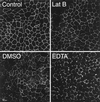
Rapid latrunculin B-induced loss of polarity was not accompanied by a loss of tight junction organization. Confocal optical sections of MDCK monolayers taken of the junctional complex-associated protein ZO-1 at the level of the tight junction in control cells (top left), after treatment with Lat B (top right), DMSO (bottom left), or EDTA (bottom right). All treatments for 30 min at 37°C.
Figure 4
Figure 4
Latrunculin B did not allow free diffusion of lipids across the tight junction. Plasma membrane of MDCK cells labeled with C6-NBD ceramide (green), which was then depleted from the apical surface with a BSA wash. Cells were then incubated in the presence or absence of various drugs for 30 min. The apical surface was labeled just before imaging with wheat germ agglutinin. In control cells (top left) the NBD-ceramide label was largely basolateral and did not colocalize with the apical marker (red arrow). Lat B treatment (top right) resulted in some redistribution to subapical endosomes and there was flattening of the apical domain, but not colocalization of the basolateral and apical markers (red arrow). In contrast DMSO (bottom left) and EDTA (bottom right) caused movement of lipids across the tight junction from basolateral-to-apical domains (green arrows).
Figure 5
Figure 5
Redistribution of plasma membrane receptors requires endocytosis. (A) Comparison of endocytosis-competent and endocytosis-defective receptors. Filter-grown MDCK cells expressing either the LDLR (top row) or a nonendocytosed mutant of the LDLR (LDLR 18YA, bottom row) were fixed and stained for surface receptor with C7 IgG. Both receptors were expressed basolaterally (left column). After Lat B treatment for 30 min (right column), the wild-type receptor was partially redistributed to the apical membrane. In contrast, the mutant receptor was not redistributed by Lat B treatment, except in a few isolated examples (bottom row, right column). (B) Quantitative assay of receptor polarity after Lat B treatment. The polarity of the TfnR, LDLR, and LDLR-18YA was assessed by binding 125I-Tfn, or 125I-C7 IgG to the apical or basolateral surfaces of filter-grown MDCK cells. Background levels were determined from binding to nontransfected cells. Values are derived by summing pairs of wells and expressing to counts bound to each surface as a percentage of the total bound to an apical/basolateral pair. All assays were in triplicate. Lat B treatment for 30 min resulted in depolarization of the TfnR and LDLR but not of LDLR-18YA.
Figure 6
Figure 6
Latrunculin B disrupts endosomal sorting of transferrin. (A) 125I-Tfn bound at 0°C to the basolateral side of MDCK cells was internalized at 37°C and the upper and lower media removed for counting at the intervals shown. In control cells, recycling into the basolateral medium (▪) approached 85% after 1 h, with minimal transcytosis to the apical medium (●). Addition of Lat B resulted in randomization of sorting of recycled Tfn to the basolateral (□) and apical (○) media. (B) 125I-Tfn bound to the apical surface recycled largely back to the apical media (●), although apical-to-basolateral transcytosis accounted for up to 25% of the bound Tfn (▪). Lat B had a lesser effect on apically internalized Tfn than on the basolaterally endocytosed Tfn. Recycling to the apical side (○) decreased to 60% and apical-to-basolateral transcytosis increased to 35% (□). All values are expressed as a percentage of the total cpm initially bound.
Figure 7
Figure 7
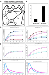
Kinetic modeling of Tfn trafficking and latrunculin B effects. (A) Diagram of possible pathways involved in endocytic traffic from the apical and basolateral surfaces. Kinetic constants are shown for each pathway, values for these constants are given in Table 1. Models including traffic along dashed pathway (k6) are unique to Lat B-treated cells. (B) Predicted ratio of IgA to Tfn in EE and RE after fitting this model to transcytosis and recycling of apically and basolaterally applied Tfn and basolaterally applied IgA in untreated cells. (C) Left, 125I Tfn was bound to the basolateral then internalized. Counts released into the apical and basolateral media are measured at intervals shown. Data points are the same as in Figure 5. The lines are generated from a mathematical kinetic model. Counts are given as a percentage of the initially bound 125I-Tfn. Control basolateral media data: ▪, model: dark blue line. Control apical media data: ●, model: dark red line. Lat B-treated basolateral media data: □, model: light blue line. Lat B-treated apical media data: ○, model: light red line. Right, trafficking of Tfn bound on the apical surface. Data points are the same as in Figure 5. Symbols are the same as in the left panels. (D) Refitting the model to the same data as described above but adding a single extra pathway (k6) to the model for Lat B-treated cells. Only the Lat B treatment data is shown. (E) Predicted passage of basolaterally applied Tfn through the BEE (blue lines) and RE (red lines) in untreated (left) and Lat B-treated cells (right).
Figure 8
Figure 8
Effects of Lat B on endosomal compartments. Top row, AEE and BEE were selectively labeled with 2-min pulses of basolaterally bound Alexa-488-Tfn and apically bound TR-Tfn. After internalization, surface bound material was stripped with acid wash. Untreated cells (A) and Lat B-treated cells (B), aberrant placement of some early endosomes into more apical and perinuclear regions indicated by arrow in B and D. Middle row, BEE and RE remain distinct after Lat B treatment. Basolaterally bound Alexa-488-Tfn internalized for 2 min (BEE) and basolaterally bound TR-Tfn internalized for 30 min (RE). Untreated cells (C) Lat B-treated cells (D). Bottom row, RE targeting is altered by Lat B. Cells expressing FcLR(5-22) (red, apical) and human TfnR (green, basolateral) were labeled to equilibration then chased for 5 min, and the surface acid stripped to remove surface label. This resulted in preferential labeling of the RE from both sides where labels colocalized (E, arrowheads) and lesser labeling of the early endosomes. Lat B treatment did not affect traffic into RE (as in D) but apical and basolaterally applied labels no longer consistently converged in the same RE (F, arrowheads).
Figure 9
Figure 9
Three-dimensional reconstruction of endosome compartments in MDCK cells (supplemental material). MDCK cells were apically labeled to equilibrium with a Texas Red-labeled antibody to the apical FcLR construct (red). They were labeled basolaterally with a 30-min pulse of Alexa-488-Tfn (green). The cells were chilled, the plasma membrane stripped with low pH to remove surface-bound Tfn and antibody, and then fixed. Green BEE and RE are filled with internalized Tfn. Red endosomes are AEE. Yellow endosomes are RE that contain material endocytosed from apical and basolateral surfaces. The reconstruction may be viewed as a QuickTime video included as supplemental material.
Figure 10
Figure 10
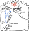
Comparison of endocytic recycling models. The two-compartment model of endocytic recycling pathways used here is illustrated in black. BEEs are portrayed tubulovesicular structures whose tubules extend medially. An additional proposed compartment, the medial common endosome (CE) between the BEE or AEE and the RE is shown with its associated pathways in blue. Another alternative additional compartment, the apical recycling endosome (ARE), between the RE and the apical surface, is shown in red.

Similar articles

Cited by

References

    1. Apodaca G. Endocytic traffic in polarized epithelial cells: role of the actin and microtubule cytoskeleton. Traffic. 2001;2:149–159. - PubMed
    1. Apodaca G, Katz LA, Mostov KE. Receptor-mediatedtranscytosis of IgA in MDCK cells is via apical recycling endosomes. J Cell Biol. 1994;125:67–86. - PMC - PubMed
    1. Balda MS, Matter K. Tight junctions. J Cell Sci. 1998;111:541–547. - PubMed
    1. Bomsel M, Parton R, Kuznetsov SA, Schroer TA, Gruenberg J. Microtubule- and motor-dependent fusion in vitro between apical and basolateral endocytic vesicles from MDCK cells. Cell. 1990;62:719–731. - PubMed
    1. Brown PS, Wang E, Areoti B, Chapin SJ, Mostov KE, Dunn KE. Definition of distinct compartments in polarized Madin-Darby canine kidney (MDCK) cells for membrane-volume sorting, polarized sorting and apical recycling. Traffic. 2000;1:124–140. - PubMed

Publication types

MeSH terms