. 2002 Feb;76(4):1839-55.
doi: 10.1128/jvi.76.4.1839-1855.2002.

Endoplasmic reticulum-Golgi intermediate compartment membranes and vimentin filaments participate in vaccinia virus assembly

Affiliations

Endoplasmic reticulum-Golgi intermediate compartment membranes and vimentin filaments participate in vaccinia virus assembly

Cristina Risco et al. J Virol. 2002 Feb.

Abstract

Vaccinia virus (VV) has a complex morphogenetic pathway whose first steps are poorly characterized. We have studied the early phase of VV assembly, when viral factories and spherical immature viruses (IVs) form in the cytoplasm of the infected cell. After freeze-substitution numerous cellular elements are detected around assembling viruses: membranes, ribosomes, microtubules, filaments, and unidentified structures. A double membrane is clearly resolved in the VV envelope for the first time, and freeze fracture reveals groups of tubules interacting laterally on the surface of the viroplasm foci. These data strongly support the hypothesis of a cellular tubulovesicular compartment, related to the endoplasmic reticulum-Golgi intermediate compartment (ERGIC), as the origin of the first VV envelope. Moreover, the cytoskeletal vimentin intermediate filaments are found around viral factories and inside the viroplasm foci, where vimentin and the VV core protein p39 colocalize in the areas where crescents protrude. Confocal microscopy showed that ERGIC elements and vimentin filaments concentrate in the viral factories. We propose that modified cellular ERGIC membranes and vimentin intermediate filaments act coordinately in the construction of viral factories and the first VV form through a unique mechanism of viral morphogenesis from cellular elements.


Figures
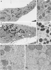
FIG. 1.
FIG. 1.
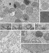
Freeze-substitution and freeze fracture of VV-infected cells. Low-magnification fields of freeze-substituted HeLa cells infected with Western Reserve (WR) VV at 10 (A and B) and 24 (C and D) h p.i. shows the characteristic accumulation of spherical IVs (marked IV) and dense brick-shaped mature viruses (arrowheads). As seen in higher-magnification views of selected areas in panels A and C, many mature viruses are IEVs at shorter p.i. times (B) while most of them are IMVs at longer p.i. times (D). Higher-magnification fields show a significant improvement in preservation of fine details in samples processed by freeze-substitution (E) compared to that with conventional processing (F). In freeze-substituted cells, numerous small structures are seen around the viroplasm foci (marked F) of the viral factories (c marks the viral crescents) and assembling IV particles. Microtubules (MT), cytoskeletal IFs, ribosomes (r), and membranes (m) are abundant around assembling IVs at 10 h p.i. (E). (F) Equivalent regions from conventionally processed cultures show few structural details around assembling IVs. Only some membranes (m) are distinguished. (G) Low-magnification views of freeze-fractured infected cells show that IV particles are surrounded by different types of membranes (arrowheads). Asterisks mark the center of cross-fractured IVs. Mitochondria (mi) are frequently located near IVs. (H) Also at 10 h p.i. IVs are frequently close to RER, with dense deposits on their periphery (arrows). (I) Tubular rigid structures (arrows) of around 50 to 60 nm in diameter are frequently seen around VV particles at 10 h p.i. (J to L) Even when assembly is blocked (during 10 h of infection with the recombinant VVindA17L virus in the absence of IPTG), many structural elements are found near the nucleus (J), around the characteristic dense masses representing truncated viral factories (asterisks). IFs are abundant: the arrowhead in panel K points to a longitudinal view, while a cross-section is marked by the arrowhead in panel L. Large areas containing structured material similar to chromatin (single arrow in panel K), and 30-nm-diameter tubular membranes (double arrows in panel L) are also seen. Double arrowheads in panel L point to a cross-section of the 50- to 60-nm-diameter rigid tubes shown in panel I. N, nucleus. Bars, 1 μm in panels A and C, 200 nm in panels B, D, G, H, and J, and 100 nm in panels E, F, I, K, and L.
FIG. 1.
FIG. 1.
Freeze-substitution and freeze fracture of VV-infected cells. Low-magnification fields of freeze-substituted HeLa cells infected with Western Reserve (WR) VV at 10 (A and B) and 24 (C and D) h p.i. shows the characteristic accumulation of spherical IVs (marked IV) and dense brick-shaped mature viruses (arrowheads). As seen in higher-magnification views of selected areas in panels A and C, many mature viruses are IEVs at shorter p.i. times (B) while most of them are IMVs at longer p.i. times (D). Higher-magnification fields show a significant improvement in preservation of fine details in samples processed by freeze-substitution (E) compared to that with conventional processing (F). In freeze-substituted cells, numerous small structures are seen around the viroplasm foci (marked F) of the viral factories (c marks the viral crescents) and assembling IV particles. Microtubules (MT), cytoskeletal IFs, ribosomes (r), and membranes (m) are abundant around assembling IVs at 10 h p.i. (E). (F) Equivalent regions from conventionally processed cultures show few structural details around assembling IVs. Only some membranes (m) are distinguished. (G) Low-magnification views of freeze-fractured infected cells show that IV particles are surrounded by different types of membranes (arrowheads). Asterisks mark the center of cross-fractured IVs. Mitochondria (mi) are frequently located near IVs. (H) Also at 10 h p.i. IVs are frequently close to RER, with dense deposits on their periphery (arrows). (I) Tubular rigid structures (arrows) of around 50 to 60 nm in diameter are frequently seen around VV particles at 10 h p.i. (J to L) Even when assembly is blocked (during 10 h of infection with the recombinant VVindA17L virus in the absence of IPTG), many structural elements are found near the nucleus (J), around the characteristic dense masses representing truncated viral factories (asterisks). IFs are abundant: the arrowhead in panel K points to a longitudinal view, while a cross-section is marked by the arrowhead in panel L. Large areas containing structured material similar to chromatin (single arrow in panel K), and 30-nm-diameter tubular membranes (double arrows in panel L) are also seen. Double arrowheads in panel L point to a cross-section of the 50- to 60-nm-diameter rigid tubes shown in panel I. N, nucleus. Bars, 1 μm in panels A and C, 200 nm in panels B, D, G, H, and J, and 100 nm in panels E, F, I, K, and L.
FIG. 2.
FIG. 2.
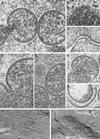
High-magnification fields of freeze-substituted and freeze-fractured samples. (A) Viral crescents have two membranes of 5 nm in thickness, as marked in the image. Depending on the plane of the section, either the three-layer profile or regularly spaced small spikes (arrows) are distinguished in the external membrane, while the internal membrane is always resolved as a typical trilamellar structure. Individual membranes within contiguous cellular double membranes exhibit a similar organization and thickness, for example, in mitochondria (arrows in panel B) or intercellular junctions (arrows in panel C). However, in viroplasm foci from conventionally processed cells (D) the internal membrane of the crescents can be distinguished (arrowhead) but the external bilayer is not preserved. The spikes are seen in some of the crescents (arrows), but the trilamellar profile is lost, probably due to a partial collapse of the structure. (E) When the section plane goes through the surface of the forming IV, a close-packing-like organization for the spikes can be distinguished (asterisk). Both the crescents attached to the viroplasm foci of the factory (c in panel F) and the crescents free in the cytoplasm (c in panel G) have the same structure. (H and I) Freeze fracture also shows the envelope of IVs as double lines in cross-fractured particles (arrows in panel H), like the double membranes of mitochondria (arrows in panel I). Bars, 50 nm.
FIG. 3.
FIG. 3.
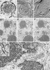
Assembling IVs have vesicles and tubular membranes. (A) Analysis of areas where IVs are forming shows that tubules and vesicles are frequently associated with them (arrows). Arrowheads point to cross-sectioned crescents. Vesicles are frequently seen attached to the pores of uncompleted IVs, as seen in thin sections (arrow in panel B) or after freeze-fracture (arrows in panel C). (D to F) Serial sections of a forming IV. Although in the first section no vesicles or tubules are seen, in the following planes they are associated with the assembling IV. The vesicles are similar to the ends of tubules recruited in the VV assembly areas (arrow in panel G). (H) Groups of tubulovesicular elements are labeled with antibodies specific for VV envelope proteins (here the p21 envelope protein [A17L gene] has been detected) and with the anti-ERGIC-53 antibody (I). (J) The ERGIC tubular elements attach to the surface of the viroplasm foci (asterisk) in a palisade-like arrangement when virus assembly is blocked by infecting cells with VVind A17L in the absence of IPTG. Confocal microscopy shows that viral factories, localized with an antibody against the VV envelope protein p21 (K) and the DNA stain To-Pro (which also stains the cell nucleus) (M), colocalize with ERGIC membranes, detected with an anti-ERGIC-53 monoclonal antibody (L). ERGIC concentrates in intense dots in the region occupied by the factory, as clearly seen in the merge picture (N). Bars, 100 nm.
FIG. 3.
FIG. 3.
Assembling IVs have vesicles and tubular membranes. (A) Analysis of areas where IVs are forming shows that tubules and vesicles are frequently associated with them (arrows). Arrowheads point to cross-sectioned crescents. Vesicles are frequently seen attached to the pores of uncompleted IVs, as seen in thin sections (arrow in panel B) or after freeze-fracture (arrows in panel C). (D to F) Serial sections of a forming IV. Although in the first section no vesicles or tubules are seen, in the following planes they are associated with the assembling IV. The vesicles are similar to the ends of tubules recruited in the VV assembly areas (arrow in panel G). (H) Groups of tubulovesicular elements are labeled with antibodies specific for VV envelope proteins (here the p21 envelope protein [A17L gene] has been detected) and with the anti-ERGIC-53 antibody (I). (J) The ERGIC tubular elements attach to the surface of the viroplasm foci (asterisk) in a palisade-like arrangement when virus assembly is blocked by infecting cells with VVind A17L in the absence of IPTG. Confocal microscopy shows that viral factories, localized with an antibody against the VV envelope protein p21 (K) and the DNA stain To-Pro (which also stains the cell nucleus) (M), colocalize with ERGIC membranes, detected with an anti-ERGIC-53 monoclonal antibody (L). ERGIC concentrates in intense dots in the region occupied by the factory, as clearly seen in the merge picture (N). Bars, 100 nm.
FIG. 4.
FIG. 4.
Redefinition of other VV-related structures. (A) The recombinant VVindA14L forms aberrant IV-like particles in the absence of p15 protein. Conventional processing shows that the envelope of these particles contains individual membranous pieces unable to complete the IV sphere. Some of these pieces are thicker (arrowhead) than the normal crescents (arrows). Freeze-substitution and freeze fracture show that the mentioned thicker pieces are indeed tubes of 30 to 40 nm in diameter (arrowheads in panels B and C). (D to F) Serial sections of the viroplasm foci formed by this virus show that IV-like particles have crescents and curved tubes in many different orientations. (G) Some of these tubular pieces (arrowhead) are connected with pieces of crescents with spikes (arrows). (H) RBs, the truncated viroplasm foci formed in cells infected with VV in the presence of the drug rifampin, exhibit different types of membranes on their periphery: dense membranes of around 18 nm (arrows), twisted dense membranes with vesicular heads (arrowheads), and less dense 30-nm-diameter tubules, some of them with vesicular ends (double arrowhead). Single, 5-nm-thick membrane units are not detected on the surface of RBs. Bars, 50 nm in panels A, B, C, G, and H and 200 nm in panels D, E, and F.
FIG. 5.
FIG. 5.
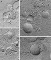
Freeze-fracture and freeze-etching of cells infected with VVindA17L (A and B), VVindA14L (C), or WR VV (D) show linear pieces interacting laterally on the surface of viroplasm foci. These pieces belong to two categories: occasional 30- to 40-nm-thick structures (arrow in panel A) or frequent 15- to 20-nm-thick structures (arrows in panels B and C). Forming IVs have short linear arrays of particles on their external surface (arrowheads in panel B, arrows in panel D), while their internal surfaces do not have a defined pattern (asterisks in panels A and B). Images in panels A and D correspond to freeze-fractured samples, while panels B and C are replicas of freeze-etched samples. F, viroplasm foci of the viral factory; c, viral crescent. Bars, 100 nm.
FIG. 6.
FIG. 6.
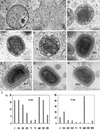
Different viral forms detected in VV-infected HeLa cells as processed by freeze-substitution. (A) IV particle packing DNA (arrow). These particles are frequently surrounded by a structured material (asterisk) similar to cellular chromatin. The open pore in the IV particle frequently exhibits a vesicle nearby (arrowhead). (B) Spherical dense particles with fibrous, DNA-like, internal material (arrows). (C and D) Potential intermediate maturation stages in the construction of the internal viral core (arrows) and the IMV. These transitional forms still have the IV envelope (arrowhead in panel D) around the forming core shell. (E) IMV shows a complex organization, with at least five differentiated layers (marked with short lines). (F) IEV with the additional double membrane (arrows). (G and H) Two different section planes of EEV, which have an external fuzzy coat (arrows). (I) Quantification of the relative amounts of the different WR VV assemblies (expressed as percentage of the total population of viral structures) from thin sections of VV-infected HeLa cells at two different p.i. times (10 and 24 h). The structures quantified were named as follows: C, individual viral crescents; IV1, incomplete immature viruses; IV2, apparently completed IVs (closed spheres); IV3, IVs with DNA spot; T1, transitional stage shown in panel B; T2, transitional stages shown in panels C and D. More than 1,000 viral particles were included in the quantification. Bars, 100 nm.
FIG. 7.
FIG. 7.
Identification of filaments recruited around viroplasm foci and IVs in cells infected with VVindA17L (A and I to K) or WR VV (B to H). (A) ERGIC membranes recruited around viroplasm foci (F) are accompanied by filaments (arrows) whose thickness (around 10 nm) corresponds to that of the cytoskeletal IFs. (B) Immunogold shows that these filaments react with antibodies specific for the IF protein vimentin. (C to F) Confocal microscopy shows that vimentin filaments are placed around viral factories, maintaining a close contact with them. In HeLa cells infected for 8 h with WR VV, viral factories were localized with an antibody specific for the VV envelope protein p21 (C) and the DNA stain To-Pro (which also stains the cell nucleus) (E). Detection of vimentin with a specific monoclonal antibody shows its concentration in a perinuclear area (D), coincident with the localization of viral factories. Vimentin filaments appear to enclose the VV factories, as can be observed in the merge picture (F). At the ultrastructural level, labeled vimentin filaments are sometimes seen entering the forming IVs (asterisk in panel G). (H) Labeling concentrates in small viroplasm foci (arrowheads) and inside IVs, while mature viruses (IMVs) are devoid of labeling. (I) In large viroplasm foci formed in HeLa cells infected with VVindA17L at long p.i. times (18 h p.i.), vimentin concentrates in the areas where crescents (c) protrude. In these areas, a VV core protein (p39, the product of the A5L gene) colocalizes with vimentin, as shown by double-labeling experiments. (J and K). The small 5-nm gold particles are associated to vimentin, while 10-nm gold particles are localized the VV core protein p39. Bars, 200 nm in panels A and H and 100 nm in panels B, G, I, J, and K.
FIG. 7.
FIG. 7.
Identification of filaments recruited around viroplasm foci and IVs in cells infected with VVindA17L (A and I to K) or WR VV (B to H). (A) ERGIC membranes recruited around viroplasm foci (F) are accompanied by filaments (arrows) whose thickness (around 10 nm) corresponds to that of the cytoskeletal IFs. (B) Immunogold shows that these filaments react with antibodies specific for the IF protein vimentin. (C to F) Confocal microscopy shows that vimentin filaments are placed around viral factories, maintaining a close contact with them. In HeLa cells infected for 8 h with WR VV, viral factories were localized with an antibody specific for the VV envelope protein p21 (C) and the DNA stain To-Pro (which also stains the cell nucleus) (E). Detection of vimentin with a specific monoclonal antibody shows its concentration in a perinuclear area (D), coincident with the localization of viral factories. Vimentin filaments appear to enclose the VV factories, as can be observed in the merge picture (F). At the ultrastructural level, labeled vimentin filaments are sometimes seen entering the forming IVs (asterisk in panel G). (H) Labeling concentrates in small viroplasm foci (arrowheads) and inside IVs, while mature viruses (IMVs) are devoid of labeling. (I) In large viroplasm foci formed in HeLa cells infected with VVindA17L at long p.i. times (18 h p.i.), vimentin concentrates in the areas where crescents (c) protrude. In these areas, a VV core protein (p39, the product of the A5L gene) colocalizes with vimentin, as shown by double-labeling experiments. (J and K). The small 5-nm gold particles are associated to vimentin, while 10-nm gold particles are localized the VV core protein p39. Bars, 200 nm in panels A and H and 100 nm in panels B, G, I, J, and K.
FIG. 8.
FIG. 8.
Hypothetical steps for the assembly of the first VV form, IV. (Step 1) The ERGIC tubulovesicular structures get deeply modified by the insertion of VV proteins coming from the RER, such as p21 and p15 (encoded by A17L and A14L VV genes, respectively) and the proteins that form the spikes (as-yet unidentified). (Step 2) Modified membranous pieces reach the periphery of viral factories, together with vimentin IFs. (Step 3) These membranes attach to each other on the surface of the viroplasm foci and form the crescents. IFs would participate in the egress of the crescents and the incorporation of VV proteins inside the IVs. (Step 4) The individual pieces originate spherical structures with open pores. DNA packaging would take place through these pores. (Step 5) By unknown mechanisms (lateral fusion of membranous pieces or attachment without fusion?) the IV spheres would finally be sealed.

Similar articles

Cited by

References

    1. Appenzeller, C., H. Andersson, F. Kappeler, and H.-P. Hauri. 1999. The lectin ERGIC-53 is a cargo transport receptor for glycoproteins. Nat. Cell Biol. 1:330-334. - PubMed
    1. Aridor, M., K. N. Fish, S. Bannykh, J. Weissman, T. H. Roberts, J. Lippincott-Schwartz, and W. E. Balch. 2001. The Sar1 GTPase coordinates biosynthetic cargo selection with endoplasmic reticulum export site assembly. J. Cell Biol. 152:213-229. - PMC - PubMed
    1. Baker, T. S., N. H. Olson, and S. D. Fuller. 1999. Adding the third dimension to virus life cycles: three-dimensional reconstruction of icosahedral viruses from cryoelectron micrographs. Microbiol. Mol. Biol. Rev. 63:862-922. - PMC - PubMed
    1. Baumeister, W., and A. C. Steven. 2000. Macromolecular electron microscopy in the era of structural genomics. Trends Biochem. Sci. 25:624-631. - PubMed
    1. Betakova, T., E. J. Wolffe, and B. Moss. 1999. Regulation of vaccinia virus morphogenesis: phosphorylation of the A14L and A17L membrane proteins and C-terminal truncation of the A17L protein are dependent on the F10L kinase. J. Virol. 73:3534-3543. - PMC - PubMed

Publication types