. 2002 Jan 15;16(2):222-34.
doi: 10.1101/gad.214202.

Intracellular trafficking by Star regulates cleavage of the Drosophila EGF receptor ligand Spitz

Affiliations

Intracellular trafficking by Star regulates cleavage of the Drosophila EGF receptor ligand Spitz

Rachel Tsruya et al. Genes Dev. .

Abstract

Spitz (Spi) is a TGFalpha homolog that is a cardinal ligand for the Drosophila EGF receptor throughout development. Cleavage of the ubiquitously expressed transmembrane form of Spi (mSpi) precedes EGF receptor activation. We show that the Star and Rhomboid (Rho) proteins are necessary for Spi cleavage in Drosophila cells. Complexes between the Spi and Star proteins, as well as between the Star and Rho proteins were identified, but no Spi-Star-Rho triple complex was detected. This observation suggests a sequential activity of Star and Rho in mSpi processing. The interactions between Spi and Star regulate the intracellular trafficking of Spi. The Spi precursor is retained in the periphery of the nucleus. Coexpression of Star promotes translocation of Spi to a compartment where Rho is present both in cells and in embryos. A Star deletion construct that maintains binding to Spi and Rho, but is unable to facilitate Spi translocation, lost biological activity. These results underscore the importance of regulated intracellular trafficking in processing of a TGFalpha family ligand.


Figures
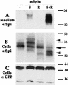
Figure 1
Figure 1
Star and Rho are required for mSpi processing in S2 cells. Schneider S2 cells were transiently transfected with combinations of different expression constructs. mSpi alone did not undergo cleavage, coexpression with Star gave rise to low levels of sSpi in the medium, whereas coexpression with Star and Rho generated high levels of sSpi. Note that in the cell lysates higher molecular weight forms of mSpi were observed in the mSpi+Star cells (arrowhead), whereas lower molecular weight forms were detected in cells expressing mSpi+Star+Rho (open arrowhead), and the levels of original mSpi (arrow) were reduced. (A) Medium probed with anti-Spi. (B) Cell lysates probed with anti-Spi. (C) Cell lysates probed with anti-GFP, to detect cotransfected UAS-CD8–GFP show similar transfection efficiencies. In this experiment we used mSpi, Star-HA (S), and Rho-HA (R).
Figure 2
Figure 2
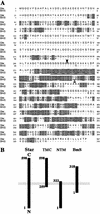
B. mori Star homolog and Star deletion constructs. (A) Alignment of the BmS and Star protein sequences. Note the shorter amino-terminal domain of BmS, and the homology to Star at the carboxyl terminus. The transmembrane domain is underlined, and the amino or carboxyl terminus of TMC or NTM are shown by filled or open arrowheads, respectively. (B) Schematic representation of the Star deletion constructs and BmS. The carboxy-terminal domain is extracellular, as Star and BmS are type II transmembrane proteins (BmS GenBank accession no. AF455272).
Figure 3
Figure 3
Biological activity of Star deletion constructs and BmS. (A) The capacity of Star constructs to promote mSpi cleavage in S2 cells was tested alone or in combination with Rho. The medium was probed with anti-Spi. sSpi was detected only in the medium of cells transfected with mSpi, TMC, and Rho. Thus, only TMC showed biological activity, which, however, is lower than that of full-length Star. (B) The activity of the constructs was also tested in transgenic flies, for their capacity to rescue Star haplo-insufficiency in the eye, and induce ectopic eye and wing phenotypes. The NTM and BmS did not show any activity (data not shown), whereas the TMC construct displayed a weak activity and was thus capable of rescuing the haplo-insufficiency, but did not elicit ectopic phenotypes. (a) wild-type; (b) Star −/+; (c) Star −/+; GMR-Gal4/UAS-TMC; (d) Star −/+; GMR-Gal4/UAS-Star; (e) MS1096-Gal4; (f) MS1096-Gal4/UAS-Star; (g) MS1096-Gal4/UAS-TMC. (Arrow) Ectopic wing veins. The TMC construct normally gave rise to wild-type wings and only rarely to small extra veins.
Figure 4
Figure 4
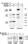
mSpi–Star protein interactions. (A) mSpi-FLAG was expressed in S2 cells with Star–HA and CD8–GFP. Immunoprecipitation of mSpi–FLAG coprecipitated Star–HA (detected by anti-HA), but not CD8–GFP (detected by anti-GFP). The presence of mSpi–GFP was verified by anti-Spi. (B) In the reciprocal experiment, mSpi–GFP was expressed with Star–TAP. Immunoprecipitation of Star–TAP led to coprecipitation of mSpi.
Figure 5
Figure 5
Dissection of mSpi–Star interaction domains. (A) The capacity of the Star constructs to interact with mSpi was analyzed by coexpression of mSpi–FLAG, immunoprecipitation of mSpi–FLAG, and monitoring of coprecipitation of Star. All constructs tested retained the binding to mSpi. (B) The domains of mSpi required for interaction with Star were dissected by expression of Star–TAP together with Spi deletion constructs fused to GFP. Following immunoprecipitation of Star–TAP, coprecipitation of Spi was tested by anti-GFP. The EGF domain of Spi was shown to be essential for binding to Star.
Figure 6
Figure 6
Star–Rho protein interactions. Interactions between Star and Rho were demonstrated by expression of Rho–TAP and Star–HA, immunoprecipitation of Rho–TAP, and detection of coprecipitating Star–HA. Examination of Star deletion constructs showed that NTM–HA and BmS–HA retained strong binding to Rho, whereas TMC–HA showed weak, borderline interaction. The cytoplasmic domain of Star is thus mediating the interaction with Rho.
Figure 7
Figure 7
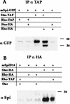
Sequential activity of Star and Rho in mSpi processing. The presence of mSpi–Star–Rho triple complexes was examined. (A) Rho–TAP is unable to precipitate detectable levels of mSpi. It was thus possible to ask whether coexpression of Star, which forms mSpi–Star and Star–Rho complexes, can promote the formation of a triple complex and allow Rho–TAP to coimmunoprecipitate mSpi–GFP, as detected by anti-GFP. The results were negative. Coprecipitation of mSpi–GFP with Star–TAP was also detected by anti-GFP, in the presence or absence of Rho. (B) To examine the possibility of a short-lived triple complex due to mSpi cleavage, a construct of mSpi devoid of a site necessary for cleavage was used (mSpiΔ16) and its coprecipitation was monitored by anti-Spi. Rho–HA or Star–HA were immunoprecipitated by anti-HA. Again, no triple complex was detected, arguing against the presence of a short-lived triple complex.
Figure 8
Figure 8
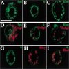
Intracellular localization of mSpi, Star, and Rho. Localization was monitored in live cells following transfection (using GFP), or in fixed cells following antibody staining. (A,D) mSpi–GFP shows a predominantly peripheral nuclear distribution (green), that does not colocalize with the Golgi marker (red); (B,E) Similarly, Star–HA shows a peripheral nuclear distribution (green) with no Golgi colocalization; (C,F) Rho–HA (green) is found in a punctate distribution, which is mostly nonoverlapping with the Golgi. (GI) Double staining of Star and Rho–HA (anti-Star green, anti-HA red) shows the distinct distributions with minimal overlap. Scale bar, 20 μm.
Figure 9
Figure 9
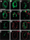
Star translocates mSpi from the ER in S2 cells. The effects of Star and Rho on mSpi intracellular distribution were examined in S2 cells. (A) mSpi–GFP showed a peripheral nuclear distribution. (B) mSpiΔIC–GFP lacking the intracellular domain was not retained and predominantly localized to plaques. (C) Following cotransfection with Star, the peripheral nuclear mSpi disappeared, and instead a punctate distribution was observed (mSpi–GFP, green, Star–HA, red). (D) mSpi–GFP showed only limited colocalization with lysosomes. (E) Similarly, the punctate distribution in mSpi–GFP+Star showed only some overlap with lysosomes (red), or endosomes (red) (F). (GI) Coexpression of mSpi–GFP and Rho did not alter the distribution of mSpi or Rho (mSpi–GFP green, Rho–HA red). (JL) Coexpression of mSpi–GFP, Star, and Rho–HA gave rise to a punctate mSpi–GFP distribution that was weaker in intensity than the transfections of mSpi+Star, because of a high level of cleavage and secretion. The mSpi–GFP vesicles predominantly colocalized with the Rho staining, indicating that Star targeted mSpi to a compartment containing Rho and consistent with sequential activity (mSpi–GFP, green; Rho–HA, red). Bar, 20 μm.
Figure 10
Figure 10
Star translocates mSpi from the ER in embryos. The effect of Star on mSpi–GFP localization was tested in embryos. (A) actin–Gal4/UAS-mSpi–GFP embryos show a peripheral nuclear localization. (B) Merge with anti-Lamin (red) marking the nuclear membrane. (C) actin–Gal4/UAS-mSpi–GFP/UAS–Star embryos show only residual peripheral nuclear Spi, and primarily a punctate localization. (D) Injection of UAS-mSpi–GFP into actin–Gal4 cyncytial embryos gave rise at stage 10 to a peripheral nuclear distribution. (E) Injection of UAS-mSpi–GFP and UAS-Star into prd–Gal4 embryos gave rise to the punctate distribution of mSpi–GFP. Similar results were obtained when UAS-mSpi–GFP was injected into prd–Gal4/UAS-Star embryos (data not shown). (F) Injection of UAS-mSpi–GFP+UAS-rho into prd–Gal4/UAS–Star embryos retained the punctate distribution of mSpi–GFP, but the intensity was lower, presumably due to secretion. Bar, 20 μm.
Figure 11
Figure 11
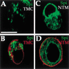
Biological activity of Star correlates with mSpi trafficking. (A,B) The TMC construct, which is biologically active, albeit less than full-length Star, retained the ability to translocate mSpi–GFP from the ER (mSpi–GFP, green, TMC–HA, red). (C,D) The NTM construct, which binds mSpi but is not biologically active, failed to alter the peripheral nuclear distribution of mSpi (mSpi–GFP, green, NTM–HA, red). Interestingly, instead of the peripheral nuclear distribution of Star and TMC, NTM was found weakly in vesicles and predominantly on the plasma membrane, suggesting that the cellular distribution of Star itself is regulated and important for mSpi shuttling. Bar, 20 μm.
Figure 12
Figure 12
Scheme for the sequential activities of Star and Rho in mSpi processing. mSpi is retained in a peripheral nuclear distribution, consistent with the ER. Expression of Star promotes binding to mSpi and shuttling to a compartment that does not colocalize with the Golgi and is possibly the trans Golgi network (TGN). In the absence of Rho, efficient cleavage does not take place. When mSpi, Star and Rho are expressed, mSpi is shuttled by Star to a TGN compartment containing Rho, thus leading to efficient cleavage and secretion of Spi.

Similar articles

Cited by

References

    1. Bang AG, Kintner C. Rhomboid and Star facilitate presentation and processing of the Drosophila TGFα homolog Spitz. Genes & Dev. 2000;14:177–186. - PMC - PubMed
    1. Bier E, Jan LY, Jan YN. rhomboid, a gene required for dorsoventral axis establishment and peripheral nervous system development in Drosophila melanogaster. Genes & Dev. 1990;4:190–203. - PubMed
    1. Chen C, Okayama H. High-efficiency transformation of mammalian cells by plasmid DNA. Mol Cell Biol. 1987;7:2745–2752. - PMC - PubMed
    1. Eggenschwiler JT, Espinoza E, Anderson KV. Rab23 is an essential negative regulator of the mouse Sonic hedgehog signalling pathway. Nature. 2001;412:194–198. - PubMed
    1. Ellgaard L, Molinari M, Helenius A. Setting the standards: Quality control in the secretory pathway. Science. 1999;286:1882–1888. - PubMed

Publication types

LinkOut - more resources