Phosphatidylserine-dependent ingestion of apoptotic cells promotes TGF-beta1 secretion and the resolution of inflammation
- PMID: 11781349
- PMCID: PMC150814
- DOI: 10.1172/JCI11638
Phosphatidylserine-dependent ingestion of apoptotic cells promotes TGF-beta1 secretion and the resolution of inflammation
Abstract
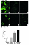
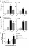
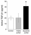
Ingestion of apoptotic cells in vitro by macrophages induces TGF-beta1 secretion, resulting in an anti-inflammatory effect and suppression of proinflammatory mediators. Here, we show in vivo that direct instillation of apoptotic cells enhanced the resolution of acute inflammation. This enhancement appeared to require phosphatidylserine (PS) on the apoptotic cells and local induction of TGF-beta1. Working with thioglycollate-stimulated peritonea or LPS-stimulated lungs, we examined the effect of apoptotic cell uptake on TGF-beta1 induction. Viable or opsonized apoptotic human Jurkat T cells, or apoptotic PLB-985 cells, human monomyelocytes that do not express PS during apoptosis, failed to induce TGF-beta1. PS liposomes, or PS directly transferred onto the PLB-985 surface membranes, restored the TGF-beta1 induction. Apoptotic cell instillation into LPS-stimulated lungs reduced proinflammatory chemokine levels in the bronchoalveolar lavage fluid (BALF). Additionally, total inflammatory cell counts in the BALF were markedly reduced 1-5 days after apoptotic cell instillation, an effect that could be reversed by opsonization or coinstillation of TGF-beta1 neutralizing antibody. This reduction resulted from early decrease in neutrophils and later decreases in lymphocytes and macrophages. In conclusion, apoptotic cell recognition and clearance, via exposure of PS and ligation of its receptor, induce TGF-beta1 secretion, resulting in accelerated resolution of inflammation.
Figures


Similar articles
-
Transcriptional and translational regulation of inflammatory mediator production by endogenous TGF-beta in macrophages that have ingested apoptotic cells.J Immunol. 1999 Dec 1;163(11):6164-72. J Immunol. 1999. PMID: 10570307
-
Particle digestibility is required for induction of the phosphatidylserine recognition mechanism used by murine macrophages to phagocytose apoptotic cells.J Immunol. 1993 Oct 15;151(8):4274-85. J Immunol. 1993. PMID: 8409401
-
Vitamin E inhibits anti-Fas-induced phosphatidylserine oxidation but does not affect its externalization during apoptosis in Jurkat T cells and their phagocytosis by J774A.1 macrophages.Antioxid Redox Signal. 2004 Apr;6(2):227-36. doi: 10.1089/152308604322899297. Antioxid Redox Signal. 2004. PMID: 15025924
-
The central role of phosphatidylserine in the phagocytosis of apoptotic thymocytes.Ann N Y Acad Sci. 2000;926:217-25. doi: 10.1111/j.1749-6632.2000.tb05614.x. Ann N Y Acad Sci. 2000. PMID: 11193037 Review.
-
An Apoptotic 'Eat Me' Signal: Phosphatidylserine Exposure.Trends Cell Biol. 2015 Nov;25(11):639-650. doi: 10.1016/j.tcb.2015.08.003. Epub 2015 Oct 1. Trends Cell Biol. 2015. PMID: 26437594 Review.
Cited by
-
Phagocytic clearance of apoptotic, necrotic, necroptotic and pyroptotic cells.Biochem Soc Trans. 2021 Apr 30;49(2):793-804. doi: 10.1042/BST20200696. Biochem Soc Trans. 2021. PMID: 33843978 Free PMC article. Review.
-
Host susceptibility to gram-negative pneumonia after lung contusion.J Trauma Acute Care Surg. 2012 Mar;72(3):614-22; discussion 622-3. doi: 10.1097/TA.0b013e318243d9b1. J Trauma Acute Care Surg. 2012. PMID: 22491544 Free PMC article.
-
Dying cell clearance and its impact on the outcome of tumor radiotherapy.Front Oncol. 2012 Sep 11;2:116. doi: 10.3389/fonc.2012.00116. eCollection 2012. Front Oncol. 2012. PMID: 22973558 Free PMC article.
-
A NET Outcome.Front Immunol. 2012 Dec 5;3:365. doi: 10.3389/fimmu.2012.00365. eCollection 2012. Front Immunol. 2012. PMID: 23227026 Free PMC article.
-
Phosphatidylserine exposure by Toxoplasma gondii is fundamental to balance the immune response granting survival of the parasite and of the host.PLoS One. 2011;6(11):e27867. doi: 10.1371/journal.pone.0027867. Epub 2011 Nov 29. PLoS One. 2011. PMID: 22140476 Free PMC article.
References
-
- Fadok VA, et al. Exposure of phosphatidylserine on the surface of apoptotic lymphocytes triggers specific recognition and removal by macrophages. J Immunol. 1992; 148:2207–2216. - PubMed
-
- Frasch SC, et al. Regulation of phospholipid scramblase activity during apoptosis and cell activation by protein kinase Cdelta. J Biol Chem. 2000; 275:23065–23073. - PubMed
-
- Bratton DL, et al. Appearance of phosphatidylserine on apoptotic cells requires calcium-mediated nonspecific flip-flop and is enhanced by loss of the aminophospholipid translocase. J Biol Chem. 1997; 272:26159–26165. - PubMed
-
- Fadok VA, et al. A receptor for phosphatidylserine-specific clearance of apoptotic cells. Nature. 2000; 405:85–90. - PubMed
-
- Savill J. Apoptosis in resolution of inflammation. Kidney Blood Press Res. 2000; 23:173–174. - PubMed
Publication types
MeSH terms
Substances
Grants and funding
LinkOut - more resources
Full Text Sources
Other Literature Sources

