. 2001 Dec;13(12):2823-39.
doi: 10.1105/tpc.010202.

The chloroplastic GrpE homolog of Chlamydomonas: two isoforms generated by differential splicing

Affiliations

The chloroplastic GrpE homolog of Chlamydomonas: two isoforms generated by differential splicing

M Schroda et al. Plant Cell. 2001 Dec.

Abstract

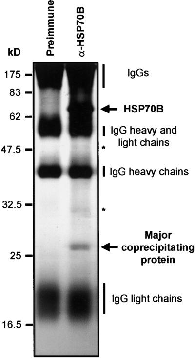
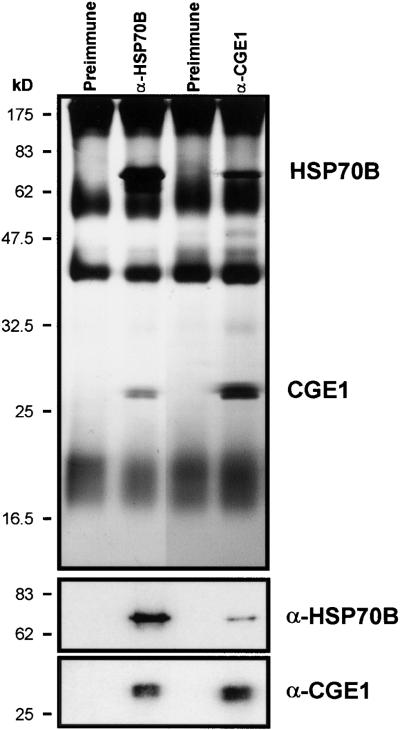
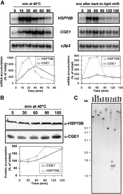
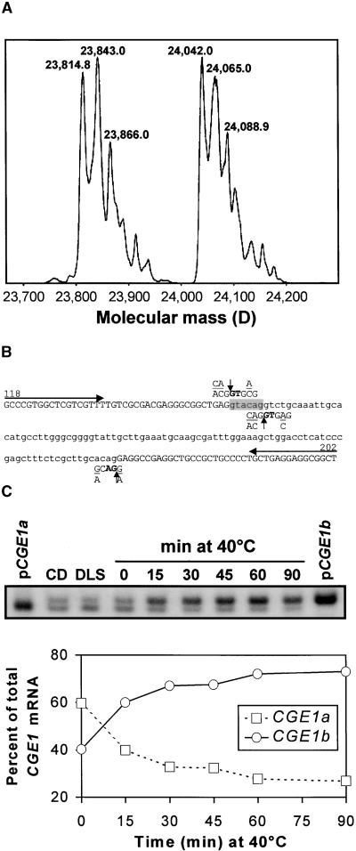
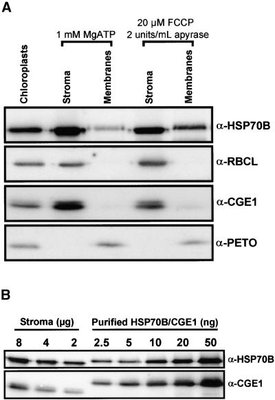
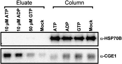
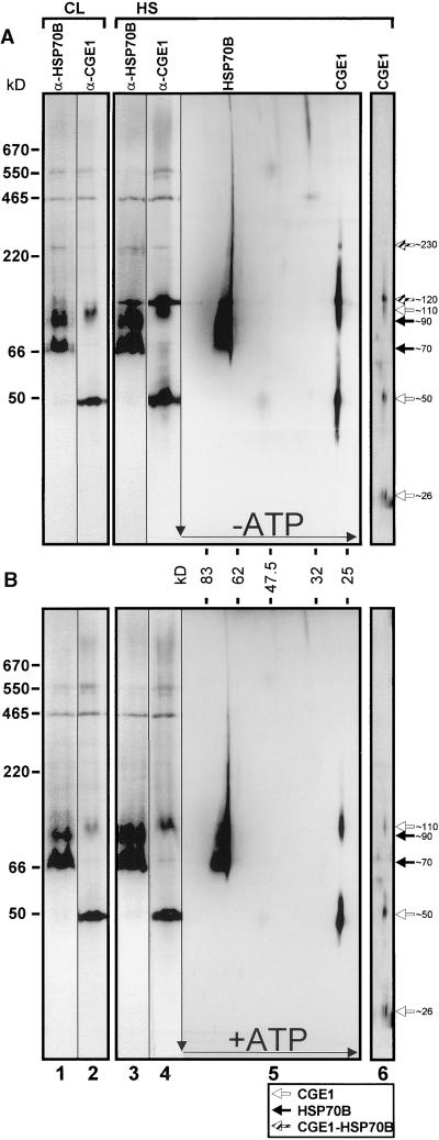
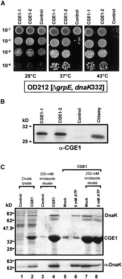
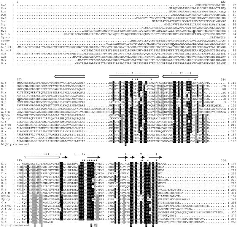
In eubacteria and mitochondria, Hsp70 chaperone activity is controlled by the nucleotide exchange factor GrpE. We have identified the chloroplastic GrpE homolog of Chlamydomonas, CGE1, as an approximately 26-kD protein coimmunoprecipitating with the stromal HSP70B protein. When expressed in Escherichia coli, CGE1 can functionally replace GrpE and interacts physically with DnaK. CGE1 is encoded by a single-copy gene that is induced strongly by heat shock and slightly by light. Alternative splicing generates two isoforms that differ only by two residues in the N-terminal part. The larger form is synthesized preferentially during heat shock, whereas the smaller one dominates at lower temperatures. Fractions of both HSP70B and CGE1 associate with chloroplast membranes in an ATP-sensitive manner. By colorless native PAGE and pulse labeling, CGE1 monomers were found to assemble rapidly into dimers and tetramers. In addition, CGE1 was found to form ATP-sensitive complexes with HSP70B of approximately 230 and approximately 120 kD, the latter increasing dramatically after heat shock.


Figures

Figure 1.
Figure 1.
Immunoprecipitation of HSP70B. Chlamydomonas chloroplasts were isolated and treated for 10 min with the uncoupler FCCP (20 μM) before lysis and with apyrase (2 units/mL) after lysis to eliminate endogenous ATP. The soluble stroma fraction was incubated with protein A–Sepharose coupled to antibodies of either preimmune serum or anti-HSP70B serum (α-HSP70B). Precipitated proteins were separated on an SDS–12% polyacrylamide gel and visualized by silver staining. Minor proteins of ∼32 and ∼48 kD coprecipitating with HSP70B are indicated by asterisks.
Figure 2.
Figure 2.
Nucleotide Sequence of the cDNA and Deduced Amino Acid Sequence of a Chloroplast-Targeted GrpE Homolog. The ATG start codon, the TAA stop codon, and the putative polyadenylation signal TGAAA are shown in boldface, and the stop codon is indicated by an asterisk. Underlined sequences are an N-terminal sequence and an internal peptide sequence generated by microsequencing. The sequence within the triangles is a 6-bp insertion giving rise to an additional valine (V) and glutamine (Q) residue found in one of the ESTs. The arrow indicates the cleavage site of the chloroplast targeting signal.
Figure 3.
Figure 3.
Coimmunoprecipitation of CGE1 and HSP70B. ATP-depleted stromal fractions were incubated with antibodies against HSP70B and CGE1 (α-HSP70B and α-CGE1) coupled to protein A–Sepharose, as described in Figure 1. Immunoprecipitated proteins were separated on an SDS–12% polyacrylamide gel and visualized by silver staining (top gel) or blotted to nitrocellulose membranes (bottom gels). Membranes were immunodecorated with antiserum against HSP70B or CGE1, incubated with 125I-protein A, and exposed to a Phosphorimager screen.
Figure 4.
Figure 4.
Induction Pattern of HSP70B and CGE1 mRNA and Protein and Analysis of the CGE1 Gene Copy Number. (A) HSP70B and CGE1 mRNA accumulation after a shift of cells from 25 to 40°C (left gels) and after a shift of cells grown for 15 hr in the dark to light of 20 μE·m−2·sec−1 (right gels). RNA gel blots were hybridized with probes made of the coding regions of HSP70B, CGE1, and the entire cDNA of the Chlamydomonas β-like protein 2 (cβlp2). Signals were quantified by phosphorimaging using cβlp2 as a loading control. (B) HSP70B and CGE1 protein accumulation after a shift of cells from 25 to 40°C. Immunoblots were incubated with 125I-protein A, and signals were quantified by phosphorimaging. (C) DNA gel blot analysis of Chlamydomonas total DNA digested with the restriction enzymes indicated. The blot was hybridized with a probe made of the coding region of CGE1.
Figure 5.
Figure 5.
Analysis of the Nature of the Two CGE1 Isoforms. (A) Mass determination of CGE1 by ESI-MS. Chlamydomonas total soluble proteins were incubated with anti-CGE1 antibodies coupled to protein A–Sepharose. Immunoprecipitated CGE1 was separated on an SDS–15% polyacrylamide gel, blotted to a PVDF membrane, eluted, and analyzed by ESI-MS. (B) Sequence of an intron (lowercase) and parts of the flanking exons (uppercase) of the CGE1 gene. The sequence shown is situated between base 118 and base 202 of the CGE1 cDNA of Figure 2. (C/A)(A/C)G↓GTG(A/C)G and (G/A)CAG↓(G/A) are the Chlamydomonas consensus sequences for the 5′ and 3′ splice sites, respectively. The 6-bp sequence shaded in gray is present only in a fraction of CGE1 mRNAs. Arrows indicate the primers used for the reverse transcriptase–mediated PCR shown in (C). (C) Reverse transcriptase–mediated PCR analysis of the two CGE1 mRNAs. Total RNA was isolated from Chlamydomonas cells kept for 15 hr in the dark (CD), shifted for 90 min from dark to light of 20 μE·m−2·sec−1 (DLS), grown at 25°C under continuous light, or heat shocked for 40 min at 40°C. Reverse transcribed mRNA, or plasmid DNA containing cloned CGE1a or CGE1b cDNAs, was used as a template for a PCR including 33P-dATP and the primers depicted in (B). The PCR products of 85 and 79 bp were separated on a Tris-borate/EDTA (TBE)–12% polyacrylamide gel, which was dried and used for phosphorimaging.
Figure 6.
Figure 6.
Determination of the Suborganellar Localization and the Concentration of HSP70B and CGE1. (A) Fractionation of chloroplasts into stroma and membranes. Chlamydomonas chloroplasts were isolated to high purity, and one-half was incubated with 20 μM FCCP. Chloroplasts were lysed in the presence of 2 units/mL apyrase or 1 mM ATP and separated into stroma and membranes. Proteins were separated on an SDS–7.5 to 15% polyacrylamide gradient gel and blotted to a nitrocellulose membrane, which was immunodecorated with antiserum (α) against HSP70B, CGE1, the large subunit of Rubisco (RBCL), or the PETO protein of the cytochrome b6f complex. Immunoblots were incubated with 125I-protein A, and signals were quantified by phosphor–imaging. (B) Determination of HSP70B and CGE1 concentrations in the stroma. Eight, 4, and 2 μg of ATP-repleted stroma prepared as described in (A) were separated on an SDS–7.5 to 15% polyacrylamide gradient gel next to the indicated amounts of double hexahistidine-tagged HSP70B and CGE1 purified from overexpressing E. coli strains and blotted to nitrocellulose. The membrane was immunodecorated with antiserum (α) against HSP70B or CGE1 and incubated with 125I-protein A, and signals were quantified by phosphorimaging.
Figure 7.
Figure 7.
Effect of Nucleotides on the Dissociation of the HSP70B-CGE1 Complex. HSP70B-CGE1 complexes were immunoprecipitated from an ATP-depleted stromal fraction with HSP70B antibodies coupled to protein A–Sepharose as described in Figure 1 and, after washing, divided into four equal fractions. The fractions were incubated with elution buffer containing 10 μM ATP, 10 μM ADP, 50 μM GTP, or no nucleotide (mock) for 15 min at 24°C. Eluted proteins (eluate) and those remaining bound to the resin (column) were separated on an SDS–12% polyacrylamide gel and blotted to nitrocellulose. Membranes were immunodecorated with antiserum (α) against HSP70B or CGE1 and incubated with 125I-protein A, and signals were quantified by phosphorimaging.
Figure 8.
Figure 8.
Analysis of Complexes Involving HSP70B and CGE1 by Two-Dimensional Gel Electrophoresis. A Chlamydomonas culture was split into two fractions, one of which was left at 25°C in continuous light (CL; lanes 1 and 2), the other heat-shocked at 40°C for 55 min (HS; lanes 3 to 6). One-half of each fraction was supplemented with 20 μM FCCP and 2 units/mL apyrase (A), and the other half was supplemented with 1 mM Mg-ATP (B). To the heat-shocked fraction, 100 μg/mL chloramphenicol was added 30 min after the temperature shift, and a 15-min pulse labeling was performed by adding 10 μCi/mL 14C-acetate 40 min after the temperature shift for 15 min. After sonication, soluble proteins were separated in the first dimension on a 6 to 18% native gel, transferred to nitrocellulose, and immunodecorated with antibodies (α) against HSP70B (lanes 1 and 3) and CGE1 (lanes 2 and 4). Alternately, gel strips were subjected to two-dimensional SDS–7.5 to 15% gradient gel electrophoresis and blotted. Before immunodetection with antibodies against HSP70B and CGE1 (lanes 5), membranes were exposed to autoradiography (lanes 6). Only the region containing 14C-labeled CGE1 is displayed. Native marker proteins used were bovine thyroglobulin (670 kD), the endogenous Rubisco (550 kD), E. coli β-galactosidase (465 kD), bovine liver catalase (220 kD), BSA (66 kD), and potato apyrase (50 kD). Black, white, and gray arrows indicate HSP70B, CGE1, and complexes containing both proteins, respectively.
Figure 9.
Figure 9.
Complementation of the E. coli grpE Deletion Strain OD212 with CGE1 and Analysis of CGE1–DnaK Interactions. (A) Temperature-sensitive E. coli strain OD212 carrying a deletion of its grpE gene was transformed with a plasmid vector for the expression of CGE1 (CGE1-1 and CGE1-2) or the same vector expressing an unrelated gene (control). Dilutions of transformant cultures were spotted onto Luria-Bertani plates and incubated overnight at 25, 37, or 43°C. (B) Comparison of the expression of CGE1 in the OD212 transformants described in (A) with Chlamydomonas (Chlamy) CGE1 by protein gel blot analysis and detection with CGE1 antiserum (α-CGE1). Each lane contained 15 μg of total soluble protein. (C) Expression of hexahistidine-tagged versions of CGE1 and an unrelated 30-kD protein (control) from plasmid vectors was induced in E. coli strain M15 by isopropylthio-β-galactoside. Cells were lysed under native conditions (crude lysate; lanes 1 and 2) and incubated with nickel–nitrilotriacetic acid agarose (Ni-NTA). After washing, proteins bound to Ni-NTA were eluted by incubation with 250 mM imidazole (lanes 3 and 4). In a parallel experiment, Ni-NTA beads binding CGE1 were first incubated for 10 min at 24°C with a buffer containing 20 mM 3-(N-morpholino)-propanesulfonic acid (Mops)-KOH, pH 7.4, 80 mM KCl, 5 mM MgCl2, and either 5 mM ATP or no nucleotide (mock) (lanes 5 and 6). The proteins that had remained on the resin then were eluted with 250 mM imidazole (lanes 7 and 8). Eluted proteins were precipitated with trichloroacetic acid, separated on an SDS–10% polyacrylamide gel, and visualized by Coomassie blue staining (top) or transferred to nitrocellulose and immunodetected with an antiserum (α) against DnaK using enhanced chemiluminescence (bottom).
Figure 10.
Figure 10.
Alignment of GrpE Protein Sequences from Mitochondria, Cyanobacteria, and Chloroplasts of Various Organisms. Aligned are deduced GrpE protein sequences from Escherichia coli (E.c; GenBank accession number P09372), Homo sapiens (H.s; AF298592), Rattus norvegicus (R.n; U62940), Drosophila melanogaster (D.m; U34903), Caenorhabditis elegans (C.e; Q18421), Saccharomyces cerevisiae (S.c; NC_001147), Schizosaccharomyces pombe (S.p; T40358), Neurospora crassa (N.c; AL355932), Nicotiana tabacum mitochondrial (N.t; AF098636), Arabidopsis thaliana mitochondrial (A.t-m; AL035440), Synechococcus sp PCC7942 (Synco; Q59984), Synechocystis sp PCC6803 (Syncy; Q59978), Chlamydomonas reinhardtii (C.r; AF406935), Arabidopsis thaliana chloroplastic 1 (A.t-c1; ATH010819), Arabidopsis thaliana chloroplastic 2 (A.t-c2; AC021199), Glycine max (G.m; assembled from ESTs BE609312 and BE658750), Zea mays (Z.m; assembled from ESTs AW355861 and AI665573), Hordeum vulgare (H.v; assembled from ESTs BF617074 and BE421818), and Oryza sativa (O.s; assembled from ESTs AU101207, C28419, and AU101208). Residues highlighted in black are conserved in at least 80% of all aligned GrpEs. Residues highlighted in light gray are conserved only in mitochondrial GrpEs, and those highlighted in dark gray are conserved only in chloroplastic/cyanobacterial GrpEs. Conserved amino acids were defined as Q/N/D/E, R/K, S/T, and V/I/L. Experimentally determined N termini of mature proteins are shown in boldface, and their corresponding transit peptides are shown in italics. Gaps are indicated by dots, and missing residues in the Oryza sativa sequence are indicated by hyphens. Roman numerals indicate the highly conserved domains described by Wu et al. (1994). α-Helices (cylinders), β-sheets (arrows), and contact sites (asterisks) of the proximal GrpE monomer with DnaK are drawn according to Harrison et al. (1997). Alignments were made with the ClustalW program, refined manually, and piled up by the GeneDoc program.

Similar articles

Cited by

References

    1. Ang, D., Chandrasekhar, G.N., Zylicz, M., and Georgopoulos, C. (1986). Escherichia coli grpE gene codes for heat shock protein B25.3, essential for both λ DNA replication at all temperatures and host growth at high temperatures. J. Bacteriol. 167, 25–29. - PMC - PubMed
    1. Asamizu, E., Nakamura, Y., Sato, S., Fukuzawa, H., and Tabata, S. (1999). A large scale structural analysis of cDNAs in a unicellular green alga, Chlamydomonas reinhardtii: Generation of 3433 non-redundant expressed sequence tags. DNA Res. 6, 369–373. - PubMed
    1. Azem, A., Oppliger, W., Lustig, A., Jenö, P., Feifel, B., Schatz, G., and Horst, M. (1997). The mitochondrial hsp70 chaperone system: Effect of adenine nucleotides, peptide substrate, and mGrpE on the oligomeric state of mhsp70. J. Biol. Chem. 272, 20901–20906. - PubMed
    1. Ballinger, C.A., Connell, P., Wu, Y., Hu, Z., Thompson, L.J., Yin, L.-Y., and Patterson, C. (1999). Identification of CHIP, a novel tetratricopeptide repeat-containing protein that interacts with heat shock proteins and negatively regulates chaperone functions. Mol. Cell. Biol. 19, 4535–4545. - PMC - PubMed
    1. Beckmann, R.P., Mizzen, L.A., and Welch, W.J. (1990). Interaction of Hsp 70 with newly synthesized proteins: Implications for protein folding and assembly. Science 248, 850–854. - PubMed

Publication types

MeSH terms

Associated data

LinkOut - more resources