. 2001 Dec;12(12):4129-38.
doi: 10.1091/mbc.12.12.4129.

Plasma membrane proton ATPase Pma1p requires raft association for surface delivery in yeast

Affiliations
Free PMC article

Plasma membrane proton ATPase Pma1p requires raft association for surface delivery in yeast

M Bagnat et al. Mol Biol Cell. 2001 Dec.
Free PMC article

Abstract

Correct sorting of proteins is essential to generate and maintain the identity and function of the different cellular compartments. In this study we demonstrate the role of lipid rafts in biosynthetic delivery of Pma1p, the major plasma membrane proton ATPase, to the cell surface. Disruption of rafts led to mistargeting of Pma1p to the vacuole. Conversely, Pma1-7, an ATPase mutant that is mistargeted to the vacuole, was shown to exhibit impaired raft association. One of the previously identified suppressors, multicopy AST1, not only restored surface delivery but also raft association of Pma1-7. Ast1p, which is a peripheral membrane protein, was found to directly interact with Pma1p inducing its clustering into a SDS/Triton X100-resistant oligomer. We suggest that clustering facilitates partition of Pma1p into rafts and transport to the cell surface.


Figures

Figure 1
Figure 1
Raft association of Pma1p during biosynthetic transport. WT (RH690-15D) cells were radiolabeled with [35S]methionine for 5 min and chased for various times. Aliquots corresponding to the different time points were subjected to TX100 extraction, 1% at 4°C, and Optiprep density gradient centrifugation. Pma1p was immunoprecipitated from detergent-resistant (R) and soluble fractions (S) and analyzed by SDS-PAGE and phosphorimaging (right panel). Raft association is expressed as the percentage of total Pma1p present in the detergent insoluble fraction.
Figure 2
Figure 2
Newly synthesized Pma1p associates with rafts, mainly, in the Golgi. (A) WT (NY179) and sec18 (H891) cells were radiolabeled for 5 min at 24 or 37°C and chased for 0 or 45 min at 24 or 37°C. After TX100 extraction (1% at 4°C) and density gradient centrifugation, Pma1p was immunoprecipitated from detergent-resistant (R) and soluble (S) fractions and analyzed by SDS-PAGE and phosphorimaging. ER-to-Golgi transport was monitored by following Gas1p processing from precursor (p) to mature (m) form (right panel). (B) WT (NY179) and sec6-4 (NY778) cells were radiolabeled for 30 min at 24 or 37°C and chased for 1 h at 24 or 37°C. Raft association of Pma1p was analyzed as in A.
Figure 3
Figure 3
PM delivery of Pma1p in lcb1-100 mutant. (A) Distribution of organelle markers in the last step of subcellular fractionation. Cells were lysed, and after clearing from cell debris, a 20,000 × g pellet (P20) was generated that was subsequently resuspended in 20% glycerol and loaded on top of a sucrose step gradient and centrifuged for 2 h at 100,000 × g. Then, six fractions were collected from the top, and the distribution of Pma1p, Sec61p, and vacuolar alkaline phophatase was analyzed by Western blotting. (B) PM delivery of Pma1p. WT (RH690-15D) and lcb1-100 (RH3804) mutant cells were radiolabeled for 10 min at 24 or 30°C and chased for 30 min at 24 or 30°C, and the lysates were fractionated as previously described. Pma1p was immunoprecipitated from fractions 2-3 (E/V) and 5-6 (PM) and analyzed by SDS-PAGE and phosphorimaging; the percentage of Pma1p in the PM fraction is indicated. ER and vacuole markers fractionated similarly in WT and lcb1-100 at 24 and 30°C. (C) Degradation of newly synthesized Pma1p in lcb1-100 cells is PEP4 dependent. WT (RH690-15D), lcb1-100 (RH3804), and lcb1-100 pep4 (MBY208) cells were radiolabeled for 10 min at 24 or 30°C and chased for 0, 30, 60, and 120 min at 24 or 30°C. Pma1p was immunoprecipitated from cell lysates and analyzed by SDS-PAGE and autoradiography. (D) Sorting of Pma1p in lcb1-100 pep4 cells. Cells (MBY208) were radiolabeled for 10 min at 24 or 30°C and chased for 30 min at 24 or 30°C. Cell lysates were fractionated as before and Pma1p was immunoprecipitated from E/V and PM fractions and analyzed by SDS-PAGE and phosphorimaging.
Figure 4
Figure 4
Raft association of free sterols in subcellular fractions. WT (RH690-15D) cells were grown in YPD medium at 24°C to mid log phase. Cell lysates were fractionated as in Figure 3, P20, E/V and PM membranes were diluted threefold in water and pelleted at 100,000 × g for 30 min. After treatment with TX100 or buffer, samples were subjected to Optiprep density gradient centrifugation. Sterols were extracted from the top fraction as described and determined with the use of the Amplex red cholesterol assay kit from Molecular Probes. The percentage of sterol in rafts was calculated as the ratio of sterol in the TX100 treated sample to the buffer treated one (n = 3).
Figure 5
Figure 5
Vacuole targeted H+-ATPase mutant has a low affinity for rafts. WT (L3852) and pma1-7 (ACY7) cells were radiolabeled for 10 min at 24°C and chased for 30 min at the same temperature. After TX100 extraction (1% at 4°C) and density gradient centrifugation the ATPase was immunoprecipitated from detergent resistant (R) and soluble (S) fractions and analyzed by SDS-PAGE and phosphorimaging.
Figure 6
Figure 6
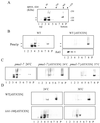
AST1 overexpression restores raft-association of mutant ATPase. (A) Raft association of ATPase in WT (L3852) and pma1-7 (ACY7) cells at 24°C. ATPase raft association, with and without high copy AST1 (pAC49), was determined after radiolabeling and immunoprecipitation from TX100 containing density gradients as before. (B) Mutant ATPase raft association in the presence of high copy AST1. pma1-7 (ACY7) cells harboring the pAC49 plasmid were radiolabeled for 10 min at 24°C and chased for 15 or 30 min at 24°C. Raft association of newly synthesized ATPase was determined as in A. (C) Protein–protein interaction between Ast1p and Pma1p. WT (L3852) cells expressing epitope-tagged AST1 (pAC64) grown at 24°C were lysed and incubated on ice with or without the cross-linker DSP for 2 h. Ast1p was immunoprecipitated with a rabbit polyclonal anti-myc antibody or coimmunoprecipitated with anti-Pma1p antibodies and subsequently detected with a mouse monoclonal anti-myc antibody. Ast1p is coimmunoprecipitated with Pma1p after treatment with DSP, the slightly lower mobility of Ast1p is probably due to cross-linking. Cells expressing Gap1-HA were grown in media containing urea as nitrogen source and subjected to the same procedure. Gap1-HA was immunoprecipitated with anti-HA–tag rabbit polyclonal antibodies or with anti-Pma1p antibodies and subsequently detected with a mouse monoclonal anti-HA–tag antibody.
Figure 7
Figure 7
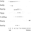
Subcellular localization of Ast1p. WT (L3852) cells expressing epitope-tagged AST1 from a centromeric plasmid (pAC64) were lysed and fractionated in a 20–60%sucrose gradient (Roberg et al., 1999). Distribution of different organelle markers was visualized by Western blotting with the use of specific antibodies. The peak of Ast1p, detected with the use of an anti-myc antibody, coincides with that of Sed5p, an early Golgi marker, lower amounts are also found in fractions containing Pep12p, a late endosome marker, and to a small extent in fractions containing Pma1p, a plasma membrane marker.
Figure 8
Figure 8
Plasma membrane rerouting of Pma1p in vps1 mutant does not restore its raft association. Pma1 and Pma1-7 association with DRMs was examined in VPS1 and vps1 background. Cells were radiolabeled for 10 min at 24 or 37°C and chased for 30 min at 24 or 37°C. ATPase distribution in R and S fractions was analyzed by immunoprecipitation and phosphorimaging.
Figure 9
Figure 9
Pma1p is present in SDS-TX100–resistant oligomers that are stabilized by overexpression of AST1. (A) PMA1 (RH690-15D) cells were grown at 24°C. Cell lysates were adjusted to 0.4% SDS and 0.2% TX100 and loaded on top of a 5–20% sucrose velocity gradient containing detergent and centrifuged for 16 h at 215,000 × g (at 4°C). Fractions were collected from the top and Pma1p distribution was analyzed by Western blotting. The approximate size was determined with the use of markers of known size (transferrin, catalase, ferritin, and thyroglobulin). The apparent molecular weight is shown on the right. (B) Overexpression of AST1 promotes the formation of a SDS/TX100-resistant ATPase oligomer. Wild-type cells (L3852) or cells expressing AST1 from a centromeric vector (pAC64) were grown at 24°C and treated as in A. The distribution of Pma1p and Ast1 was determined by Western blotting. (C) Overexpression of AST1 induces oligomer formation in pma1-7 mutant. pma1-7 (ACY7) and pma1-7 [AST1CEN] cells were grown at 24°C and incubated for 2 h at 24°C or 37°C and subjected to fractionation in velocity gradients as before. Distribution of mutant ATPase was determined by Western blotting. (D) Lipid rafts are required for Pma1p oligomer formation. WT [AST1CEN] and lcb1-100 [AST1CEN] cells were grown at 24°C and incubated for 1 h at 24 or 30°C and subjected to fractionation in velocity gradients as before. Oligomer formation is impaired in the lcb1-100 mutant at 24°C and completely abolished at 30°C.
Figure 10
Figure 10
Ast1p mediates clustering of Pma1p during biosynthetic transport. PMA1 (L3852) and PMA1[AST1CEN] cells were pulse-labeled with [35S]methionine for 10 min at 24°C and chased for 0, 15, or 30 min at the same temperature. Cells lysates were subjected to SDS/TX100 velocity gradient fractionation as in Figure 9. Fractions 1-2-3 (A), 4-5-6 (B), and 7-8-9 (C) were pooled and Pma1p and Ast1-myc were immunoprecipitated and analyzed as before. Pma1p becomes oligomerized already after 15 min of chase. Note that the slower migrating form of Pma1p (see Figure 9) is not preserved during immunoprecipitation.

Similar articles

Cited by

References

    1. Ambesi A, Miranda M, Petrov VV, Slayman CW. Biogenesis and function of the yeast plasma-membrane H(+)-ATPase. J Exp Biol. 2000;203(Pt 1):155–160. - PubMed
    1. Bagnat M, Keranen S, Shevchenko A, Simons K. Lipid rafts function in biosynthetic delivery of proteins to the cell surface in yeast. Proc Natl Acad Sci USA. 2000;97:3254–3259. - PMC - PubMed
    1. Benito B, Moreno E, Lagunas R. Half-life of the plasma membrane ATPase and its activating system in resting yeast cells. Biochim Biophys Acta. 1991;1063:265–268. - PubMed
    1. Black MW, Pelham HR. A selective transport route from Golgi to late endosomes that requires the yeast GGA proteins. J Cell Biol. 2000;151:587–600. - PMC - PubMed
    1. Brown DA, Rose JK. Sorting of GPI-anchored proteins to glycolipid-enriched membrane subdomains during transport to the apical cell surface. Cell. 1992;68:533–544. - PubMed

Publication types

MeSH terms

Substances