. 2002 Jan;22(1):182-95.
doi: 10.1128/MCB.22.1.182-195.2002.

Tyrosine phosphorylation of protein kinase Cdelta is essential for its apoptotic effect in response to etoposide

Affiliations

Tyrosine phosphorylation of protein kinase Cdelta is essential for its apoptotic effect in response to etoposide

Michal Blass et al. Mol Cell Biol. 2002 Jan.

Abstract

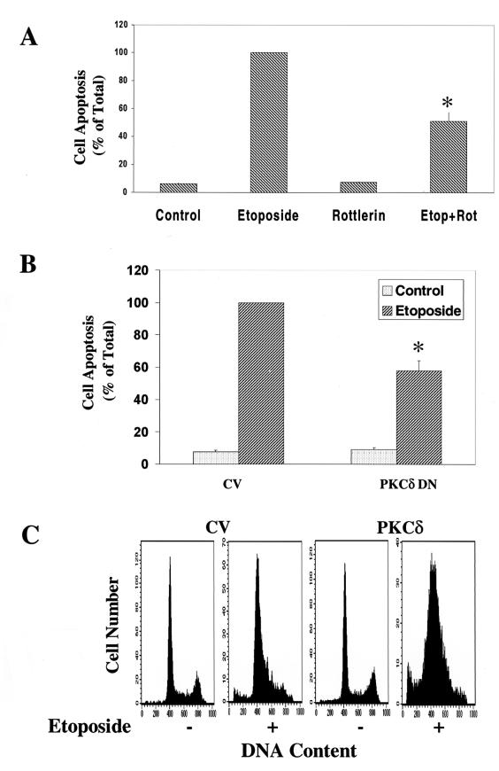
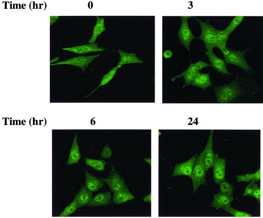
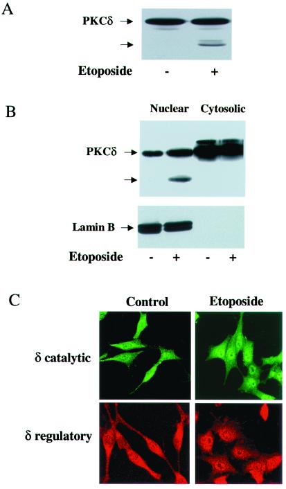
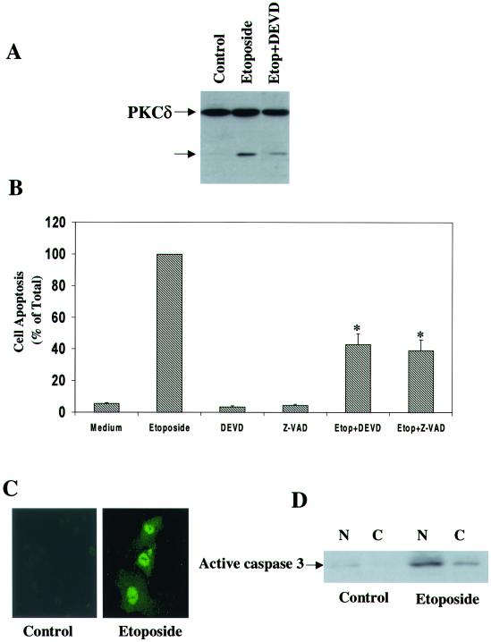
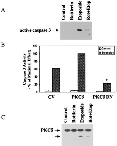
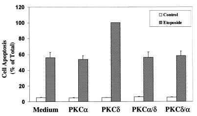
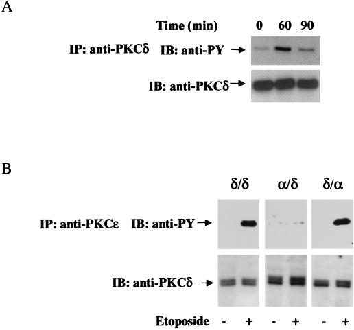
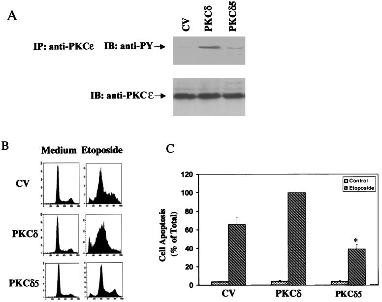
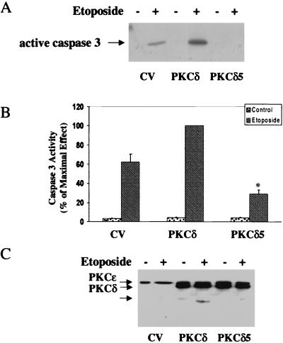
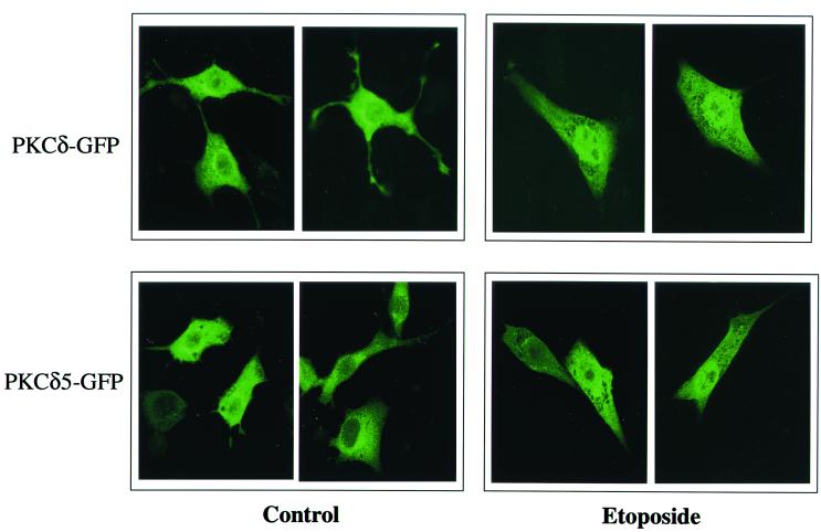
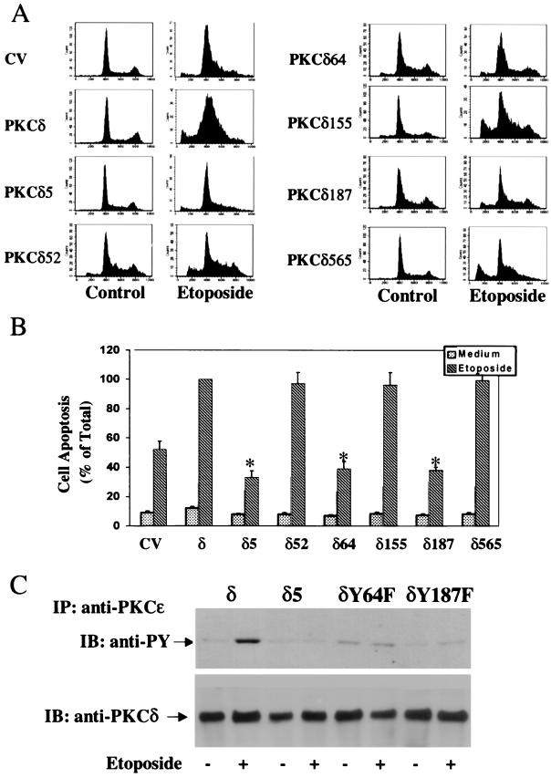
Protein kinase Cdelta (PKCdelta) is involved in the apoptosis of various cells in response to diverse stimuli. In this study, we characterized the role of PKCdelta in the apoptosis of C6 glioma cells in response to etoposide. We found that etoposide induced apoptosis in the C6 cells within 24 to 48 h and arrested the cells in the G(1)/S phase of the cell cycle. Overexpression of PKCdelta increased the apoptotic effect induced by etoposide, whereas the PKCdelta selective inhibitor rottlerin and the PKCdelta dominant-negative mutant K376R reduced this effect compared to control cells. Etoposide-induced tyrosine phosphorylation of PKCdelta and its translocation to the nucleus within 3 h was followed by caspase-dependent cleavage of the enzyme. Using PKC chimeras, we found that both the regulatory and catalytic domains of PKCdelta were necessary for its apoptotic effect. The role of tyrosine phosphorylation of PKCdelta in the effects of etoposide was examined using cells overexpressing a PKCdelta mutant in which five tyrosine residues were mutated to phenylalanine (PKCdelta5). These cells exhibited decreased apoptosis in response to etoposide compared to cells overexpressing PKCdelta. Likewise, activation of caspase 3 and the cleavage of the PKCdelta5 mutant were significantly lower in cells overexpressing PKCdelta5. Using mutants of PKCdelta altered at individual tyrosine residues, we identified tyrosine 64 and tyrosine 187 as important phosphorylation sites in the apoptotic effect induced by etoposide. Our results suggest a role of PKCdelta in the apoptosis induced by etoposide and implicate tyrosine phosphorylation of PKCdelta as an important regulator of this effect.


Figures

FIG. 1.
FIG. 1.
Etoposide induces apoptosis in C6 glioma cells. C6 cells were treated with etoposide (50 μM) for 24 or 48 h. Cell apoptosis was determined using PI staining and FACS analysis (A) or anti-histone ELISA (B). The results are from one representative experiment out of six similar experiments. (A) The optical densities of etoposide-treated cells (48 h) were designated 100% (total apoptosis), and all other values are presented as percent of this total. (B) The results represent the means ± SE of triplicate measurements in each of three experiments. (C) The morphology of the cells (48 h) was monitored under a phase contrast light microscope. The results are representative of four similar experiments.
FIG. 2.
FIG. 2.
Effects of rottlerin, PKCδ DN, and PKCδ overexpression on the apoptosis of C6 cells induced by etoposide. C6 cells were treated with etoposide (50 μM) in the absence and presence of rottlerin (5 μM) for 48 h (A) or cells overexpressing control vector (CV), PKCδ DN (B), or PKCδ (C) were treated with etoposide. Cell apoptosis was determined using anti-histone ELISA (A and B) or PI staining and FACS analysis (C). The optical densities of etoposide-treated cells (A) or of etoposide-treated CV cells (B) were designated 100% (total apoptosis), and all other values are presented as percent of this total. (A and B) The results represent the means ± SE of triplicate measurements in each of three experiments. (C) Distributions are from a representative experiment. ∗, P < 0.001, compared to control cells.
FIG. 3.
FIG. 3.
Translocation of PKCδ in C6 cells treated with etoposide. C6 cells were treated with etoposide (50 μM) for 0, 3, 6, and 24 h, and the translocation of PKCδ was assessed using immunofluorescence staining. Following fixation with 4% PFA, cells were incubated with a rabbit anti-PKCδ antibody for 1 h and with an anti-rabbit antibody conjugated to fluorescein isothiocyanate. Cells were visualized by confocal microscopy. The results are from one representative experiment out of five similar experiments.
FIG. 4.
FIG. 4.
Nuclear cleavage and translocation of PKCδ in etoposide-treated cells. C6 cells were treated with etoposide (50 μM) for 24 h, and the cleavage of PKCδ was determined by Western blotting using an anti-PKCδ antibody (rabbit; Santa Cruz). (A) A 40-kDa cleaved form is marked by an arrow. For the detection of PKCδ in the nucleus, extracts from control and etoposide-treated C6 cells (24 h) were fractionated and the nuclear and cytoplasmic fractions (50 μg/ml) were examined for the presence of PKCδ and for its cleaved form by using Western blot analysis. (B) The extracts were also examined for the expression of the nuclear marker lamin B. (C) The nuclear translocation of PKCδ in C6 cells treated with etoposide for 6 h was monitored by immunofluorescence using anti-PKCδ antibody directed against the catalytic domain (fluorescein isothiocyanate) or against the regulatory domain (Alexa FluorTM 546 goat anti-mouse IgG). The results shown represent one of three separate experiments, which yielded similar results.
FIG. 5.
FIG. 5.
Role of caspase 3 in cleavage of PKCδ and C6 cell apoptosis induced by etoposide. C6 cells were treated with etoposide for 24 h in the absence and presence of DEVD (20 μM) and Z-VAD (25 μM). The cleavage of PKCδ was determined using Western blot analysis (A), and cell apoptosis was determined using anti-histone ELISA (B). The results are representative of four similar experiments (A) or represent the means ± SE of three separate experiments (B). The expression of active caspase 3 in the nucleus of etoposide-treated cells (24 h) was determined using immunofluorescence staining (C) and Western blot analysis of cytosolic (C) and nuclear (N) fractions (D). ∗, P < 0.001
FIG. 6.
FIG. 6.
The PKCδ inhibitor rottlerin and PKCδ DN decrease the activation of caspase 3 and the cleavage of PKCδ in etoposide-treated cells. C6 cells were treated with etoposide (50 μM) in the absence or presence of rottlerin (5 μM) for 24 h. The cleavage of caspase 3 (A) and of PKCδ (C) was detected using Western blot analysis. The membranes were probed with antibodies against active caspase 3 (17 kDa) (A) or PKCδ (C). The results represent one of three separate experiments which yielded similar results. (B) Cells overexpressing PKCδ DN were treated with etoposide for 24 h, and the activity of caspase 3 was measured using the Caspase 3 Cellular Activity Assay Kit as described in Materials and Methods. Caspase 3 activity was calculated and expressed as picomoles/minute/microgram of protein, and the percent of maximal effect was determined. The results represent the means ± SE of four experiments. ∗, P < 0.001
FIG. 7.
FIG. 7.
Apoptosis of C6 cells overexpressing different PKC chimeras in response to etoposide. C6 cells overexpressing PKCα, PKCδ, and the chimeras PKCα/δ and PKCδ/α were treated with etoposide for 48 h. Cell apoptosis was determined using anti-histone ELISA. The optical densities of etoposide-treated PKCδ-overexpressing cells were designated 100% (total apoptosis), and all other values are presented as a percent of this total. The results represent the means ± SE of triplicate measurements in each of three experiments.
FIG. 8.
FIG. 8.
Etoposide induces tyrosine phosphorylation of PKCδ in the regulatory domain. Parental C6 cells or cells stably transfected with PKCδ or the chimeras PKCδ/α and PKCα/δ were treated with etoposide (50 μM) for various periods of time. Cells were then harvested, and immunoblotting (IB) and immunoprecipitation (IP) of PKCδ were performed using anti-PKCδ (A) or anti-PKCɛ (B) antibodies as described in Materials and Methods. Following SDS-PAGE, membranes were stained with anti-phosphotyrosine antibody (anti-PY) or with anti-PKCδ antibodies. The results are from one representative experiment out of four separate experiments.
FIG. 9.
FIG. 9.
Tyrosine phosphorylation and cell apoptosis in response to etoposide in cells overexpressing PKCδ and PKCδ5. C6 cells overexpressing control vector (CV), PKCδ, or PKCδ5 were treated with etoposide (50 μM) for 60 min (A), 24 h (B), or 48 h (C). (A) Cells were then harvested, and immunoprecipitation of PKCδ was performed using anti-PKCɛ antibody as described in Materials and Methods. Following SDS-PAGE, membranes were stained with anti-phosphotyrosine antibody (anti-PY) or with an anti-PKCδ antibody (rabbit; Santa Cruz). The results represent one of three separate experiments, which yielded similar results. For the measurement of apoptosis, cells were harvested after 24 h (B) or 48 h (C) of treatment and were analyzed using PI staining and FACS analysis (B) or by anti-histone ELISA (C). The optical densities of etoposide-treated PKCδ-overexpressing cells were designated 100% (total apoptosis), and all other values are presented as a percent of this total. The results represent the means ± SE of triplicate measurements in each of three experiments. ∗, P < 0.001.
FIG. 10.
FIG. 10.
Activation of caspase 3 and cleavage of PKCδ in cells overexpressing PKCδ and PKCδ5. C6 cells overexpressing control vector (CV), PKCδ, or PKCδ5 were treated with etoposide (50 μM) for 24 h, and the activation of caspase 3 (A and B) and cleavage of PKCδ (C) were determined. Cells were harvested and subjected to SDS-PAGE and Western blot analysis. The membranes were probed with active caspase 3 antibody (A) or with anti-PKCɛ that recognizes the ɛ tag which is located in the catalytic domain of the PKCδ constructs (C). The results represent one of three separate experiments which yielded similar results. The activity of caspase 3 was measured using the Caspase 3 Cellular Activity Assay Kit as described in Materials and Methods (B). Caspase 3 activity was calculated and expressed as picomoles/minute/microgram of protein, and the percent of maximal effect was determined. The results represent the means ± SE of three experiments.
FIG. 11.
FIG. 11.
Translocation of PKCδ and PKCδ5 in etoposide-treated C6 cells. Cells were transiently transfected with GFP-PKCδ or GFP-PKCδ5. After 48 h, cells were treated with etoposide (50 μM) for 6 h and cells were viewed using confocal microscopy. Cells shown are representative of four independent experiments
FIG. 12.
FIG. 12.
Apoptosis and tyrosine phosphorylation in C6 cells overexpressing different PKCδ tyrosine mutants. C6 cells overexpressing PKCδ or the PKCδ mutants were plated in the absence and presence of etoposide (50 μM). (A) Cell apoptosis was measured by PI staining and FACS analysis after 24 h. The results are from one representative experiment out of four separate experiments. (B) Cell apoptosis was also determined after 48 h using anti-histone ELISA. The optical densities of etoposide-treated PKCδ-overexpressing cells were designated 100% (total apoptosis), and all other values are presented as percent of this total. The results represent the means ± SE of triplicate measurements in each of five experiments. Tyrosine phosphorylation of PKCδ, PKCδ5, PKCδY64F, and PKCδY187F was determined following 1 h of etoposide treatment. (C) Cells were harvested, and immunoprecipitation of PKCδ was performed using anti-PKCɛ antibody as described in Materials and Methods. Following SDS-PAGE, membranes were stained with anti-phosphotyrosine antibody (anti-PY) or with an anti-PKCδ antibody. The results represent one of three separate experiments which yielded similar results.

Similar articles

Cited by

References

    1. Acs, P., K. Bogi, P. S. Lorenzo, A. M. Marquez, T. Biro, Z. Szallasi, and P. M. Blumberg. 1997. The catalytic domain of protein kinase C chimeras modulates the affinity and targeting of phorbol ester-induced translocation. J. Biol. Chem. 272:22148–22153. - PubMed
    1. Acs, P., Q. J. Wang, K. Bogi, A. M. Marquez, P. S. Lorenzo, T. Biro, Z. Szallasi, J. F. Mushinski, and P. M. Blumberg. 1997. Both the catalytic and regulatory domains of protein kinase C chimeras modulate the proliferative properties of NIH 3T3 cells. J. Biol. Chem. 272:28793–28799. - PubMed
    1. Bharti, A., S. K. Kraeft, M. Gounder, P. Pandey, S. Jin, Z. M. Yuan, S. P. Lees-Miller, R. Weichselbaum, D. Weaver, L. B. Chen, D. Kufe, and S. Kharbanda. 1998. Inactivation of DNA-dependent protein kinase by protein kinase Cδ: implications for apoptosis. Mol. Cell. Biol. 18:6719–6728. - PMC - PubMed
    1. Blake, R. A., P. Garcia-Paramio, P. J. Parker, and S. A. Courtneidge. 1999. Src promotes PKCδ degradation. Cell Growth Differ. 10:231–241. - PubMed
    1. Brodie, C., K. Bogi, P. Acs, P. S. Lorenzo, L. Baskin, and P. M. Blumberg. 1998. Protein kinase Cδ (PKCδ) inhibits the expression of glutamine synthetase in glial cells via the PKCδ regulatory domain and its tyrosine phosphorylation. J. Biol. Chem. 273:30713–30718. - PubMed

Publication types

MeSH terms

Substances