. 2001 Dec 1;21(23):9291-303.
doi: 10.1523/JNEUROSCI.21-23-09291.2001.

A functional role for intra-axonal protein synthesis during axonal regeneration from adult sensory neurons

Affiliations

A functional role for intra-axonal protein synthesis during axonal regeneration from adult sensory neurons

J Q Zheng et al. J Neurosci. .

Abstract

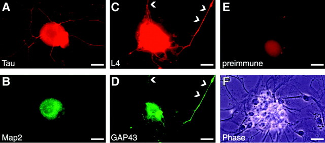
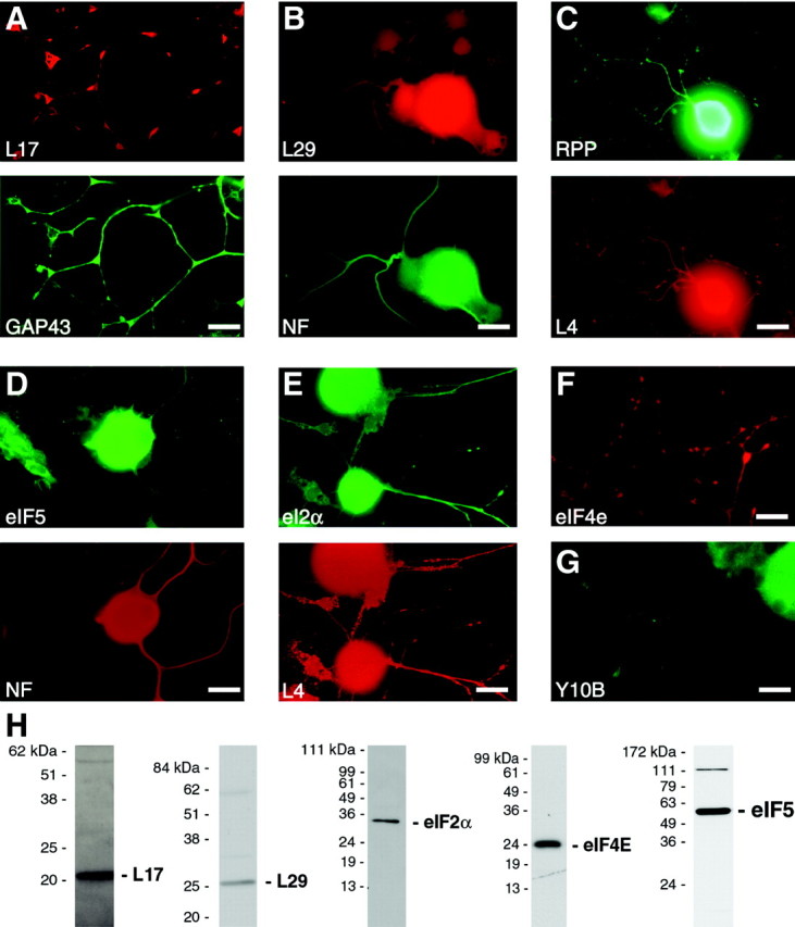
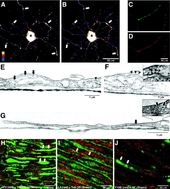
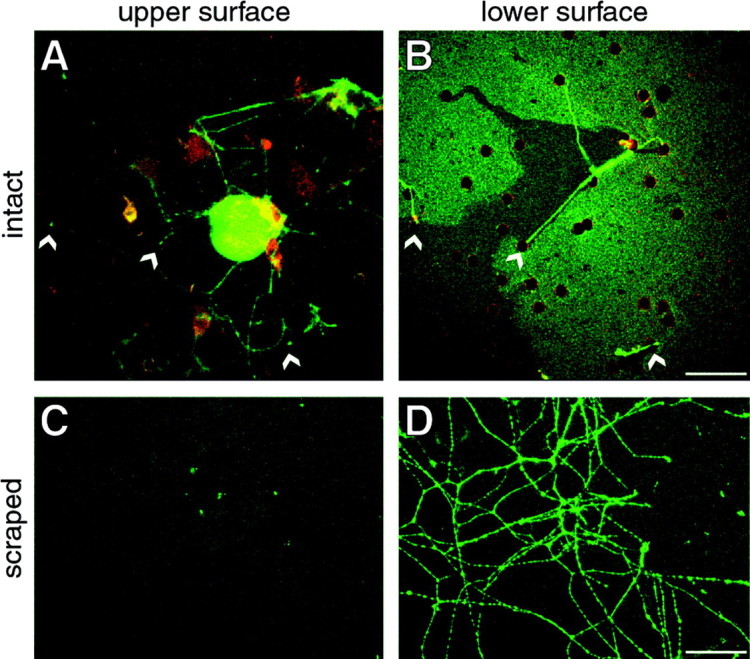
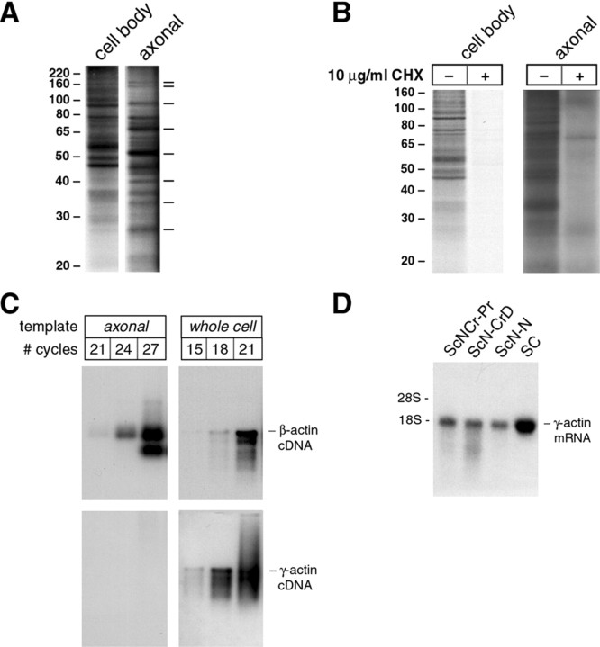
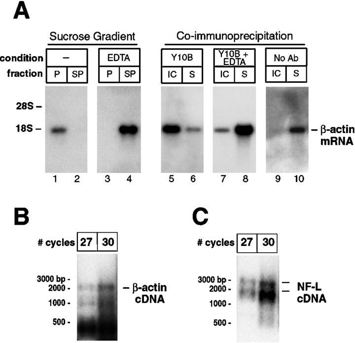
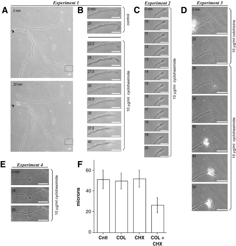
Although intradendritic protein synthesis has been documented in adult neurons, the question of whether axons actively synthesize proteins remains controversial. Adult sensory neurons that are conditioned by axonal crush can rapidly extend processes in vitro by regulating the translation of existing mRNAs (Twiss et al., 2000). These regenerating processes contain axonal but not dendritic proteins. Here we show that these axonal processes of adult sensory neurons cultured after conditioning injury contain ribosomal proteins, translational initiation factors, and rRNA. Pure preparations of regenerating axons separated from the DRG cell bodies can actively synthesize proteins in vitro and contain ribosome-bound beta-actin and neurofilament mRNAs. Blocking protein synthesis in these regenerating sensory axons causes a rapid retraction of their growth cones when communication with the cell body is blocked by axotomy or colchicine treatment. These findings indicate that axons of adult mammalian neurons can synthesize proteins and suggest that, under some circumstances, intra-axonal translation contributes to structural integrity of the growth cone in regenerating axons. By immunofluorescence, translation factors, ribosomal proteins, and rRNA were also detected in motor axons of ventral spinal roots analyzed after 7 d in vivo after a peripheral axonal crush injury. Thus, adult motor neurons are also likely capable of intra-axonal protein synthesis in vivo after axonal injury.


Figures

Fig. 1.
Fig. 1.
Rapidly growing axons of conditioned DRG neurons contain ribosomal protein L4. Four days after sciatic nerve crush, conditioned L4–5 DRG neurons were dissociated and cultured on coated coverslips for 22 hr in medium containing Ara-C and DRB. The long processes extended by these DRG neurons are strongly immunoreactive for Tau (A). Colabeling with monoclonal antibody, the dendritic protein MAP2 shows signals that are limited to the cell body and do not extend into the Tau-reactive processes (B). Polyclonal anti-ribosomal protein L4 antibody showed signals in both in the neuronal cell body and the axonal-like processes (C, arrowheads). Colabeling with a monoclonal antibody to GAP43 confirmed that the L4 immunoreactivity was within the neuronal processes (D,arrowheads). Preimmune serum for L4 showed a faint fluorescence in the cell body after long exposure times (2 min inE vs 10 sec in C) and no signal in the axonal processes (E). Phase-contrast image of the same neuron in shown in E shows abundant axons extending from this sensory neuron cell body (F). Scale bars, 20 μm.
Fig. 2.
Fig. 2.
Regenerating sensory axons contain ribosomal proteins, rRNA, and translation factors. Conditioned DRG cultures were colabeled with antibodies to ribosomal proteins, translation factors, and rRNA. Processes that react with GAP43 and NF contain immunoreactivity for ribosomal proteins L17 and L29 (Aand B, respectively). RPP shows a signal in the same neuronal processes that show immunoreactivity for L4 (C). NF-positive processes also contain eIF5 immunoreactivity (D), and eIF2α shows colocalization with immunoreactivity for L4 (E). Antibodies to eIF4e showed similar signals within the regenerating sensory axons (F). Finally, immunostaining with Y10B anti-28S rRNA antibody also shows a signal within the axons (G). Scale bars, 20 μm. Specificity of the anti-L17, -L29, -eIF2α, -eIF4E, and -eIF5 antibodies was evaluated by immunoblotting (H; 12% gel for L17, L29, eIF2α, and eIF4E, and 10% gel for eIF5). Each of these antibody preparations recognized one major band of the expected molecular weight. Similar data have been published for anti-L4 and -RPP antibodies (Elkon et al., 1985; Twiss et al., 2000), and Figure 6 shows immunoprecipitation using the Y10B antibody.
Fig. 3.
Fig. 3.
Abundance of translational machinery in regenerating axons. A, B, DRG cultures costained with antisera to ribosomal protein L4 and RPP were analyzed by laser scanning confocal microscopy. Neurons were scanned at 1 μm intervals over 24 optical (Z) planes. Digital three-dimensional images are displayed as a spectrum as indicated in the bottom left corner of A. The signal intensity for L4 and RPP in the cell body is saturated (asterisk); however, high signal intensity is also seen in the axons at >100 μm distance from the cell body, and the bright intra-axonal signals for L4 and RPP colocalize (arrows). Scale bar, 50 μm.C, D, A region of high intra-axonal fluorescence for ribosomal protein L17 and 28S rRNA (recognized with Y10b antibody) similar to those shown in A andB is illustrated as a single Z-plane confocal image through the center of the axon. Note that the fluorescent signal for L17 and rRNA is granular rather than diffuse in these highly immunoreactive regions of the axon. Scale bar, 10 μm.EG, Electron micrographs of DRG cultures stained with uranyl acetate show electron-dense particles in the proximal (E) and distal axonal segments (G) that are of the similar size to those seen along the RER in the cell body (F,arrowheads) and likely represent ribosomes. The proximal segment of the axon in E extends right toleft from the cell body in F and continues on right to left as the distal axonal segment shown in G. Ribosome-like particles are seen on the RER (arrowheads) and free within the axoplasm (arrows) in the proximal axonal segment (E). Ribosome-like particles are noted in the distal segment of the axon (G, arrows). At higher magnification, these electron-dense particles in the distal axon (G, inset) are approximately the same diam-eter as those along the RER in the cell body (F, inset). Scale bars:insets, 100 nm. H–J, Confocal images of ventral L4 spinal cord root (HI) and sciatic nerve (J) illustrate signals for L4 (H), eIF5 (I), and 28S rRNA (J) that colocalize with intra-axonal signals for mouse anti-Tub βIII (HI) and rabbit anti-NF (J). These confocal images represent a three-dimensional reconstruction of four Z-plane images taken at 0.4 μm intervals to provide optical sections through individual axons and exclude the myelin sheath and Schwann cell cytoplasm. The arrows in each panel indicate such optically isolated axons where non-neuronal components are excluded from consideration and show punctate intra-axonal signals for a ribosomal protein (H), translation factor (I), and rRNA (J). Scale bars, 20 μm.
Fig. 4.
Fig. 4.
Culture system to isolate regenerating DRG axons. Dissociated cultures of conditioned DRG neurons were plated into a tissue culture insert containing a PET membrane with 8 μm pores. After 24 hr, the cultures were fixed, and indirect immunofluorescence was performed with antibodies to GAP-43 (shown in green) and S100 (shown in red). Confocal microscopy was used to image the membranes. A shows a three-dimensional reconstructed composite digital image of 19 × 1 μm Z planes along the top surface of the membrane. The arrowheadsindicate axons that enter pores of the membrane. Bdisplays a single X–Y plane image along the bottom surface of the membrane directly below that illustrated in A.Arrowheads indicate the pores where axons cross the membrane. Note that no S100-reactive cellular elements are seen along the bottom surface of the membrane. Non-neuronal elements were also not visible by phase-contrast images (data not shown). To isolate axons, the top surface of the membrane was scraped repetitively with a cotton-tipped applicator (see Materials and Methods). Scraped membranes were processed for immunostaining with antibodies to GAP43. Scraping removed all cellular elements from the top membrane surface (C; reconstructed composite digital image of 19 × 1 μm Z planes). Axons that had traversed the membrane remained adherent to the bottom surface of the membrane after scraping (D; single X–Y plane on bottom membrane surface). Scale bars, 50 μm.
Fig. 5.
Fig. 5.
Regenerating sensory axons synthesize proteins and differentially localize β- and γ-actin mRNAs. A, DRG cultures were performed in tissue culture inserts as described above. After 18 hr in culture, the top or bottom surface of the membrane was scraped to yield an axonal or cell body preparation, respectively (note that the cell body preparation contains non-neuronal cells and neuronal processes that have not traversed the membrane pores). Membranes were then incubated in medium containing 4 mCi/ml 35S-Met/Cys for 4 hr. Axonal and cell body preparation lysates were fractionated on 10% SDS-PAGE gels and processed for fluorography. The axonal preparation required a much longer exposure time than the cell body preparation (1 vs 6 d). Autoradiograms show proteins of ∼167, 160, 95, 68, 52, 40, 34, and 28 kDa that appear enriched in the axonal preparations (dashes to right of autoradiogram). These data are representative of three independent metabolic labeling experiments. We cannot state that these proteins are uniquely synthesized in the axons because the high specific activity of the cell body lysates compared with the axonal lysates does not allow for matched exposure times. B, Axonal and cell body preparations were generated as above and incubated in 10 μg/ml cycloheximide for 20 min before metabolic labeling in 1 mCi/ml35S-Met/Cys for 4 hr and analyzed as in A. Note that cycloheximide completely inhibited incorporation of35S-Met/Cys into proteins in the cell body preparation (1 d exposure) and greatly diminished protein synthesis in the axonal preparation (6 d exposure). The labeled band at ∼70 kDa in the cycloheximide-treated axonal preparation may represent a protein derived from intra-axonal mitochondria. The lower isotope levels used for labeling compared with A account for different band intensities in the axonal preparation of this and A.C, RNA was extracted from axonal and cell body preparations from DRG neurons that had been plated for 18 hr and used for RT-PCR (see Materials and Methods). Aliquots were removed from the PCR at 21, 24, 27, and 30 cycles and used for virtual Northern blotting. Blots probed with β-actin cDNA showed a prominent band after a short exposure that corresponds to the full-length β-actin mRNA (3 hr exposure). In contrast, no signal for γ-actin could be detected in the axonal cDNA even with long exposure times (72 hr exposure), but γ-actin cDNA was readily detected in RT-PCR-amplified RNA of the DRG preparations that included the cell body (i.e., RNA from nonfractionated cultures) (3 hr exposure). Note that with matched exposure times, γ-actin cDNA appears to be even more abundant than β-actin cDNA in the cell body RT-PCR samples. D, To exclude this possibility of differential actin isoform expression by non-neuronal cells contaminating the axonal RNA preparation, total RNA was isolated from 7 d crushed [distal and proximal (ScNCrPr and ScN-CrD, respectively)] and naive rat sciatic nerve (ScN-N) and Schwann cell cultures (SC) and processed for standard Northern blotting. γ-actin mRNA was easily detected in all RNA preparations of sciatic nerve and in the RNA from purified Schwann cell cultures (24 hr exposure).
Fig. 6.
Fig. 6.
Intra-axonal mRNAs are translationally active in cultured DRG neurons. Coimmunoprecipitation of mRNAs with rRNA using Y10B antibody was used to determine whether intra-axonal mRNAs are translationally active. Lysates from PC12 cells were used to test the validity of this coimmunoprecipitation. A shows Northern blot analysis of polysomal RNAs and Y10B coimmunoprecipitated RNAs. Fractionation of β-actin mRNA in discontinuous 20% sucrose gradients is shown in lanes 1–4. Note that β-actin mRNA resides in the polysome fraction (P) rather than the subpolysome fraction (SP) (A,lanes 1–2). Addition of 50 mm EDTA before ultracentrifugation causes β-actin mRNA to shift from theP to SP fraction (A,lanes 3–4). For Y10B immunoprecipitation, RNA was extracted from the Y10B immunocomplex (IC) and supernatant from immunoprecipitate (S) and equivalent proportions of these IC and S RNA fractions were used for Northern blotting. β-actin mRNA coimmunoprecipitated with Y10B (A, lanes 5–6). In lysates that were treated with EDTA to disrupt the 40S and 60S ribosome subunits, β-actin mRNA resided in the S fraction (A,lanes 7–8). Without addition of Y10B to the lysate, β-actin mRNA also resided in the S fraction (A,lanes 9–10). The Y10B coimmunoprecipitation is not limited to β-actin mRNA (data not shown). B andC show virtual Northern blots of Y10B immunoprecipitates from the DRG axonal preparations. For this, axons were isolated from the DRG cultures (Fig. 3), and RNA from axonal Y10B immunocomplexes was used for RT-PCR. The Y10B immunoprecipitates contained β-actin and NF-L mRNAs (B and C, respectively). This suggests that the axonal mRNAs encoding β-actin and NF-L are actively translated in these regenerating sensory axons.
Fig. 7.
Fig. 7.
Intra-axonal protein synthesis maintains the growth cone. To address the functional significance of intra-axonal protein synthesis, we used video microscopy to monitor changes in axons during inhibition of protein synthesis. Axons were anucleated using a glass capillary tube that had been pulled to a closed tip. Elapsed time is indicated in the top left corner of each image of a time-lapse sequence (AE).A and B show the results of one experiment. A shows anucleated axons that were incubated for 20 min in complete medium. Scale bar, 20 μm. Thearrowhead indicates where the axon was severed with the axon coursing from left to right. Note that although the proximal portion of the axon retracts slightly (arrowheads), the distal axon remains intact. Theboxed region is shown at higher magnification in the time-lapse sequence in B. Scale bar, 10 μm. In the first two images the distal axon remains stable over 20 min. After addition of 10 μg/ml cycloheximide, the distal tip of the axon begins to retract over the 30–40 min of the time-lapse sequence (10–20 min after treatment). C shows high-magnification time-lapse sequence of a second experiment in which an axon was treated with cycloheximide immediately after severing or anucleation. Scale bar, 10 μm. Note that this anucleated axon is stable over the first 10 min but then begins to retract, similar to that seen in B. In a third series of experiments, the relevance of local protein synthesis in intact neurons was addressed by treating cultures with colchicine to decrease the influence of cell body-synthesized proteins by impeding axonal transport (D). A low-magnification time-lapse sequence of a culture preparation that was treated with 10 μg/ml colchicine for 25 min followed by 10 μg/ml cycloheximide for 25 min as indicated is shown in D. Most axonal branches were stable over the course of colchicine treatment (D, top two panels). However, during treatment with cycloheximide, many of the axonal branches retracted (D, bottom five panels). Scale bar, 60 μm. This axonal retraction required pretreatment with colchicine or axotomy because treatment of intact neurons with 10 μg/ml cycloheximide alone was without effect (E). Scale bar, 20 μm. The above series of time-lapse experiments have been repeated in three different DRG preparations in at least five neurons per preparation, using the protein synthesis inhibitor anisomycin, and yielded similar results (data not shown). To quantitate the axonal retraction in colchicine plus cycloheximide-treated neurons, 16 hr DRG cultures were treated with colchicine for 60 min (COL), cycloheximide for 30 min (CHX), or colchicine for 60 min plus cycloheximide for 30 min (COL + CHX). Control (Cntl) for these studies consisted of cultures that were allowed to grow in normal medium for 17 hr. The average length of most terminal axon branches was measured for each treatment paradigm. Axonal branch lengths in the CHX + COL were approximately half that of control, COL, or CHX paradigms (F) (average ± 2 × SEM). The differences between the COL + CHX, COL, and Cntl samples was statistically significant (p ≤ 0.0001 for COL + CHX vs Cntl, COL, and CHX samples).

Similar articles

Cited by

References

    1. Alvarez J, Giuditta A, Koenig E. Protein synthesis in axons and terminals: significance for maintenance, plasticity and regulation of phenotype. With a critique of slow transport theory. Prog Neurobiol. 2000;62:1–62. - PubMed
    1. Bassell GJ, Zhang H, Byrd AL, Femino AM, Singer RH, Taneja KL, Lifshitz LM, Herman IM, Kosik KS. Sorting of beta-actin mRNA and protein to neurites and growth cones in culture. J Neurosci. 1998;18:251–265. - PMC - PubMed
    1. Baum E, Wormington W. Coordinate expression of ribosomal protein genes during Xenopus development. Dev Biol. 1985;111:488–498. - PubMed
    1. Biberman Y, Meyuhas O. Substitution of just five nucleotides at and around the transcription start site of rat beta-actin promoter is sufficient to render the resulting transcript a subject for translational control. FEBS Lett. 1997;405:333–336. - PubMed
    1. Bittner GD. Long-term survival of anucleate axons and its implications for nerve regeneration. Trends Neurosci. 1991;14:188–193. - PubMed

Publication types

MeSH terms

LinkOut - more resources