. 2001 Nov 19;194(10):1485-95.
doi: 10.1084/jem.194.10.1485.

CD8beta endows CD8 with efficient coreceptor function by coupling T cell receptor/CD3 to raft-associated CD8/p56(lck) complexes

Affiliations

CD8beta endows CD8 with efficient coreceptor function by coupling T cell receptor/CD3 to raft-associated CD8/p56(lck) complexes

A Arcaro et al. J Exp Med. .

Abstract

The extraordinary sensitivity of CD8+ T cells to recognize antigen impinges to a large extent on the coreceptor CD8. While several studies have shown that the CD8beta chain endows CD8 with efficient coreceptor function, the molecular basis for this is enigmatic. Here we report that cell-associated CD8alphabeta, but not CD8alphaalpha or soluble CD8alphabeta, substantially increases the avidity of T cell receptor (TCR)-ligand binding. To elucidate how the cytoplasmic and transmembrane portions of CD8beta endow CD8 with efficient coreceptor function, we examined T1.4 T cell hybridomas transfected with various CD8beta constructs. T1.4 hybridomas recognize a photoreactive Plasmodium berghei circumsporozoite (PbCS) peptide derivative (PbCS (4-azidobezoic acid [ABA])) in the context of H-2K(d), and permit assessment of TCR-ligand binding by TCR photoaffinity labeling. We find that the cytoplasmic portion of CD8beta, mainly due to its palmitoylation, mediates partitioning of CD8 in lipid rafts, where it efficiently associates with p56(lck). In addition, the cytoplasmic portion of CD8beta mediates constitutive association of CD8 with TCR/CD3. The resulting TCR-CD8 adducts exhibit high affinity for major histocompatibility complex (MHC)-peptide. Importantly, because CD8alphabeta partitions in rafts, its interaction with TCR/CD3 promotes raft association of TCR/CD3. Engagement of these TCR/CD3-CD8/lck adducts by multimeric MHC-peptide induces activation of p56(lck) in rafts, which in turn phosphorylates CD3 and initiates T cell activation.


Figures
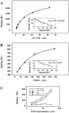
Figure 1.
Figure 1.
Soluble CD8αβ binds to Kd-PbCS(ABA) without affecting its interaction with T1 TCR. For affinity measurements sT1 TCR (A) and sCD8αβ (B) were injected for 30 s at the indicated concentrations over surfaces expressing Kd-SYIPSAEK(ABA)I (4,500 RU) or sCD4 (4,200 RU). Binding was calculated as the difference in the observed equilibrium response between the Kd-SYIPSAEK(ABA)I and the control sCD4 flow cells. The solid lines represent nonlinear fits of the Langmuir binding isotherm to the data, which yielded the indicated KD values. The maximal binding was 2,600 RU for sT1 TCR and 1,200 RU for sCD8αβ. The insets show Scatchard transformations of the same data. (C) The indicated concentrations of sT1 TCR were injected for 30 s over Kd-SYIPSAEK(ABA)I (2,000 RU) or CD4 (2,300 RU) coated sensor chips in the absence (▪) or presence (⋄) of added sCD8αβ (80 μM). The calculated difference in the responses observed with sT1 alone versus sT1 plus sCD8αβ are also shown (▴). The binding level observed with sCD8αβ alone at 80 μM is shown as dashed line, indicating that the effect of adding sCD8αβ on sT1 binding was never more than additive.
Figure 2.
Figure 2.
TCR and CD8 expression of cells under study.
Figure 2.
Figure 2.
TCR and CD8 expression of cells under study.
Figure 3.
Figure 3.
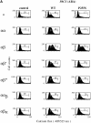
Efficient calcium mobilization of T1.4 hybridomas requires that they express CD8β transmembrane and cytoplasmic portions. (A) Indo-1–labeled hybridomas with the indicated CD8 expression were incubated with P815 cells either untreated (control) or previously sensitized with 1 μM of IASA-YIPSAEK(ABA)I (PbCS(ABA) wt) or IASA-YISSAEK(ABA)I (PbCS(ABA) P255S) at an E/T ratio of 1/3 for 3 min at 37°C and calcium-dependent Indo-1 fluorescence was measured by FACS®. (B) Alternatively the indicated Indo-1–labeled cells were incubated or not (control) with Kd-PbCS(ABA) tetramer (50 nM) and calcium flux was measured likewise after 2 min of incubation at 37°C. One out of three experiments is shown.
Figure 3.
Figure 3.
Efficient calcium mobilization of T1.4 hybridomas requires that they express CD8β transmembrane and cytoplasmic portions. (A) Indo-1–labeled hybridomas with the indicated CD8 expression were incubated with P815 cells either untreated (control) or previously sensitized with 1 μM of IASA-YIPSAEK(ABA)I (PbCS(ABA) wt) or IASA-YISSAEK(ABA)I (PbCS(ABA) P255S) at an E/T ratio of 1/3 for 3 min at 37°C and calcium-dependent Indo-1 fluorescence was measured by FACS®. (B) Alternatively the indicated Indo-1–labeled cells were incubated or not (control) with Kd-PbCS(ABA) tetramer (50 nM) and calcium flux was measured likewise after 2 min of incubation at 37°C. One out of three experiments is shown.
Figure 4.
Figure 4.
Palmitoylation of CD8β is essential for CD8 localization in rafts and efficient lck association. (A) T1.4 hybridomas with the indicated CD8 expression were surface-biotinylated, lysed in cold TX-100 (1%), and fractionated in M and DIM fractions. These were immunoprecipitated with anti-CD8α mAb 53.6.72. The samples were analyzed by SDS-PAGE and Western blotting with streptavidin. (B–E) T1.4 hybridomas with the indicated CD8 expression were lysed in Brij96 (1%) and the lysates immunoprecipitated with mAb 53.6.72 (B and D) or anti-CD8β mAB H35–17 (C and E). The immunoprecipitates were analyzed by SDS-PAGE and Western blotting with anti-lck mAb 3A5 (B and C) or anti-CD8α antiserum (D and E). Note that anti-CD8β mAb H35 is unable to precipitate CD8αα and CD8ααβ. One out of two experiments is shown.
Figure 5.
Figure 5.
The cytoplasmic tail of CD8β is required for efficient CD8-mediated increase in TCR photoaffinity labeling. T1.4 hybridoma cells (5 × 10⁶/ml) with the indicated CD8 expression were incubated for 2 h at 0–4°C with soluble monomeric Kd -125”IASA”-YIPSAEK(ABA)I (2–6 × 10⁶ cpm/ml) in the absence or presence of anti-CD8β mAb H35–17 (10 μg/ml). After UV irradiation cells were lysed in RIPA buffer, TCR immunoprecipitated, and analyzed by SDS-PAGE and PhosphorImaging. Mean values and SD were calculated from six experiments.
Figure 6.
Figure 6.
The CD8β tail mediates CD8 association with TCR/CD3. (A–C) T1.4 hybridomas with the indicated CD8 expression were lysed in 0.3% NP-40, and lysates immunoprecipitated with anti-TCR mAb H57 (A and B), anti-CD8α mAb 53.6.72 (C, left), or with anti-CD8β mAb H35–17 (C, right). The samples were analyzed by SDS-PAGE and Western blotting with anti-CD8α antiserum (A and C) or anti-CD3ε antiserum (B). (D) T1.4 T cell hybridomas expressing CD8αα or CD8αβ were incubated with monomeric Kd-SYIPSAEK(ABA)I complexes (1.16 μM) for 2 h at 0–4°C. After UV irradiation the cells were lysed in 0.3% NP-40. Lysates were immunoprecipitated with anti-TCR mAb H57 or anti-CD8α mAb 53.6.72, as indicated and the immunoprecipitates analyzed by SDS-PAGE and Western blotting with anti-CD8α antiserum.
Figure 6.
Figure 6.
The CD8β tail mediates CD8 association with TCR/CD3. (A–C) T1.4 hybridomas with the indicated CD8 expression were lysed in 0.3% NP-40, and lysates immunoprecipitated with anti-TCR mAb H57 (A and B), anti-CD8α mAb 53.6.72 (C, left), or with anti-CD8β mAb H35–17 (C, right). The samples were analyzed by SDS-PAGE and Western blotting with anti-CD8α antiserum (A and C) or anti-CD3ε antiserum (B). (D) T1.4 T cell hybridomas expressing CD8αα or CD8αβ were incubated with monomeric Kd-SYIPSAEK(ABA)I complexes (1.16 μM) for 2 h at 0–4°C. After UV irradiation the cells were lysed in 0.3% NP-40. Lysates were immunoprecipitated with anti-TCR mAb H57 or anti-CD8α mAb 53.6.72, as indicated and the immunoprecipitates analyzed by SDS-PAGE and Western blotting with anti-CD8α antiserum.
Figure 7.
Figure 7.
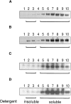
CD8αβ mediates raft association of TCR/CD3. T1.4 hybridomas (5 × 10⁷) expressing no CD8 (A), CD8αβ (B), CD8αβ′ (C), or CD8αβ′′ (D) were photoaffinity labeled at 26°C with Kd -125”IASA”-YIPSAEK(ABA)I (0.5–1.5 × 10⁸ cpm/7 ml). After UV irradiation the washed cells were lysed in 0.5% Brij58 and the lysates fractionated on sucrose density gradients. Fractions were collected from the top and immunoprecipitated with anti-TCRCβ mAb H57. The immunoprecipitates were analyzed by SDS-PAGE and PhosphorImaging.
Figure 8.
Figure 8.
T1.4 cells expressing CD8αβ exhibit strong lck and CD3ζ tyrosine phosphorylation in DIM upon incubation with Kd-PbCS(ABA) tetramer. (A–C) T1.4 hybridomas with the indicated CD8 expression were incubated with or without Kd-SYIPSAEK(ABA)I tetramer (50 nM) for 3 min at 37°C. The cells were lysed in 1% TX-100 and fractionated in M and DIM. DIM fractions were immunoprecipitated with anti-lck (A and B) antiserum or anti-CD3ζ mAb HAM 146 (C). The samples were analyzed by SDS-PAGE and Western blotting with anti-pY mAb 4G10 (A and C), or anti-lck mAb 3A5 (B). A representative experiment out of three is shown.
Figure 9.
Figure 9.
Membrane proximal cytoplasmic sequences of CD3, CD8, lck and LAT. The indicated sequences were taken from Swiss-Prot (http://www. expasy.ch/cgi-bin/sprotsearch-de). Cysteines labeled with an asterisk are known to be palmitoylated and hence are membrane integrated. The numbers of the displayed residues are indicated in parenthesis. Basic residues are shown in oval, acidic ones in boxes, tyrosines in diamonds, and serines and threonines in shaded gray.

Similar articles

Cited by

References

    1. Janeway, C.A., Jr. 1992. The T cell receptor as a multicomponent signaling machine: CD4/CD8 coreceptors and CD45 in T cell activation. Annu. Rev. Immunol. 10:645–674. - PubMed
    1. Zamoyska, R. 1994. The CD8 coreceptor revisited: one chain good, two chains better. Immunity. 1:243–246. - PubMed
    1. Wheeler, C.J., J.-Y. Chen, T.A. Potter, and J.R. Potter. 1998. Mechanism of CD8β-mediated T cell response enhancement: interaction with MHC class I-β2-microglobulin and functional coupling to TCR/CD3. J. Immunol. 160:4199–4207. - PubMed
    1. Renard, V., P. Romero, E. Vivier, B. Malissen, and I.F. Luescher. 1996. CD8β increases CD8 coreceptor function and participation in TCR-ligand binding. J. Exp. Med. 184:2439–2444. - PMC - PubMed
    1. Bosselut, R., S. Kubo, T. Guinter, J.L. Kopacz, J.D. Altman, L. Feigenbaum, and A. Singer. 2000. Role of CD8beta domains in CD8 coreceptor function: importance for MHC I binding, signaling, and positive selection of CD8+ T cells in the thymus. Immunity. 12:409–418. - PubMed