

The invasion front of human colorectal adenocarcinomas shows co-localization of nuclear beta-catenin, cyclin D1, and p16INK4A and is a region of low proliferation
- PMID: 11696421
- PMCID: PMC1867079
- DOI: 10.1016/s0002-9440(10)63007-6
The invasion front of human colorectal adenocarcinomas shows co-localization of nuclear beta-catenin, cyclin D1, and p16INK4A and is a region of low proliferation
Abstract
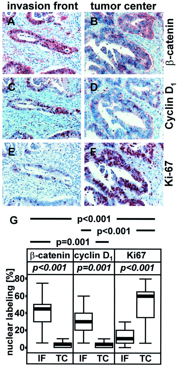
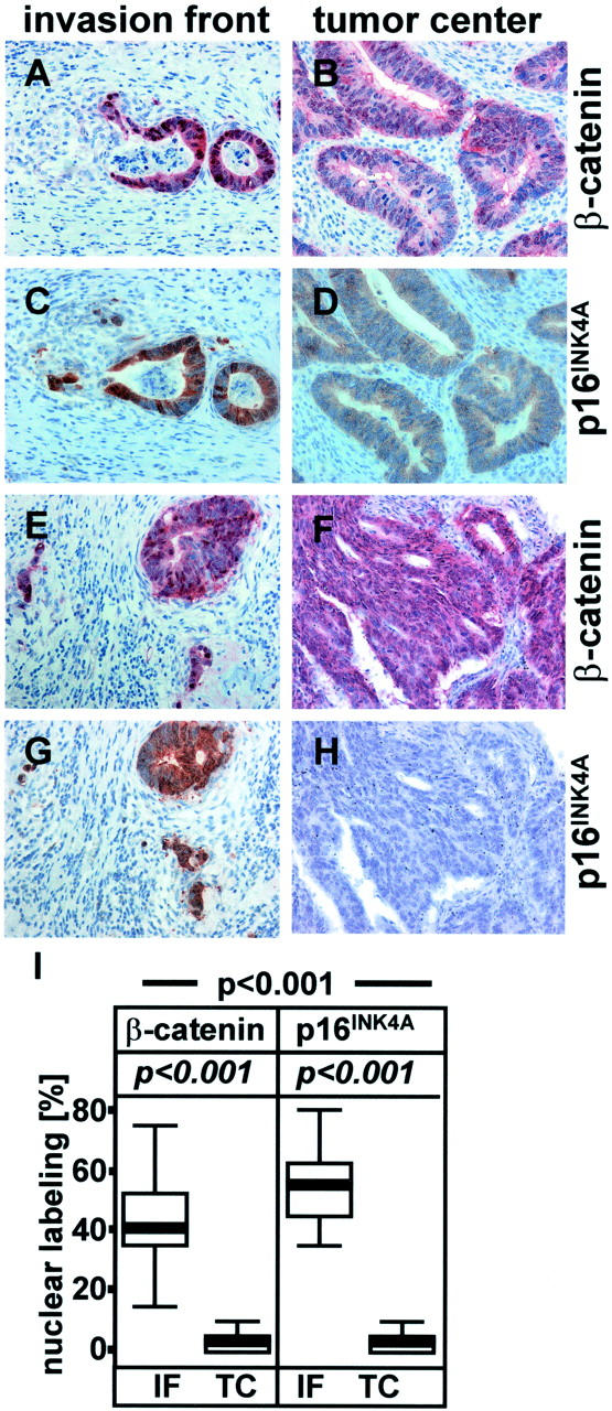
At the invasion front of well-differentiated colorectal adenocarcinomas, the oncogene beta-catenin is found in the nuclear compartment of tumor cells. Under these conditions, beta-catenin can function as a transcription factor and thus activate target genes. One of these target genes, cyclin D1, is known to induce proliferation. However, invasion front of well-differentiated colorectal adenocarcinomas are known to be zones of low proliferation and express the cell cycle inhibitor p16INK4A. Therefore, we investigated the expression profiles of nuclear beta-catenin, cyclin D1, p16INK4A, and the Ki-67 antigen, a marker for proliferation, in serial sections of well-differentiated colorectal adenocarcinomas. Invasion fronts with nuclear beta-catenin were compared with areas from central parts of the tumors without nuclear beta-catenin, for the expression of cyclin D1, p16INK4A, and Ki-67. It was observed that expression of nuclear beta-catenin, cyclin D1, and p16INK4A at the invasion front are significantly correlated. Such areas exhibit low Ki-67 expression indicating a low rate of proliferation. Thus, in colorectal carcinogenesis the function of beta-catenin and its target gene cyclin D1 does not appear to be the induction of tumor cell proliferation. In particular, the function of cyclin D1 should be reconsidered in view of these observations.
Figures
Similar articles
-
Elevated protein expression of cyclin D1 and Fra-1 but decreased expression of c-Myc in human colorectal adenocarcinomas overexpressing beta-catenin.Int J Cancer. 2002 Oct 1;101(4):301-10. doi: 10.1002/ijc.10630. Int J Cancer. 2002. PMID: 12209953
-
A survival-stratification model of human colorectal carcinomas with beta-catenin and p27kip1.Cancer. 2002 Dec 15;95(12):2479-86. doi: 10.1002/cncr.10986. Cancer. 2002. PMID: 12467060
-
Nuclear localization of beta-catenin is correlated with the expression of cyclin D1 in endometrial carcinomas.Anticancer Res. 2003 Sep-Oct;23(5A):3749-54. Anticancer Res. 2003. PMID: 14666673
-
Beta-catenin--a linchpin in colorectal carcinogenesis?Am J Pathol. 2002 Feb;160(2):389-401. doi: 10.1016/s0002-9440(10)64856-0. Am J Pathol. 2002. PMID: 11839557 Free PMC article. Review.
-
Beta-catenin and cyclin D1: connecting development to breast cancer.Cell Cycle. 2004 Feb;3(2):145-8. Cell Cycle. 2004. PMID: 14712077 Review.
Cited by
-
To differentiate or not--routes towards metastasis.Nat Rev Cancer. 2012 May 11;12(6):425-36. doi: 10.1038/nrc3265. Nat Rev Cancer. 2012. PMID: 22576165 Review.
-
Enhanced expression of hepatocyte growth factor activator inhibitor type 2-related small peptide at the invasive front of colon cancers.Gut. 2007 Feb;56(2):215-26. doi: 10.1136/gut.2005.084079. Epub 2006 Jun 29. Gut. 2007. PMID: 16809422 Free PMC article.
-
Converging signals synergistically activate the LAMC2 promoter and lead to accumulation of the laminin gamma 2 chain in human colon carcinoma cells.Biochem J. 2003 Apr 1;371(Pt 1):211-21. doi: 10.1042/BJ20021454. Biochem J. 2003. PMID: 12519076 Free PMC article.
-
Disruption of β-Catenin-Dependent Wnt Signaling in Colon Cancer Cells Remodels the Microenvironment to Promote Tumor Invasion.Mol Cancer Res. 2022 Mar 1;20(3):468-484. doi: 10.1158/1541-7786.MCR-21-0349. Mol Cancer Res. 2022. PMID: 34799404 Free PMC article.
-
Loss of enhancer of zeste homologue 2 (EZH2) at tumor invasion front is correlated with higher aggressiveness in colorectal cancer cells.J Cancer Res Clin Oncol. 2019 Sep;145(9):2227-2240. doi: 10.1007/s00432-019-02977-1. Epub 2019 Jul 17. J Cancer Res Clin Oncol. 2019. PMID: 31317325 Free PMC article.
References
-
- Kinzler KW, Vogelstein B: Lessons from hereditary colorectal cancer. Cell 1996, 87:159-170 - PubMed
-
- Polakis P: Wnt signaling and cancer. Genes Dev 2000, 14:1837-1851 - PubMed
-
- Korinek V, Barker N, Moerer P, van Donselaar E, Huls G, Peters PJ, Clevers H: Depletion of epithelial stem-cell compartments in the small intestine of mice lacking Tcf-4. Nat Genet 1998, 19:379-383 - PubMed
Publication types
MeSH terms
Substances
LinkOut - more resources
Full Text Sources
Medical
Research Materials
Miscellaneous
