. 2001 Nov;12(11):3573-88.
doi: 10.1091/mbc.12.11.3573.

GGAs: roles of the different domains and comparison with AP-1 and clathrin

Affiliations
Free PMC article

GGAs: roles of the different domains and comparison with AP-1 and clathrin

J Hirst et al. Mol Biol Cell. 2001 Nov.
Free PMC article

Abstract

We have previously identified a novel family of proteins called the GGAs (Golgi-localized, gamma-ear-containing, ADP-ribosylation factor-binding proteins). These proteins consist of an NH(2)-terminal VHS domain, followed by a GAT domain, a variable domain, and a gamma-adaptin ear homology domain. Studies from our own laboratory and others, making use of both yeast and mammals cells, indicate that the GGAs facilitate trafficking from the trans-Golgi network to endosomes. Here we have further investigated the function of the GGAs. We find that GGA-deficient yeast are not only defective in vacuolar protein sorting but they are also impaired in their ability to process alpha-factor. Using deletion mutants and chimeras, we show that the VHS domain is required for GGA function and that the VHS domain from Vps27p will not substitute for the GGA VHS domain. In contrast, the gamma-adaptin ear homology domain contributes to GGA function but is not absolutely required, and full function can be restored by replacing the GGA ear domain with the gamma-adaptin ear domain. Deleting the gamma-adaptin gene together with the two GGA genes exacerbates the phenotype in yeast, suggesting that they function on parallel pathways. In mammalian cells, the association of GGAs with the membrane is extremely unstable, which may account for their absence from purified clathrin-coated vesicles. Double- and triple-labeling immunofluorescence experiments indicate that the GGAs and AP-1 are associated with distinct populations of clathrin-coated vesicles budding from the trans-Golgi network. Together with results from other studies, our findings suggest that the GGAs act as monomeric adaptors, with the four domains involved in cargo selection, membrane localization, clathrin binding, and accessory protein recruitment.


Figures

Figure 1
Figure 1
GGA-deficient yeast have a defect in α-factor processing. (a) Colonies of wild-type (wt) cells, single-deletion mutants (gga1Δ and gga2Δ), or double-deletion mutants (gga1Δ/gga2Δ) were spotted onto a lawn of mating type a cells to test for the secretion of active α-factor. The reduced halo size surrounding the gga1Δ/gga2Δ colony when compared with the other three colonies indicates that less active α-factor is secreted in the double-deletion mutant. (b) Secreted α-factor was immunoprecipitated from the medium of metabolically labeled cells using an antibody that preferentially recognizes the precursor form. The gga1Δ/gga2Δ cells secrete mainly α-factor precursor.
Figure 2
Figure 2
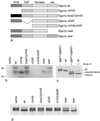
(a) Diagrams of the Gga1p deletion mutants and chimeras. These constructs were all transformed into gga1Δ/gga2Δ cells. (b) Stability of the various constructs in yeast. Total extracts of cells expressing each of the constructs shown in a, as well as cells transformed with empty vector, were subjected to SDS-PAGE, loading equal amounts of protein in each lane, and Western blots were probed with anti-Gga1p. Labeled bands with the expected mobility are seen in most of the cells, with the exception of the cells transformed with the ΔGAT and ΔVHSΔGAT constructs, indicating that these constructs are unstable. The lower molecular weight band seen in some of the lanes presumably corresponds to a breakdown product. (c) CPY is aberrantly processed in gga1Δ/gga2Δ cells. Cells were labeled with 35S for 10 min and chased for 30 min, and then cell extracts were immunoprecipitated with anti-CPY. In the absence of endo H digestion, CPY in the gga1Δ/gga2Δ cells runs as a triplet. In the wild-type cells, most of the CPY comigrates with the lowest molecular weight band of the triplet, indicating that it has been processed to the mature form, although there is still a small amount of higher molecular weight (p2) CPY that has not yet been processed. In the presence of endo H, which removes N-linked oligosaccharides, the CPY in the gga1Δ/gga2Δ cells still runs as a triplet, indicating that the middle (pseudomature) band is the result of partial proteolysis rather than incomplete glycosylation. (d) CPY in cells expressing the constructs shown in a. The CPY in the cells transformed with empty vector runs as a triplet, whereas in the cells rescued with wild-type Gga1p most of the CPY runs at the mature position. The ΔVHS, ΔGAT, and ΔVHSΔGAT deletion mutants all fail to rescue the phenotype, although the mobility of the CPY is somewhat different in the cells expressing the ΔVHS construct. Unlike the other deletion mutants, the Δear mutant partially rescues the phenotype. The Vps27pVHS domain chimera does not rescue the phenotype; however, the γ-ear domain chimera appears to give full rescue.
Figure 3
Figure 3
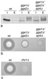
a. Extracts from intracellular (I) and extracellular (E) fractions of pulse-chased gga1Δ/gga2Δ cells transformed with either empty vector, wild-type Gga1p (wt), the Δ-ear deletion mutant, or the γ-ear chimera were immunoprecipitated with anti-CPY. The γ-ear chimera gives full rescue of CPY sorting, whereas the Δ-ear deletion mutant gives partial rescue. (b) Secretion of active α-factor was investigated using the halo assay. Again, the γ-ear chimera gives full rescue of the wild-type phenotype, and the Δ-ear deletion mutant gives partial rescue
Figure 4
Figure 4
Effect of deleting other coat components. (a) Wild-type (wt) cells, cells lacking both GGA genes (gga1Δ/gga2Δ), and cells lacking both GGA genes as well as γ-adaptin (gga1Δ/gga2Δ/apl4Δ) were pulse-chased, and CPY was immunoprecipitated from both intracellular (I) and extracellular (E) fractions. Although the ratios of extracellular to intracellular CPY are similar in the gga1Δ/gga2Δ and gga1Δ/gga2Δ/apl4Δ cells, the electrophoretic mobility of the secreted CPY is different in the triple mutant, suggesting that more proteolytic degradation has occurred. (b) Colonies of wild-type, gga1Δ/gga2Δ, and gga1Δ/gga2Δ/apl4Δ cells were spotted onto a lawn of mating type a cells and secretion of active α-factor was monitored using the halo assay. Deleting the APL4 gene together with the two GGA genes exacerbates the α-factor misprocessing phenotype. (c) The clathrin heavy chain (CHC1) gene was deleted in the same strain as the cells shown in b, and the halo assay was performed at the same time under identical conditions. The chc1Δ cells have a more severe phenotype than either the gga1Δ/gga2Δ cells or the gga1Δ/gga2Δ/apl4Δ cells.
Figure 5
Figure 5
The gga1Δ/gga2Δ/apl4Δ cells were transformed with either empty vector, wild-type Gga1p (wt), the Δ-ear deletion mutant, or the γ-ear chimera and then assayed for both CPY processing and active α-factor secretion. (a) Total CPY was immunoprecipitated from pulse-chased cells. The γ-ear chimera produces the same phenotype as the wild-type construct, whereas the Δ-ear deletion mutant produces an intermediate phenotype. (b) Secretion of active α-factor was monitored using the halo assay. Again, the γ-ear chimera gives full rescue and the Δ-ear deletion mutant gives partial rescue. Thus, even in the absence of γ-adaptin, the Δ-ear deletion mutant is partially functional.
Figure 6
Figure 6
Epitope tagging of Gga1p. (a) An HA tag was engineered onto the COOH terminus of Gga1p and expressed at either endogenous levels (HA[CEN]) or higher than normal levels (HA[2μ]). Cells were pulse-chased and total CPY was immunoprecipitated. The tagged construct gives full rescue at both endogenous and high levels. (b) Cells expressing HA-tagged Gga1p on a 2μ plasmid were metabolically labeled and then immunoprecipitated with anti-HA, together with cells expressing wild-type Gga1p (wt) as a control. The autoradiograph shows that the tagged construct is recognized by anti-HA. (c) Localization of the tagged Gga1p in a vps4 mutant. Cells were double labeled for HA-tagged Gga1p (top) and myc-tagged Vps10p (bottom). Vps10p is known to go to the class E compartment in a vps4 mutant; therefore, the colocalization of Gga1p with Vps10p indicates that it also goes to the class E compartment. The reason for the use of a class E mutant is that late Golgi, endosomal, and vacuolar proteins all accumulate in an exaggerated prevacuolar compartment, the class E compartment, making the membrane localization of tagged Gga1p much easier to see. Scale bar, 5 μm.
Figure 7
Figure 7
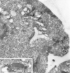
Localization of Gga1p at the electron microscope level. (a) vps4 cells expressing HA-tagged Gga1p on a 2μ plasmid were labeled with anti-HA followed by protein A coupled to 10 nm gold. Most of the label is associated with a multilamellar compartment with the characteristic appearance of the class E compartment. A coated vesicular profile is also labeled (arrowheads). (b) vps4 cells coexpressing HA-tagged Gga1p and myc-tagged Vps10p were double labeled for HA (10 nm gold, arrowheads) and myc (15 nm gold, arrows). The colocalization of the two proteins on the same multilamellar compartment confirms that in a vps4 mutant GGAs are targeted to the class E compartment. Scale bars, 200 nm.
Figure 8
Figure 8
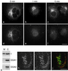
GGAs in mammalian cells. (a–f) COS cells were double labeled for the γ-adaptin subunit of AP-1 (a–c) and GGA1 (d–f). In cells that were fixed immediately after freezing and thawing (a and d), both proteins have a punctate perinuclear distribution, although the two patterns are distinct. In cells that had been incubated in a physiological buffer for either 1 min (b and e) or 5 min (c and f) after freezing and thawing, much of the γ-adaptin is still associated with the membrane (b and c), but membrane-associated GGA1 is no longer detectable (e and f). Scale bar, 20 μm. (g) HeLa cells were homogenized, unbroken cells and nuclei were pelleted, and the remaining postnuclear supernatant was centrifuged at 100,000 × g for 45 min. Blots of the membrane-containing pellets (M) and cytosol-containing supernatants (C) were then probed with antibodies against γ-adaptin, GGA1, and GGA2. Nearly half of the γ-adaptin remains in the membrane-containing pellet; however, at least 95% of the GGA1 and GGA2 are in the cytosol. H–j. HeLa cells were double labeled with antibodies against GGA1 (h) and clathrin (i) and then photographed with a confocal microscope. There is significant colocalization between the two proteins, as shown in the merged image (j; GGA1 is labeled green, clathrin is labeled red). Scale bar, 10 μm.
Figure 9
Figure 9
Triple labeling of COS cells. COS cells were fixed in methanol/acetate and then labeled sequentially with rabbit anti-GGA1 followed by Alexa488 protein A (a), rabbit anti-clathrin followed by Alexa594 protein A (b), and mouse anti-γ-adaptin followed by Alexa350 goat anti-mouse. (d) Merging of the three images, with GGA1 labeled in green, clathrin labeled in red, and γ-adaptin labeled in blue. Overlap between clathrin and GGA1 appears yellow, and overlap between clathrin and γ-adaptin appears purple-pink. The presence of discrete structures labeled either for clathrin and GGA1 or for clathrin and γ-adaptin indicates that GGA1 and AP-1 are segregated from each other but both are associated with clathrin. Scale bars, 20 μm.
Figure 10
Figure 10
Schematic diagram showing the four distinct GGA domains and their predicted functions. The VHS domain has been shown to bind to the cytoplasmic tails of proteins that traffic from the TGN to endosomes (Nielsen et al., 2001; Puertollano et al., 2001a; Zhu et al., 2001), indicating that it is involved in cargo selection. The GAT domain both binds ARF and targets the protein to the Golgi apparatus (Boman et al., 2000; Dell'Angelica et al., 2000; Puertollano et al., 2001b). The variable domain binds clathrin (Costaguta et al., 2001; Puertollano et al., 2001b). The ear domain is likely to recruit accessory proteins, by analogy with the α-adaptin and γ-adaptin ear domains (Page et al., 1999; Slepnev and De Camilli, 2000).

Similar articles

Cited by

References

    1. Austin C, Hinners I, Tooze SA. Direct and GTP-dependent interaction of ADP-ribosylation factor 1 with clathrin adaptor protein AP-1 on immature secretory granules. J Biol Chem. 2000;275:21862–21869. - PubMed
    1. Barlowe C, Orci L, Yeung T, Hosobuchi M, Hamamoto S, Salama N, Rexach MF, Ravazzola M, Amherdt M, Schekman R. COPII: a membrane coat formed by Sec proteins that drive vesicle budding from the endoplasmic reticulum. Cell. 1994;77:895–907. - PubMed
    1. Black MW, Pelham HR. A selective transport route from Golgi to late endosomes that requires the yeast GGA proteins. J Cell Biol. 2000;151:587–600. - PMC - PubMed
    1. Boman AL, Zhang CJ, Zhu X, Kahn RA. A family of ADP-ribosylation factor effectors that can alter membrane transport through the trans-Golgi. Mol Biol Cell. 2000;11:1241–1255. - PMC - PubMed
    1. Brodsky FM. Clathrin structure characterized with monoclonal antibodies. I. Analysis of multiple antigenic sites. J Cell Biol. 1985;101:2047–2054. - PMC - PubMed

Publication types

MeSH terms

LinkOut - more resources