. 2001 Nov 1;15(21):2837-51.
doi: 10.1101/gad.937401.

Reconstitution of recombinant chromatin establishes a requirement for histone-tail modifications during chromatin assembly and transcription

Affiliations

Reconstitution of recombinant chromatin establishes a requirement for histone-tail modifications during chromatin assembly and transcription

A Loyola et al. Genes Dev. .

Abstract

The human ISWI-containing factor RSF (remodeling and spacing factor) was found to mediate nucleosome deposition and, in the presence of ATP, generate regularly spaced nucleosome arrays. Using this system, recombinant chromatin was reconstituted with bacterially produced histones. Acetylation of the histone tails was found to play an important role in establishing regularly spaced nucleosome arrays. Recombinant chromatin lacking histone acetylation was impaired in directing transcription. Histone-tail modifications were found to regulate transcription from the recombinant chromatin. Acetylation of the histone tails by p300 was found to increase transcription. Methylation of the histone H3 tail by Suv39H1 was found to repress transcription in an HP1-dependent manner. The effects of histone-tail modifications were observed in nuclear extracts. A highly reconstituted RNA polymerase II transcription system was refractory to the effect imposed by acetylation and methylation.


Figures

Figure 1
Figure 1
RSF mediates the assembly of nucleosome arrays. (A) Ethidium bromide staining of micrococcal nuclease digestions of chromatin assembled by RSF. Chromatin assembly was performed in the presence (left) and absence (right) of NAP-1, with increasing amounts of RSF. The highest amount of RSF was also assayed in the absence of ATP, as indicated on the panel. Products of the assembly reaction were partially digested with two different concentrations of micrococcal nuclease. Each lane number, at the bottom of the panel, denotes two different concentrations of micrococcal nuclease used in the assays. (B) Fractionation of RSF by gel filtration. Fractions of the Superose 6 column were assayed by Coomassie blue staining (top), Western blot (middle), and chromatin assembly (bottom). Each lane number on the micrococcal nuclease assay is between lanes and denotes two different concentrations of micrococcal nuclease used in the assay. RSF fractionated between fractions 32 and 36, which corresponds to a complex of ∼450 kD. (C) Southern blot of micrococcal nuclease digestions of RSF-assembled chromatin. Supercoiled (pG5MLP) DNA was linearized with EcoRI and used as a substrate in the RSF-assembly reaction (RFIII). As a control, supercoiled DNA was assayed in parallel (RFI). Lane numbers, at the bottom of the panel, denote two different concentrations of micrococcal nuclease used in the assays.
Figure 2
Figure 2
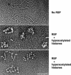
Electron microscopic analysis of RSF-assembled chromatin. (Top) An electron microscopic visualization of the RSF-assembled chromatin performed in the absence of RSF. DNA size is 3 kb. (Middle) The RSF-assembled chromatin performed with hypoacetylated histones purified from HeLa cells. (Bottom) The reaction with RSF and hyperacetylated histones purified in the presence of the histone deacetylase inhibitors TSA and Na-butyrate. The pictures are representatives of the majority of the molecules visualized on the grid. Bar, 116 nm.
Figure 3
Figure 3
RSF interacts with the histone tetramer, and its association with DNA requires the octamer. (A) Silver staining of an immunoprecipitation of the RSF–histone complexes. (Top) A schematic representation of the assay used to analyze the interaction of RSF with histones is depicted. The reaction was performed in the presence and absence of RSF, with HeLa core histones, H3/H4 tetramers, or H2A/H2B dimers, as indicated above the figure. H3/H4 tetramers and H2A/H2B dimers were separated over a Mono S column as described (Chang and Luse 1997). Immunoprecipitation reactions were performed as described in Materials and Methods. The immunocomplexes were washed with a buffer containing 0.5 M KCl and 0.01% NP-40. Immunoprecipitates were separated by SDS-PAGE followed by silver staining. (Right) The migration of each of the histones. (B) Ethidium bromide staining of an agarose gel showing a requirement for core histones for the establishment of an RSF–DNA complex.(Top) Schematic representation of the steps used to analyze binding of RSF to DNA. EcoRI-linearized DNA, RSF and core histone octamers were incubated in the presence and absence of ATP (0.6 μM) and ATP-γS (6 mM ATP), as indicated. H3/H4 tetramers, H2A/H2B dimers, and H3/H4 tetramers plus H2A/H2B dimers were incubated with or without RSF for 30 min at 30°C. The complexes were immunoprecipitated, and the immunoprecipitates were washed with a buffer containing 0.5 M KCl and 0.01% NP40 as described in Materials and Methods. Products of the immunoprecipitation reaction were separated by electrophoresis on an agarose gel. The arrow indicates the migration of DNA. (C) Electron microscopic visualization of intermediates in the RSF-chromatin assembly reaction. RSF was incubated with EcoRI-linearized plasmid and core histones in the presence of 6 mM ATP-γS plus 0.6 μM ATP for 30 min at 30°C. (Left) The products of the reaction visualized by electron microscopy. (Middle, right) RSF was incubated with EcoRI-linearized DNA and histones as above. The products of the reactions were first incubated with antibodies against histones H3 (monoclonal) and H4 (polyclonal; middle) or against the RSF subunit hSNF2h (monoclonal; right). This was followed by the incubation with a secondary antibody conjugated with gold (10 nm for H3 and H4 antibodies, 5 nm for hSNF2h antibody), as described in Material and Methods. The samples were visualized by electron microscopy. The arrow shows the secondary antibody gold particle. The big protein complex that appears in the reaction with antibodies against H3 and H4 (middle) represents multiples secondary antibodies bound to the primary antibody. The pictures are representatives of the majority of the molecules visualized on the grid. Bar, 85 nm.
Figure 4
Figure 4
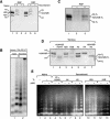
Reconstitution of RSF-mediated chromatin assembly with recombinant histones. (A) RSF interacts with recombinant histones. Silver staining of coimmunoprecipitates obtained using antibodies against hSNF2h. (Lanes 4–6) Results with RSF and recombinant histones. (Lanes 1–3) As a control, HeLa purified core histones were used. The immunoprecipitates were washed with a buffer containing 0.5 M KCl and 0.01% NP-40. Indicated are the migration of recombinant histones (right) and the migration of native histones (left). (B) Southern blot analysis of partial micrococcal nuclease digestion of RSF assembly with native and recombinant histones, as indicated. Each lane number, at the bottom of the panel, denotes two different concentrations of micrococcal nuclease used in the assays. (C) Silver staining of RSF immunoprecipitates from reactions using recombinant histone octamer deleted of N-terminal tails. Immunoprecipitation was performed using antibodies against hSNF2h. The immunoprecipitates were washed with a buffer containing 0.5 M KCl and 0.01% NP-40. (Right) The migration of recombinant tailless histones. (D) Coomassie blue staining of a 15% SDS-PAGE containing different octamers formed with histone polypeptides deleted of the tails, as indicated at the top of the panel. The migration of wild-type histones (left) and tailless histones (right). (E) Ethidium bromide staining of partial micrococcal nuclease digestion of products obtained in the RSF-assembled chromatin using recombinant octamers with different combinations of histone tails (indicated at top). The octamers used were those analyzed in D. Reactions were performed in the presence and the absence of p300 plus 10 μM acetyl-CoA as indicated. As a control, the first lane represents chromatin assembled with native histones. Each lane number, at the bottom of the panel, denotes two different concentrations of micrococcal nuclease used in the assays.
Figure 5
Figure 5
Transcription on RSF-assembled chromatin. (A) Southern blot of micrococcal nuclease digestions of RSF-assembled and remodeled chromatin. Chromatin was assembled with RSF in the presence and absence of the activator Gal4VP16, added during the assembly or after the assembly as indicated. The Southern blot was probed with a promoter probe (left), followed by stripping off the probe and reprobing with a distal probe (right). The illustration (bottom) shows schematically the location of the probes (for details, see Orphanides et al. 1998). Each lane number, at the bottom of the panel, denotes two different concentrations of micrococcal nuclease used in the assays. (B) Transcription on RSF-assembled chromatin. (Top) A scheme of the procedure used to deposit chromatin and its analysis on transcription. Transcription was performed with nuclear extract in the presence and absence of the activator Gal4VP16. As a control, transcription from naked DNA is shown (lanes 1–3). The same amount of DNA was used in all lanes. Products of the transcription reactions were separated by electrophoresis on a denaturing polyacrylamide gel. (Right) Full-length transcripts (390 nt). (C) Reconstituted transcription on RSF-assembled chromatin. Transcription was performed using a reconstituted system, in the presence and absence of FACT. All reactions contained the activator Gal4VP16. Short products are observed as a result of transcription without FACT. Products of the transcription reactions were separated by electrophoresis on a denaturing polyacrylamide gel. (Right) Full-length transcripts obtained in the presence of FACT (390 nt). (D) Transcription on nuclear extract using chromatin templates reconstituted with hypo- and hyperacetylated histones isolated from HeLa cells. Increasing amounts of hypo- and hyperacetylated chromatin (equal molar amounts) were added to nuclear extract in the presence and absence of the activator Gal4VP16. Products of the transcription reactions were separated by electrophoresis on a denaturing polyacrylamide gel. (Right) Full-length transcripts (390 nt). (E) Analysis of the chromatin reconstituted with hypo- and hyperacetylated histones using a reconstituted transcription system. Transcription reactions were performed in the presence of Gal4–VP16, but with and without FACT, as described in Materials and Methods. Products of the transcription reactions were separated by electrophoresis on a denaturing polyacrylamide gel. (Right) Full-length transcripts (390 nt).
Figure 6
Figure 6
Transcription using chromatin reconstituted with RSF and recombinant histones. (A) p300 stimulates transcription in nuclear extract but not in the reconstituted system. Chromatin templates were incubated (top, bottom) with recombinant p300 and 1 μM acetylCoA/2 mM Na-butyrate for 30 min before performing transcription. Assays performed in the reconstituted system (top) received Gal4VP16 and were performed in the presence or absence of FACT, as indicated. Transcriptions performed in extracts (bottom) received Gal4VP16 as indicated. Products of the transcription reactions were separated by electrophoresis on a denaturing polyacrylamide gel. (Right) Full-length transcripts (390 nt). (B) SUV39H1 methylates octamer and nucleosome. Baculovirus-expressed Suv39H1 was assayed for methylation on recombinant core histone octamers and recombinant nucleosomes. The substrates were incubated with 3H-SAM in the presence of Suv39H1 for 30 min at 30°C. Products were separated by SDS-PAGE, and labeled polypeptides were detected. (Right) Methylated histone H3. (C) Suv39H1 methylation inhibits transcription in an HP1β-dependent manner. Chromatin was incubated with recombinant Suv39H1 and 2.5 μM SAM for 30 min followed by 30-min incubation with HP1β. The reaction was further incubated under transcription conditions using nuclear extracts in the presence or absence of Gal4VP16, as indicated. Products of the transcription reactions were separated by electrophoresis on a denaturing polyacrylamide gel. (Right) Full-length transcripts (390 nt). (D) Suv39H1/HP1β-mediated repression is not observed in the reconstituted transcription system. Chromatin was incubated with recombinant Suv39H1 and 2.5 μM SAM for 30 min followed by 30-min incubation with HP1β followed by transcription in a reconstituted system. Reactions were performed in the presence of Gal4VP16 in the presence or absence of FACT, as indicated. Products of the transcription reactions were separated by electrophoresis on a denaturing polyacrylamide gel. (Right) Full-length transcripts (390 nt).

Similar articles

Cited by

References

    1. Agalioti T, Lomvardas S, Parekh B, Yie J, Maniatis T, Thanos D. Ordered recruitment of chromatin modifying and general transcription factors to the IFN-β promoter. Cell. 2000;103:667–678. - PubMed
    1. Angelov D, Vitolo JM, Mutskov V, Dimitrov S, Hayes JJ. Preferential interaction of the core histone tail domain with linker DNA. Proc Natl Acad Sci. 2001;98:6599–6604. - PMC - PubMed
    1. Bannister AJ, Zegerman P, Partridge JF, Miska EA, Thomas JO, Allshire RC, Kouzarides T. Selective recognition of methylated lysine 9 on histone H3 by the HP1 chromo domain. Nature. 2001;410:120–124. - PubMed
    1. Berger SL. Molecular biology: The histone modification circus. Science. 2001;292:64–65. - PubMed
    1. Bochar DA, Savard J, Wang W, Lafleur DW, Moore P, Cote J, Shiekhattar R. A family of chromatin remodeling factors related to Williams syndrome transcription factor. Proc Natl Acad Sci. 2000;97:1038–1043. - PMC - PubMed

Publication types

LinkOut - more resources