. 2001 Oct 29;155(3):447-58.
doi: 10.1083/jcb.200105017. Epub 2001 Oct 29.

EGF-R signaling through Fyn kinase disrupts the function of integrin alpha6beta4 at hemidesmosomes: role in epithelial cell migration and carcinoma invasion

Affiliations

EGF-R signaling through Fyn kinase disrupts the function of integrin alpha6beta4 at hemidesmosomes: role in epithelial cell migration and carcinoma invasion

A Mariotti et al. J Cell Biol. .

Abstract

We have examined the mechanism and functional significance of hemidesmosome disassembly during normal epithelial cell migration and squamous carcinoma invasion. Our findings indicate that a fraction of EGF receptor (EGF-R) combines with the hemidesmosomal integrin alpha6beta4 in both normal and neoplastic keratinocytes. Activation of the EGF-R causes tyrosine phosphorylation of the beta4 cytoplasmic domain and disruption of hemidesmosomes. The Src family kinase inhibitors PP1 and PP2 prevent tyrosine phosphorylation of beta4 and disassembly of hemidesmosomes without interfering with the activation of EGF-R. Coimmunoprecipitation experiments indicate that Fyn and, to a lesser extent, Yes combine with alpha6beta4. By contrast, Src and Lck do not associate with alpha6beta4 to a significant extent. A dominant negative form of Fyn, but not Src, prevents tyrosine phosphorylation of beta4 and disassembly of hemidesmosomes. These observations suggest that the EGF-R causes disassembly of hemidesmosomes by activating Fyn, which in turn phosphorylates the beta4 cytoplasmic domain. Neoplastic cells expressing dominant negative Fyn display increased hemidesmosomes and migrate poorly in vitro in response to EGF. Furthermore, dominant negative Fyn decreases the ability of squamous carcinoma cells to invade through Matrigel in vitro and to form lung metastases following intravenous injection in nude mice. These results suggest that disruption of hemidesmosomes mediated by Fyn is a prerequisite for normal keratinocyte migration and squamous carcinoma invasion.


Figures

Figure. 1.
Figure. 1.
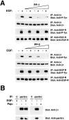
A fraction of EGF-R is associated with α6β4 and induces tyrosine phosphorylation of the β4 cytoplasmic domain. The indicated cells were serum starved and then treated with EGF (25 ng/ml, 4 min) or left untreated. After solubilization in TX-DOC lysis buffer, the samples were immunoprecipitated with normal mouse IgGs (C) or the anti-β4 monoclonal antibody 3E1 (β4) followed by immunoblotting with the rabbit anti–β4-exo antiserum, the monoclonal antibody to P-Tyr RC-20, and rabbit antibodies to the COOH-terminus of the EGF-R or ErbB-2.
Figure 2.
Figure 2.
A Src family kinase is associated with α6β4 and phosphorylates β4 in response to activation of the EGF-R. (A) After serum starvation, HaCaT cells were incubated for 15 min with the Src family kinase inhibitor PP-1 or -2 (1, 2, and 4 μM), then treated with 25 ng/ml EGF for 4 min in presence of the inhibitors. The samples were immunoprecipitated with the anti-β4 monoclonal antibody 3E1 and probed by immunoblotting with the monoclonal antibody to P-Tyr RC-20 or the rabbit anti–β4-cyto serum. As a control, the samples were immunoprecipitated with the anti-EGFR monoclonal antibody 528 and probed by immunoblotting with the monoclonal antibody to P-Tyr RC-20 or the rabbit antiserum to the COOH terminus of the EGF-R. (B) Lysates from serum-starved HaCaT cells treated with EGF or left untreated (25 ng/ml, 4 min) were immunoprecipitated with normal rabbit IgGs (C) or the affinity-purified rabbit antibodies SRC2, which recognize Src, Fyn, and Yes (panSrc). The samples were probed by immunoblotting with rabbit anti–β4-cyto or anti-panSrc antibodies. The coimmunoprecipitation of α6β4 and Src kinases was blocked in presence of 20 μg of the peptide used as antigen to generate the anti-panSrc antibodies (pep).
Figure 3.
Figure 3.
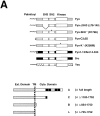
Mutational analysis of the association of Fyn with α6β4. (A) Structure of the Src family kinases (top) and wild-type and mutant β4 subunits (bottom) used to define the association of Fyn with α6β4. *, point mutations. β4 constructs: black boxes, Fn type III modules; grey box, connecting segment. (B) 293 T cells were transiently transfected with plasmids encoding wild-type α6 and β4 in combination with the indicated wild-type and mutant Src family kinases. After extraction in RIPA buffer, the lysates were immunoprecipitated with the monoclonal antibody 3E1 covalently linked to Sepharose beads. The top part of the blot was probed with the rabbit antiserum β4-exo and the bottom with affinity purified anti-panSrc antibodies (SRC2). Total lysates were probed by immunoblotting with affinity-purified anti-Fyn (FYN3) antibodies to verify the level of expression of wild-type and mutant Fyn proteins (with the exception of the Fyn–Src chimera) and anti-panSrc (SRC2) antibodies to verify the level of expression of Src, the chimera Fyn–Src, and Yes; IgG H, immunoglobulin heavy chain. (C) 293 T cells were transiently transfected with plasmids encoding wild-type Fyn and α6 in combination with the indicated wild-type and mutant β4 subunits. After extraction in RIPA buffer, the lysates were immunoprecipitated with the monoclonal antibody 3E1 covalently linked to Sepharose beads. The top part of the blot was probed with the rabbit antiserum β4-exo and the bottom with affinity-purified antibodies to Fyn (Fyn3). Total lysates were probed by immunoblotting with the same antibodies to verify equal expression of Fyn in the four samples.
Figure 3.
Figure 3.
Mutational analysis of the association of Fyn with α6β4. (A) Structure of the Src family kinases (top) and wild-type and mutant β4 subunits (bottom) used to define the association of Fyn with α6β4. *, point mutations. β4 constructs: black boxes, Fn type III modules; grey box, connecting segment. (B) 293 T cells were transiently transfected with plasmids encoding wild-type α6 and β4 in combination with the indicated wild-type and mutant Src family kinases. After extraction in RIPA buffer, the lysates were immunoprecipitated with the monoclonal antibody 3E1 covalently linked to Sepharose beads. The top part of the blot was probed with the rabbit antiserum β4-exo and the bottom with affinity purified anti-panSrc antibodies (SRC2). Total lysates were probed by immunoblotting with affinity-purified anti-Fyn (FYN3) antibodies to verify the level of expression of wild-type and mutant Fyn proteins (with the exception of the Fyn–Src chimera) and anti-panSrc (SRC2) antibodies to verify the level of expression of Src, the chimera Fyn–Src, and Yes; IgG H, immunoglobulin heavy chain. (C) 293 T cells were transiently transfected with plasmids encoding wild-type Fyn and α6 in combination with the indicated wild-type and mutant β4 subunits. After extraction in RIPA buffer, the lysates were immunoprecipitated with the monoclonal antibody 3E1 covalently linked to Sepharose beads. The top part of the blot was probed with the rabbit antiserum β4-exo and the bottom with affinity-purified antibodies to Fyn (Fyn3). Total lysates were probed by immunoblotting with the same antibodies to verify equal expression of Fyn in the four samples.
Figure 4.
Figure 4.
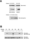
Dominant negative Fyn, but not Src, suppresses EGF-mediated tyrosine phosphorylation of β4. (A) Total lysates of 804G clones stably transfected with empty vector (C1) or dominant negative Fyn (F1, 2, and 3) were probed by immunoblotting with affinity purified anti-Fyn (FYN3) or anti-panSrc antibodies (SRC2). Total lysates of 804G clones stably transfected with empty vector (C1) or dominant negative Src (S1 and 2) were probed by immunoblotting with affinity purified anti-Src (N-16) or anti-panSrc antibodies (SRC2). (B) After serum starvation, the indicated 804G clones were treated with 50 ng/ml EGF for 5 min or left untreated. The lysates were immunoprecipitated with the rabbit antiserum β4 cyto and probed by immunoblotting with the anti–P-Tyr monoclonal antibody RC-20 or the rabbit antiserum β4 cyto.
Figure 5.
Figure 5.
Dominant negative Fyn promotes assembly and/or inhibits disassembly of hemidesmosomes. The indicated 804G clones were plated on glass coverslips for 24 h and then treated for 12 h with 50 ng/ml EGF in serum-free medium or left untreated. After a mild extraction with Triton X-100, the cells were fixed and stained with affinity purified antibodies to BPAG-2 to visualize hemidesmosomal structures. Results similar to those shown here were obtained with all three clones expressing dominant negative Fyn and the two clones expressing dominant negative Src.
Figure 6.
Figure 6.
Dominant negative Fyn inhibits disassembly of hemidesmosomes and suppresses cell migration. The indicated 804G clones were grown until confluent and then serum starved. After wounding, the monolayers were treated with EGF (50 ng/ml) for 5 or 10 h or left untreated for 5 h, and then either stained with Crystal violet or subjected to immunofluorescent staining with affinity purified anti–BPAG-2 antibodies. Cell migration results were quantitated as described in the Materials and methods section and plotted graphically. The graph shows the mean and SD of values from a representative experiment performed in triplicate. Results similar to those shown here were obtained with all three clones expressing dominant negative Fyn and the two clones expressing dominant negative Src. Bar, 250 μm.
Figure 7.
Figure 7.
Src-family kinases are required for squamous carcinoma invasion through Matrigel. A431 and MSK-QLL-1 cells were plated on Matrigel together with the Src family kinase inhibitors Herbimycin A (100 ng/ml) and PP-2 (1, 5, and 10 μM), the PI-3K inhibitor LY294002 (25 μM), or the p38 kinase inhibitor SB203580 (10 μM). Serum-free medium containing EGF (25 ng/ml) was added to the lower chamber. After 24 h, cells that had invaded through Matrigel and remained attached to the lower side of the filter were stained with Crystal violet and counted under a microscope. The graphs show the mean and SD of values from a representative experiment performed in triplicate.
Figure 8.
Figure 8.
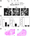
Dominant negative Fyn restores hemidesmosomes and inhibits both Matrigel invasion and experimental metastasis in A431 cells. (A) Two A431 clones expressing dominant negative Fyn (F35 and F25) and a control clone (C1) were starved and then treated with EGF (25 ng/ml) for 10 min or left untreated. RIPA lysates were immunoprecipitated with the anti-β4 monoclonal antibody 3E1 and probed by immunoblotting with the anti–P-Tyr monoclonal antibody RC-20 or the rabbit antiserum β4 cyto. The indicated A431 clones were plated on laminin 5 matrix–coated glass coverslips for 48 h. Cells were mildly permeabilized with Triton X-100, fixed, and stained with the anti-β4 monoclonal antibody 3E1. (C) The indicated A431 clones were tested for their ability to migrate in an in vitro wound assay, to invade through Matrigel in response to EGF (25 ng/ml) as described in the legends to Figs. 6 and 7, and to form lung metastases upon injection in the tail vein of nude mice. The number of metastatic over total number of lungs examined is shown at the top of each bar. (D) Hematoxylin and eosin staining of lung sections from mice injected with C1 or F25 cells. The arrows point to a large and a smaller lung metastasis in a mouse injected with C1 cells; no metastases are visible in the lung section from a mouse injected with F25 cells. Bar, 500 μm.
Figure 9.
Figure 9.
Relationship between α6β4 signaling and assembly of hemidesmosomes. Hypothetical model of the underlying pathways. We have observed that the palmitoylated fraction of α6β4 is localized in lipid rafts and is preferentially associated with Fyn and Yes (unpublished data). We hypothesize that this fraction of α6β4 mediates recruitment of Shc and activation of Ras and PI-3K. The EGF-R induces α6β4 signaling by activating Fyn and Yes and causing phosphorylation of the β4 cytoplasmic domain. It is likely that the signaling fraction of α6β4 exists in dynamic equilibrium with the cytoskeletal fraction involved in assembly of hemidesmosomes. We hypothesize that assembly of hemidesmosomes requires dephosphorylation of the β4 tail (Dans et al., 2001).

Similar articles

Cited by

References

    1. Alland, L., S.M. Peseckis, R.E. Atherton, L. Berthiaume, and M.D. Resh. 1994. Dual myristylation and palmitylation of Src family member p59fyn affects subcellular localization. J. Biol. Chem. 269:16701–16705. - PubMed
    1. Andra, K., H. Lassmann, R. Bittner, S. Shorny, R. Fassler, F. Propst, and G. Wiche. 1997. Targeted inactivation of plectin reveals essential function in maintaining the integrity of skin, muscle, and heart cytoarchitecture. Genes Dev. 11:3143–3156. - PMC - PubMed
    1. Baldari, C.T., J.L. Telford, and O. Acuto. 2000. Lymphocyte antigen receptor and coreceptor signaling. Embo J. 19:4857–4865. - PMC - PubMed
    1. Biscardi, J.S., D.A. Tice, and S.J. Parsons. 1999. c-Src, receptor tyrosine kinases, and human cancer. Adv. Cancer Res. 76:61–119. - PubMed
    1. Borradori, L., and A. Sonnenberg. 1999. Structure and function of hemidesmosomes: more than simple adhesion complexes. J. Invest. Dermatol. 112:411–418. - PubMed

Publication types

MeSH terms