. 2001 Oct 15;15(20):2720-9.
doi: 10.1101/gad.908401.

Coilin forms the bridge between Cajal bodies and SMN, the spinal muscular atrophy protein

Affiliations

Coilin forms the bridge between Cajal bodies and SMN, the spinal muscular atrophy protein

M D Hebert et al. Genes Dev. .

Abstract

Spinal muscular atrophy (SMA) is a genetic disorder caused by mutations in the human survival of motor neuron 1 gene, SMN1. SMN protein is part of a large complex that is required for biogenesis of various small nuclear ribonucleoproteins (snRNPs). Here, we report that SMN interacts directly with the Cajal body signature protein, coilin, and that this interaction mediates recruitment of the SMN complex to Cajal bodies. Mutation or deletion of specific RG dipeptide residues within coilin inhibits the interaction both in vivo and in vitro. Interestingly, GST-pulldown experiments show that coilin also binds directly to SmB'. Competition studies show that coilin competes with SmB' for binding sites on SMN. Ectopic expression of SMN and coilin constructs in mouse embryonic fibroblasts lacking endogenous coilin confirms that recruitment of SMN and splicing snRNPs to Cajal bodies depends on the coilin C-terminal RG motif. A cardinal feature of SMA patient cells is a defect in the targeting of SMN to nuclear foci; our results uncover a role for coilin in this process.


Figures

Figure 1
Figure 1
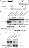
Coilin contains a conserved RG box and interacts with SMN. (A) Schematic representation of human coilin. The self-interaction domain and positively charged nuclear and nucleolar localization signals (NLS, NoLS) are indicated (Hebert and Matera 2000). The two stippled regions represent potentially negatively charged regions and reside upstream of the bipartite RG box, spanning residues 392–420 in human. For comparison, similar RG-box motifs from other vertebrate coilins as well as from the human SmD3 and SmD1 proteins are aligned. Carats (^) mark sites where two to three amino acids were excluded to facilitate the alignment. Gaps are denoted by a dash. The top line of the alignment shows the position of a mutation of the RG box (substitution of four arginines to glycines) used in this study (mtRG). (B) Coilin interacts with SMN in HeLa lysate. Immunoprecipitation of the lysate by either an anti-SMN antibody or normal mouse serum (NMS) was conducted as described in Materials and Methods, followed by SDS-PAGE and Western blotting with anticoilin antibodies. The input lane shows 8% of the lysate used in the immunoprecipitation reactions.
Figure 2
Figure 2
The coilin RG box mediates interaction with SMN. (A) Schematic of coilin constructs. The RG box is indicated, as is the tag used for each construct. GFP–C214 contains the C-terminal 214 amino acids of coilin, residues 362–576. Myc-tagged coilinΔNΔC spans residues 94–482, as shown. The RG box of each construct was mutated (mtRG), as shown in Fig. 1A. Furthermore, the entire RG box (392–420) was deleted in myc–ΔNΔC (myc–ΔNΔCΔRG). (B) Coilin interaction with SMN is dependent on the RG box. HeLa cells were transfected with the various coilin constructs, and the lysates were immunoprecipitated with anti-GFP (lanes 2–5) or anti-myc (lanes 6,7) antibodies, as described in Materials and Methods. The beads were washed and subjected to SDS-PAGE and Western blotting with anti-SMN antibodies (top). The immunoglobulin (IgG) heavy (H) and light (L) chains are marked. The input lane shows 5% of the lysate used in the immunoprecipitation reactions. The same immunoprecipitates were electrophoresed and blotted with anticoilin antibodies to show that equal amounts of protein were pulled down (bottom). (C) Deletion of the RG box in coilin reduces SMN interaction. HeLa cells were transfected with myc–ΔNΔC or myc–ΔNΔCΔRG, and the lysates were immunoprecipitated with monoclonal anti-myc antibodies, followed by SDS-PAGE and Western blotting with anti-SMN antibodies (top). The same blot was reprobed with polyclonal anti-myc antibodies (bottom). The input lanes show 5% of the lysate used in the reactions.
Figure 3
Figure 3
Coilin directly interacts with SMN via the RG box. (A) Binding assays of His–SMN and GST–C214 or GST–C214(mtRG) were conducted as described in Materials and Methods, followed by SDS-PAGE and Western blotting with anti-SMN antibodies (top). The same blot was reprobed with anticoilin antibodies (bottom) to show that equal amounts of GST–C214 and GST–C214(mtRG) were used. (B) Mapping of the coilin interaction site on SMN. Various GST–SMN fusions were tested for their ability to bind His–C214. GST–SMNΔEx7 does not contain exon 7 sequences. GST–SMN(Ex1–3) and GST–SMN(Ex3) contain the first three or only the third exon of SMN, respectively. The Western blot was probed with an anticoilin antibody (top). The same blot was reprobed with anti-GST antibodies to verify that equal amounts of beads were used in the assay (bottom). (C) Control pulldowns with the same GST–SMN constructs used in B were assayed using His–T7–SmB′. Western blotting was performed with an anti-T7 antibody (top) or an anti-GST antibody (bottom). The input lanes for all reactions are equivalent to 10% of that used in the binding assay. (D,E) The Tudor domain of SMN mediates binding to coilin and SmB′. GST–SMN(Ex3), encompassing SMN residues 91–158, and GST–SMN(Tudor), spanning residues 83–173, were used in pulldown assays with purified coilin-C214 or SmB′. A less stringent buffer was used in these binding assays compared with the buffer used in A–C (see Materials and Methods). The blots were reprobed with an antibody to GST to verify that equal amounts of beads were used in the assay. The input lane for D was equivalent to 10% of the C214 used in the binding assay, whereas the input lane for E represents 20% of the SmB′ used in the binding reaction.
Figure 4
Figure 4
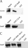
Coilin interacts with SmB′. (A) Direct interaction of His–C214 with GST–SmB′, but not with GST alone. His–C214 was detected by anticoilin antibodies. (B) His–T7–SmB′ interacts with GST–C214. The blot was probed with anti-T7 antibodies (top) to detect His–T7–SmB′. Reprobing with anticoilin antibodies showed equal levels of GST (data not shown). (C) The SmB′ used in this study is properly folded because it interacts with GST–SmD3 and GST–SMN(Ex3). The input lanes account for 20% of the His-tagged reagents in A and B and 33% of the amount in C.
Figure 5
Figure 5
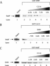
Coilin competes with SmB′ and SMN for binding. Competition experiments were conducted using GST–SMN (A), GST–C214 (B), or GST–SmB′ (C) beads incubated with a constant amount of His–T7–SmB′ (A,B) or SMN (C). Increasing amounts of His–C214 (A,C) or SMN (B) were added in separate reactions at the indicated fold excess, based on protein level, relative to the amount of His–T7–SmB′ (or SMN) present (lanes 3–6). The beads were washed, as described in Materials and Methods, and subjected to SDS-PAGE and Western blotting with an anti-T7 antibody (for detection of SmB′ and C214) or an anti-SMN antibody. The input lane (lane 1) shows an amount of protein equivalent to 20% of that used in lanes 2 and 4. Lane 2 in all panels shows the amount of His–T7–SmB′ (A,B) or SMN (C) recovered in the absence of competitor. It is clear that addition of increasing amounts of competitor did not poison the GST beads because companion blots showed increasing amounts of either C214 (A,C) or SMN (B) were bound as binding to the other component decreased (data not shown).
Figure 6
Figure 6
The RG box of coilin is required for recruitment of the SMN complex to the Cajal body. A mouse embryonic fibroblast cell line derived from coilin knockout mice (Tucker et al. 2001) was cotransfected with GFP-mouse coilin (GFP–mcoilin) and human myc-tagged SMN (top). SMN was visualized by an anti-myc antibody whereas Sm proteins were detected using the antibody Y12. A white signal in the merged image shows that all three signals are coincident. Additionally, mouse coilin deleted of the RG box (GFP–mΔRG) was cotransfected into the KO line with myc–SMN (bottom). The presence of both red and green signals, without overlap, can be observed in merged image. Cells were processed as described in Materials and Methods.

Similar articles

Cited by

References

    1. Andrade LEC, Tan EM, Chan EKL. Immunocytochemical analysis of the coiled body in the cell cycle and during cell proliferation. Proc Natl Acad Sci. 1993;90:1947–1951. - PMC - PubMed
    1. Bauer DW, Gall JG. Coiled bodies without coilin. Mol Biol Cell. 1997;8:73–82. - PMC - PubMed
    1. Brahms H, Raymackers J, Union A, de Keyser F, Meheus L, Lührmann R. The C-terminal RG dipeptide repeats of the spliceosomal Sm proteins D1 and D3 contain symmetrical dimethylarginines, which form a major B-cell epitope for anti-Sm autoantibodies. J Biol Chem. 2000;275:17122–17129. - PubMed
    1. Bühler D, Raker V, Lührmann R, Fischer U. Essential role for the Tudor domain of SMN in spliceosomal U snRNP assembly: Implications for spinal muscular atrophy. Hum Mol Genet. 1999;8:2351–2357. - PubMed
    1. Burghes AHM. When is a deletion not a deletion? When it is converted. Am J Hum Genet. 1997;61:9–15. - PMC - PubMed

Publication types

MeSH terms