. 2001 Oct 15;20(20):5769-78.
doi: 10.1093/emboj/20.20.5769.

Herpes simplex virus ICP27 protein provides viral mRNAs with access to the cellular mRNA export pathway

Affiliations

Herpes simplex virus ICP27 protein provides viral mRNAs with access to the cellular mRNA export pathway

M D Koffa et al. EMBO J. .

Erratum in

Abstract

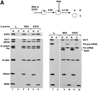
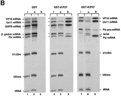
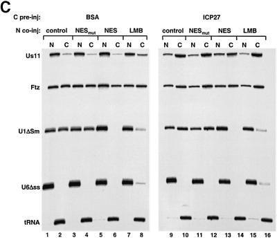
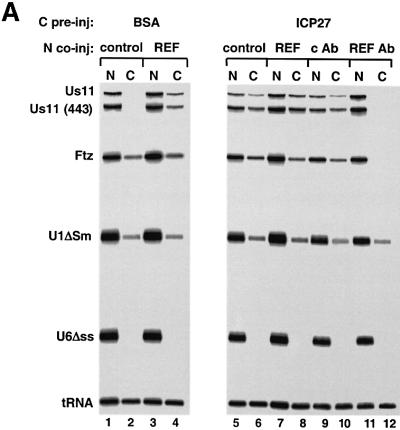
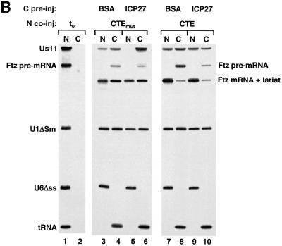
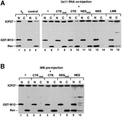
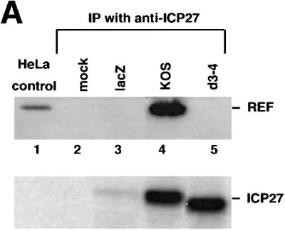
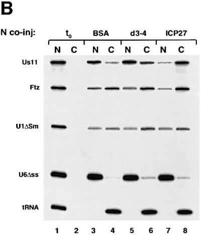
The role of herpes simplex virus ICP27 protein in mRNA export is investigated by microinjection into Xenopus laevis oocytes. ICP27 dramatically stimulates the export of intronless viral mRNAs, but has no effect on the export of cellular mRNAs, U snRNAs or tRNA. Use of inhibitors shows, in contrast to previous suggestions, that ICP27 neither shuttles nor exports viral mRNA via the CRM1 pathway. Instead, ICP27-mediated viral RNA export requires REF and TAP/NXF1, factors involved in cellular mRNA export. ICP27 binds directly to REF and complexes containing ICP27, REF and TAP are found in vitro and in virally infected cells. A mutant ICP27 that does not interact with REF is inactive in viral mRNA export. We propose that ICP27 associates with viral mRNAs and recruits TAP/NXF1 via its interaction with REF proteins, allowing the otherwise inefficiently exported viral mRNAs to access the TAP-mediated export pathway. This represents a novel mechanism for export of viral mRNAs.


Figures

None
Fig. 1. ICP27 interacts with REF in vitro. (A) [35S]methionine-labelled ICP27 (wt), and fragments of ICP27 were incubated with GST–REF1-I beads or GST beads as indicated. The bound (lanes 1–9) and unbound fractions (lanes 10–18) were analysed by SDS–PAGE followed by fluorography. (B) ICP27 associates with the central domain (RBD) of REF. ICP27 synthesized in vitro was incubated with beads pre-coated with GST–REF2-I (wt) or the truncations indicated. One-tenth of the input (lanes 1 and 9) and one-third of the bound fractions (lanes 2–8 and 10–14) were analysed by SDS–PAGE. RNase (10 µg/ml) was added to the reactions shown in lanes 12 and 14.
None
Fig. 2. ICP27 forms a complex with REF and TAP proteins. (A) HeLa cells were mock-infected or infected with wt HSV-1 virus (KOS) or the 27-lacZ mutant virus and immunoprecipitation was performed using the monoclonal anti-ICP27 antibodies H1119 and H1113. Western blot analysis was performed using anti-REF and anti-TAP polyclonal antibodies. The control lane represents western blot analysis of KOS-infected HeLa cells. In the upper panel, the ∼50 kDa bands correspond to the heavy chain of the antibody used for immunoprecipitation. (B) ICP27 synthesized in vitro was incubated with 5 µg of immobilized GST or GST–TAP. Five micrograms of histidine-tagged REF2-I, histidine-tagged thioredoxin (as control) or RNase were added to the binding reactions as indicated. Bound proteins were eluted and one-tenth of the input (lane 1) or one-third of the bound fractions (lanes 2–6) were analysed by SDS–PAGE.
None
Fig. 3. ICP27 protein shuttles in Xenopus oocytes. (AXenopus laevis oocytes were microinjected into the cytoplasm with a mix of [35S]methionine-labelled CBP80, ICP27 and GST–M10 proteins. Oocytes were either pre-injected into the cytoplasm with truncated (30 mg/ml, lanes 5 and 6) or full-length (30 mg/ml, lanes 7 and 8) IBB. Protein samples from nuclear (N) and cytoplasmic (C) fractions were collected immediately (lanes 1 and 2) or after 6 h incubation (lanes 3–8). Labelled proteins were visualized by SDS–PAGE and fluorography. CBP80, ICP27 and GST–M10 are indicated. (B) A mix of [35S]methionine-labelled ICP27 and GST–M10 proteins was microinjected into oocyte nuclei alone (lanes 1–4, 7 and 8) or in the presence of 7.5 µM Us11 mRNA (lanes 5 and 6). In lanes 7 and 8 the protein mix was injected into oocytes pre-injected into the cytoplasm with IBB. Protein samples from nuclear (N) and cytoplasmic (C) fractions were collected immediately (lanes 1 and 2) or after 3 h incubation (lanes 3–10).
None
Fig. 4. ICP27 protein stimulates viral mRNA export. (A) Oocytes were pre-injected into the cytoplasm with purified recombinant GST–ICP27–his (5.5 µM) or BSA and incubated for 6–8 h at 18°C. Subsequently, a mix of 32P-labelled RNAs was injected into the nuclei, as indicated. Oocytes were dissected into nuclear (N) and cytoplasmic (C) fractions immediately (lanes 1, 2 and 7, 8) or 2 h after injection (lanes 3–6 and 9–12). RNAs were purified and analysed on denaturing 8% polyacrylamide gels followed by autoradiography. The injected RNAs as well as the intron lariat and spliced product of the Ftz pre-mRNA are indicated. (B) Recombinant GST–ICP27–his (lanes 4–9) or GST alone (lanes 1–3) was injected into oocyte nuclei. Subsequently, a mix containing either VP16 and Us11 viral RNAs, DHFR, β-globin and Ftz mRNAs, U1ΔSm, U6Δss and tRNA (lanes 1–6), or VP16, Us11, Ftz pre-mRNA, U1ΔSm, U6Δss and tRNA was injected into the oocyte nuclei (lanes 7–9). RNA samples from total oocytes were collected 20 min after injection and analysed directly (input, i, lanes 1, 4 and 7), or immunoprecipitated using anti-GST antibodies: unbound fractions (u, lanes 2, 5 and 8) and bound fractions (b, lanes 3, 6 and 9). (C) Oocytes were pre-injected with BSA (lanes 1–8) or recombinant ICP27 (lanes 9–16), followed by nuclear injection of RNA mix. The RNA mix was injected alone (lanes 1, 2, 7–10, 15 and 16), in the presence of 5 mg/ml mutant NES conjugate (NESmut) (lanes 3, 4 and 11, 12) or 5 mg/ml wt NES conjugate (NES) (lanes 5, 6 and 13, 14), or oocytes were pre-incubated in the presence of 200 nM LMB for 2 h, followed by nuclear injection of the RNAs (lanes 7, 8, 15 and 16). RNA samples from nuclear (N) and cytoplasmic (C) fractions were collected 2 h after injection.
None
Fig. 5. REF and TAP are involved in viral mRNA export. (A) Oocytes were pre-injected with BSA (lanes 1–4) or ICP27 (lanes 5–12) followed by nuclear injection of RNA mix. The RNA mix was injected alone (lanes 1, 2, 5 and 6), with 14 µM GST–REF (lanes 3, 4, 7 and 8) or with affinity-purified anti-REF antibodies (5 mg/ml) (lanes 11 and 12) or control antibodies (lanes 9 and 10). RNA samples from nuclear (N) and cytoplasmic (C) fractions were collected 1 h after injection. (B) Oocytes were pre-injected with BSA (lanes 3, 4, 7 and 8) or ICP27 (lanes 5, 6, 9 and 10), followed by nuclear injection of RNAs in the presence of 0.1 pmol CTE M36 mutant (lanes 3–6) or 0.1 pmol wild-type CTE RNA (lanes 7–10). RNA samples from nuclear (N) and cytoplasmic (C) fractions were collected immediately (lanes 1 and 2) or 3.5 h after injection (lanes 3–10). In this gel (10%), the lariat and the Ftz mRNA co-migrate.
None
Fig. 6. ICP27 protein is exported via TAP/NXF1 in the presence of viral mRNA. (A) Labelled ICP27, GST–M10 and Rev proteins were injected into oocyte nuclei alone (lanes 1–6, 15 and 16), in the presence of 0.1 pmol M36 mutant CTE or wt CTE RNA (lanes 7–10) or 5 mg/ml mutant or wt NES conjugate (lanes 11–14). In lanes 15 and 16, the oocytes were pre-incubated in the presence of 200 nM LMB. Oocytes were dissected into nuclear (N) and cytoplasmic (C) fractions immediately (lanes 1 and 2) or after a 3 h incubation (lanes 3–16). (B) Oocytes pre-injected into the cytoplasm with IBB, were injected into the nuclei with labelled ICP27, GST–M10 and Rev proteins alone (lanes 1 and 2), in the presence of 0.1 pmol M36 mutant CTE or wt CTE (lanes 3–6), or 5 mg/ml mutant or wt NES conjugate (lanes 7–10). Oocytes were dissected into nuclear (N) and cytoplasmic (C) fractions after a 3 h incubation.
None
Fig. 7. REF binding to ICP27 is required for viral RNA export stimulation. (A) HeLa cells were mock-infected or infected with wt HSV-1 (KOS), the 27-lacZ mutant or the d3-4 ICP27 deletion mutant virus, and immunoprecipitation was performed using the mAb H1119. Western blot analysis was performed using anti-REF polyclonal antibodies (upper panel), or the anti-ICP27 mAb H1119 (lower panel). (B) Oocytes were injected into the nucleus with BSA (lanes 3 and 4), d3-4 mutant ICP27 (lanes 5 and 6) or wt ICP27 (lanes 7 and 8), followed by nuclear injection of the RNA mix. RNA samples from nuclear (N) and cytoplasmic (C) fractions were collected 3 h after injection.

Similar articles

Cited by

References

    1. Ace C.I., Dalrymple,M.A., Ramsay,F.H., Preston,V.G. and Preston,C.M. (1988) Mutational analysis of the herpes simplex virus type 1 trans-inducing factor Vmw65. J. Gen. Virol., 69, 2595–2605. - PubMed
    1. Arts G.J., Fornerod,M. and Mattaj,I.W. (1998) Identification of a nuclear export receptor for tRNA. Curr. Biol., 8, 305–314. - PubMed
    1. Bennett M., Pinol-Roma,S., Staknis,D., Dreyfuss,G. and Reed,R. (1992) Differential binding of heterogeneous nuclear ribonucleoproteins to mRNA precursors prior to spliceosome assembly in vitro. Mol. Cell. Biol., 12, 3165–3175. - PMC - PubMed
    1. Boyle S.M., Ruvolo,V., Gupta,A.K. and Swaminathan,S. (1999) Association with the cellular export receptor CRM1 mediates function and intracellular localization of Epstein–Barr virus SM protein, a regulator of gene expression. J. Virol., 73, 6872–6881. - PMC - PubMed
    1. Braun I.C., Rohrbach,E., Schmitt,C. and Izaurralde,E. (1999) TAP binds to the constitutive transport element (CTE) through a novel RNA-binding motif that is sufficient to promote CTE-dependent RNA export from the nucleus. EMBO J., 18, 1953–1965. - PMC - PubMed

MeSH terms