. 2001 Oct 1;155(1):53-63.
doi: 10.1083/jcb.200103071.

Reorganization of multivesicular bodies regulates MHC class II antigen presentation by dendritic cells

Affiliations

Reorganization of multivesicular bodies regulates MHC class II antigen presentation by dendritic cells

M Kleijmeer et al. J Cell Biol. .

Abstract

Immature dendritic cells (DCs) sample their environment for antigens and after stimulation present peptide associated with major histocompatibility complex class II (MHC II) to naive T cells. We have studied the intracellular trafficking of MHC II in cultured DCs. In immature cells, the majority of MHC II was stored intracellularly at the internal vesicles of multivesicular bodies (MVBs). In contrast, DM, an accessory molecule required for peptide loading, was located predominantly at the limiting membrane of MVBs. After stimulation, the internal vesicles carrying MHC II were transferred to the limiting membrane of the MVB, bringing MHC II and DM to the same membrane domain. Concomitantly, the MVBs transformed into long tubular organelles that extended into the periphery of the cells. Vesicles that were formed at the tips of these tubules nonselectively incorporated MHC II and DM and presumably mediated transport to the plasma membrane. We propose that in maturing DCs, the reorganization of MVBs is fundamental for the timing of MHC II antigen loading and transport to the plasma membrane.


Figures

Figure 1.
Figure 1.
LPS-induced expression of MHC II and costimulatory molecules at the plasma membrane. (A) D1 cells were cultured in 96-well plates and incubated for 0, 1, 3, 6, or 48 h with LPS. The surface expression of MHC II, CLIP-associated MHC II (MHC II/CLIP), CD40, and B7.2 was analyzed by FACS®. (B) Cells were cultured as above for 0, 1, 2, 3, 4, 5, 6, and 7 h with LPS either in the presence or absence of cycloheximide. Surface expression of B7.2 and MHC II was analyzed by FACS®. (C) Cells were lysed after 48 h of culture in the presence or absence of LPS in SDS sample buffer. Samples were incubated at room temperature, 37, 60, or 100°C and analyzed by Western blotting for MHC II β-chain. The Western blot shows monomeric MHC II β-chain and SDS stable MHC II. Upon stimulation with LPS, the amount of SDS stable MHC II/peptide complexes at room temperature strongly increased on expense of SDS unstable MHC II molecules. In contrast, the amount of SDS stable complexes at 100°C decreased after maturation. (D) Immature D1 cells secreted ∼7% of their β-hexosaminidase in a linear fashion during 18 h. The release increased only slightly after 4 and 8 h of LPS treatment but was unchanged at 18 h.
Figure 2.
Figure 2.
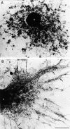
LPS-induced remodeling of the endocytic system shown by whole-mount EM. For the inspection of endosomes and lysosomes (MIICs) in overview at the EM level, D1 cells were allowed to endocytose HRP as a fluid phase marker for 1 h (A) or were loaded for 1 h with HRP after which LPS was added for 6 h (B). Cells were chased for 10 min in the absence of HRP and then processed for whole-mount viewing of HRP-containing compartments. (A) Unstimulated cell with numerous vacuolar MIICs. (B) Stimulated cell with drastically remodeled long MIIC tubules extending into the dendrites of the cell (arrows). n, nucleus. Bar, 4 μm.
Figure 3.
Figure 3.
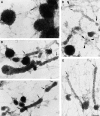
MIICs in immature and maturing D1 cells. Cells were loaded with HRP for 1 h in the absence of LPS as in the legend to Fig. 2 A (A) or 1 h in the presence of HRP plus 6 h HRP + LPS (B and C) or pulsed for 1 h with HRP and chased for 3 h in the presence of LPS (D and E) and processed for whole-mount EM. (A) Vacuolar MIICs in immature D1 cells labeled for MHC II (10 nm gold) and DM (15 nm gold). Tubular MIICs have formed after 3 (D and E) or 6 h (A and B) of LPS treatment. (B) Double labeling for MHC II (10 nm gold) and DM (15 nm gold). A tubule seems to form out of the vacuole in the left bottom corner. (C) Tubular MIICs labeled for MHC II (10 nm gold) and LAMP-1 (15 nm gold), demonstrating the late endosomal/lysosomal character of these organelles. (D) Tubular MIICs and free vesicles (arrowheads) with diameters ranging from 80 to 200 nm were double labeled for MHC II (10 nm gold) and DM (15 nm gold). Note the vesicle that seems to be formed at the tip of a tubule (thick arrow). (E) Tubular MIICs, showing DM (10 nm gold) and LAMP-1 (15 nm gold). Note the cytoskeletal elements in which the MIICs are embedded.
Figure 4.
Figure 4.
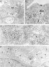
LPS-induced depletion of the MIICs internal membranes. Ultrathin cryosections of untreated D1 cells (A and C) or D1 cells treated with LPS for 48 (B) or 3 h (D) were immunolabeled for MHC II with 10 nm gold particles. In A, MHC II is primarily present in vacuolar MIICs (stars) comprised of a limiting membrane surrounding internal vesicles and membranous sheets. Only a few gold particles are present at the plasma membrane (PM). In contrast, in the LPS-treated D1 cell in B the plasma membrane is labeled strongly for MHC II, whereas only little MHC II localizes to MIICs, which have few internal membranes (stars). (C) Vacuolar MIICs (V) display many internal membrane vesicles that are labeled for MHC II, whereas the limiting membrane is labeled scarcely. (D) MHC II–positive tubules (T) and vesicles (arrowheads) after 3 h of stimulation with LPS. The dense vacuolar part is marked by a star. N, nucleus. Bars: (A and B) 250 nm; (C and D) 200 nm.
Figure 5.
Figure 5.
Morphometric analysis of MIIC membrane domains and subcellular distribution of MHC II. Random pictures (20×) were taken of ultrathin cryosections of D1 cells incubated for 0, 2, 5, or 24 h with LPS. The sections were immunolabeled for MHC II. (A) Surface densities of internal and limiting membranes of MIICs. Significant differences were found for the surface density of internal vesicles after 5 and 24 h. (B) The change in relative distribution of MHC II gold particles over PM, MIICs, ER + Golgi complex, and others (n.d. + cytosol + mitochondria + nucleus) was determined on random pictures (*p < 0.05; **P < 0.005).
Figure 6.
Figure 6.
Labeling characteristics of MHC II and DM in MIICs. (A) Ultrathin cryosections of D1 cells incubated for 0, 2, 5, or 24 h with LPS were labeled for MHC II or DM. The relative amount of labeling on the limiting membrane was determined and expressed as the percentage of total labeling on MIICs. Treatment with LPS induced a clear redistribution of MHC II towards the limiting membrane, whereas the distribution of DM did not change. (B) The population of 200 DAB-containing MHC II and/or DM-labeled transport vesicles indicated in Table I were grouped according to their MHC II and DM content. Each bar represents the number of vesicles having a certain labeling characteristic. Only a single population of vesicles, containing both MHC II and DM, was observed. (C) The labeling densities of MHC II and DM on limiting and internal membranes of MIICs were measured in electron micrographs of cells treated for 3 h with LPS. Labeling densities on the limiting membranes of V and T/Ve are expressed as the ratios of gold particles over membrane surface areas (as described in Materials and methods). The labeling density of MHC II on the limiting membrane of V is less than two times that of T/Ve. (D) Labeling densities of MHC II and DM on internal membranes do not alter after LPS treatment (*p < 0.05; **P < 0.005).
Figure 7.
Figure 7.
Localization of DM and YAe on the limiting membrane of MIIC. (A) Double immunolabeling of MIICs (stars) in immature D1 cells, showing DM (15 nm) primarily on the limiting membrane (arrowheads) of MIICs and most of the MHC II (10 nm) on internal membranes. N, nucleus; PM, plasma membrane. (B) Immunolabeling showing YAe on the limiting membrane of MIICs (stars) in BMDCs. Bars, 200 nm.
Figure 8.
Figure 8.
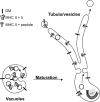
Tubulation of multivesicular MIICs during DC maturation. Multivesicular MIICs in DCs undergo a dramatic shape change from vacuolar to tubular upon stimulation, most likely by fusion of the MIIC internal membrane vesicles with the limiting membrane. This implies that MHC II–rich internal membranes stored in the lumen of the vacuolar MIIC relocate to the limiting membrane of the tubular MIICs, allowing egress of MHC II from these tubules to the plasma membrane. The final transport step to the cell surface is probably mediated by transport vesicles, which bud from the tubular MIICs and nonselectively incorporate MHC II. As a consequence of MHC II translocation to the DM-rich limiting membrane, contact between DM and MHC II is increased, which may facilitate peptide loading and editing during maturation.

Similar articles

Cited by

References

    1. Alfonso, C., and L. Karlsson. 2000. Nonclassical MHC class II molecules. Annu. Rev. Immunol. 18:113–142. - PubMed
    1. Amigorena, S., J.R. Drake, P. Webster, and I. Mellman. 1994. Transient accumulation of new class II MHC molecules in a novel endocytic compartment in B lymphocytes. Nature. 369:113–120. - PubMed
    1. Banchereau, J., and R.M. Steinman. 1998. Dendritic cells and the control of immunity. Nature. 392:245–252. - PubMed
    1. Barois, N., F. Forquet, and J. Davoust. 1997. Selective modulation of the major histocompatibility complex class II antigen presentation pathway following B cell receptor ligation and protein kinase C activation. J. Biol. Chem. 272:3641–3647. - PubMed
    1. Barois, N., F. Forquet, and J. Davoust. 1998. Actin microfilaments control the MHC class II antigen presentation pathway in B cells. J. Cell Sci. 111:1791–1800. - PubMed

Publication types

Substances