

Mutations in the human orthologue of the mouse underwhite gene (uw) underlie a new form of oculocutaneous albinism, OCA4
- PMID: 11574907
- PMCID: PMC1274374
- DOI: 10.1086/324340
Mutations in the human orthologue of the mouse underwhite gene (uw) underlie a new form of oculocutaneous albinism, OCA4
Abstract
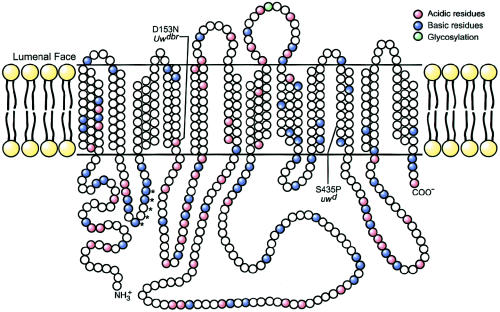
Oculocutaneous albinism (OCA) affects approximately 1/20,000 people worldwide. All forms of OCA exhibit generalized hypopigmentation. Reduced pigmentation during eye development results in misrouting of the optic nerves, nystagmus, alternating strabismus, and reduced visual acuity. Loss of pigmentation in the skin leads to an increased risk for skin cancer. Two common forms and one infrequent form of OCA have been described. OCA1 (MIM 203100) is associated with mutations of the TYR gene encoding tyrosinase (the rate-limiting enzyme in the production of melanin pigment) and accounts for approximately 40% of OCA worldwide. OCA2 (MIM 203200), the most common form of OCA, is associated with mutations of the P gene and accounts for approximately 50% of OCA worldwide. OCA3 (MIM 203290), a rare form of OCA and also known as "rufous/red albinism," is associated with mutations in TYRP1 (encoding tyrosinase-related protein 1). Analysis of the TYR and P genes in patients with OCA suggests that other genes may be associated with OCA. We have identified the mouse underwhite gene (uw) and its human orthologue, which underlies a new form of human OCA, termed "OCA4." The encoded protein, MATP (for "membrane-associated transporter protein") is predicted to span the membrane 12 times and likely functions as a transporter.
Figures


Similar articles
-
Mutations in the MATP gene in five German patients affected by oculocutaneous albinism type 4.Hum Mutat. 2004 Feb;23(2):106-110. doi: 10.1002/humu.10311. Hum Mutat. 2004. PMID: 14722913
-
Tyrosinase processing and intracellular trafficking is disrupted in mouse primary melanocytes carrying the underwhite (uw) mutation. A model for oculocutaneous albinism (OCA) type 4.J Cell Sci. 2003 Aug 1;116(Pt 15):3203-12. doi: 10.1242/jcs.00598. J Cell Sci. 2003. PMID: 12829739
-
[A new form of Oculocutaneous albinism, OCA4].Yi Chuan. 2006 Sep;28(9):1149-52. doi: 10.1360/yc-006-1149. Yi Chuan. 2006. PMID: 16963427 Review. Chinese.
-
SLC45A2 mutation frequency in Oculocutaneous Albinism Italian patients doesn't differ from other European studies.Gene. 2014 Jan 1;533(1):398-402. doi: 10.1016/j.gene.2013.09.053. Epub 2013 Oct 3. Gene. 2014. PMID: 24096233
-
Oculocutaneous albinism.Orphanet J Rare Dis. 2007 Nov 2;2:43. doi: 10.1186/1750-1172-2-43. Orphanet J Rare Dis. 2007. PMID: 17980020 Free PMC article. Review.
Cited by
-
Oculocutaneous albinism type 1: link between mutations, tyrosinase conformational stability, and enzymatic activity.Pigment Cell Melanoma Res. 2017 Jan;30(1):41-52. doi: 10.1111/pcmr.12546. Pigment Cell Melanoma Res. 2017. PMID: 27775880 Free PMC article.
-
Mutations in SLC45A2 cause plumage color variation in chicken and Japanese quail.Genetics. 2007 Feb;175(2):867-77. doi: 10.1534/genetics.106.063107. Epub 2006 Dec 6. Genetics. 2007. PMID: 17151254 Free PMC article.
-
Meningococccal meningitis and complement component 6 deficiency associated with oculocutaneous albinism.Eur J Pediatr. 2005 Mar;164(3):177-9. doi: 10.1007/s00431-004-1582-y. Epub 2004 Nov 23. Eur J Pediatr. 2005. PMID: 15565285 No abstract available.
-
Rescue from oculocutaneous albinism type 4 using medaka slc45a2 cDNA driven by its own promoter.Genetics. 2008 Feb;178(2):761-9. doi: 10.1534/genetics.107.073387. Epub 2008 Feb 1. Genetics. 2008. PMID: 18245373 Free PMC article.
-
Membrane transport proteins in melanosomes: Regulation of ions for pigmentation.Biochim Biophys Acta Biomembr. 2020 Dec 1;1862(12):183318. doi: 10.1016/j.bbamem.2020.183318. Epub 2020 Apr 22. Biochim Biophys Acta Biomembr. 2020. PMID: 32333855 Free PMC article. Review.
References
Electronic-Database Information
-
- Celera, http://www.celera.com/ (for human chromosome 5p sequences)
-
- GenBank, http://www.ncbi.nlm.nih.gov/GenBank/ (for mouse Matp cDNA from present study [accession number AY034377] and from Fukamachi et al. [2001] [accession number AF360357], human MATP cDNA [previously known as “AIM1” {accession number AF172849}], and medaka-fish MATP-b [accession number AF332510])
-
- Human Genome, The, http://www.ncbi.nlm.nih.gov/genome/guide/human/ (for human chromosome 5p sequences)
-
- Online Mendelian Inheritance in Man (OMIM), http://www.ncbi.nlm.nih.gov/Omim (for OCA1 [MIM 203100], OCA2 [MIM 203200], and OCA3 [MIM 203290])
-
- TMHMM (v. 2.0), http://www.cbs.dtu.dk/services/TMHMM/ (for membrane-topology predictions)
References
-
- Boissy RE, Zhao H, Oetting WS, Austin LM, Wildenberg SC, Boissy YL, Zhao Y, Sturm RA, Hearing VJ, King RA, Nordlund JJ (1996) Mutation in and lack of expression of tyrosinase-related protein-1 (TRP-1) in melanocytes from an individual with Brown oculocutaneous albinism: a new type of albinism classified as “OCA3.” Am J Hum Genet 58:1145–1156 - PMC - PubMed
-
- Cooksey CJ, Garratt PJ, Land EJ, Pavel S, Ramsden CA, Riley PA, Smit NP (1997) Evidence of the indirect formation of the catecholic intermediate substrate responsible for the autoactivation kinetics of tyrosinase. J Biol Chem 272:26226–26235 - PubMed
-
- Frohman MA (1994) On beyond classic RACE (rapid amplification of cDNA ends). PCR Methods Appl 4:S40–S58 - PubMed
-
- Fukamachi S, Shimada A, Shima A (2001) Mutations in the gene encoding B, a novel transporter protein, reduce melanin content in medaka. Nat Genet 28:381–385 - PubMed
-
- Gardner JM, Nakatsu Y, Gondo Y, Lee S, Lyon MF, King RA, Brilliant MH (1992) The mouse pink-eyed dilution gene: association with human Prader-Willi and Angelman syndromes. Science 257:1121–1124 - PubMed
Publication types
MeSH terms
Substances
Associated data
- Actions
- Actions
- Actions
- Actions
- Actions
- Actions
- Actions
Grants and funding
LinkOut - more resources
Full Text Sources
Other Literature Sources
Medical
Molecular Biology Databases
Research Materials
