. 2001 Oct 1;20(19):5373-82.
doi: 10.1093/emboj/20.19.5373.

A bacterial type III secretion system inhibits actin polymerization to prevent pore formation in host cell membranes

Affiliations

A bacterial type III secretion system inhibits actin polymerization to prevent pore formation in host cell membranes

G I Viboud et al. EMBO J. .

Abstract

The bacterial pathogen Yersinia pseudotuberculosis uses type III secretion machinery to translocate Yop effector proteins through host cell plasma membranes. A current model suggests that a type III translocation channel is inserted into the plasma membrane, and if Yops are not present to fill the channel, the channel will form a pore. We examined the possibility that Yops act within the host cell to prevent pore formation. Yop- mutants of Y.pseudotuberculosis were assayed for pore-forming activity in HeLa cells. A YopE- mutant exhibited high levels of pore-forming activity. The GTPase-downregulating function of YopE was required to prevent pore formation. YopE+ bacteria had increased pore-forming activity when HeLa cells expressed activated Rho GTPases. Pore formation by YopE- bacteria required actin polymerization. F-actin was concentrated at sites of contact between HeLa cells and YopE- bacteria. The data suggest that localized actin polymerization, triggered by the type III machinery, results in pore formation in cells infected with YopE- bacteria. Thus, translocated YopE inhibits actin polymerization to prevent membane damage to cells infected with wild-type bacteria.


Figures

None
Fig. 1. Infection of HeLa cells with a multi-Yop mutant of Y.pseudotuberculosis results in membrane blebbing and EtdBr uptake. HeLa cells grown on coverslips were infected with a wild-type strain (YP126) or a YopEHJ mutant (YP27). After 3 h, coverslips were inverted onto a 5 µl drop of staining solution (25 µg/ml EtdBr and 5 µg/ml AO in PBS) placed on the surface of a glass slide. Infected cells were analyzed immediately by phase contrast (A and C) or epifluorescence (B and D) microscopy using a 40× objective. Images were recorded using a digital camera. The arrows in (C) and (D) point to a large bleb on a HeLa cell.
None
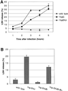
Fig. 2. Release of LDH by HeLa cells infected with Y.pseudo tuberculosis. (A) Kinetics of LDH release after infection with a wild-type strain (YP126), a YopEHJ mutant (YP27) or a YopE mutant (YP6). Culture supernatants were collected from wells containing HeLa cells at indicated time points after infection. LDH in culture supernatants was measured using a CytoTox 96 assay kit (Promega). The percentage of LDH release was calculated by dividing the amount of LDH released from infected cells by the amount of LDH released from uninfected cells that were lysed by a freeze–thaw cycle. Error bars represent the standard deviation of the mean values obtained from triplicate samples. (B) YopB is required for LDH release. HeLa cells were infected with a wild-type strain (YP126), a YopEHJ mutant (YP27), a YopEHJB mutant (YP29) or a YopEHJB mutant complemented with wild-type yopB+ (YP29/pYopB). Culture supernatants were collected 3 h post infection and assayed for LDH activity as described above.
None
Fig. 3. Effect of Dextran 6000 on release of LDH from HeLa cells infected with Y.pseudotuberculosis. HeLa cells were infected with a YopEHJ mutant (YP27) or a YopE mutant (YP6) in the presence or absence of 30 mM Dextran 6000 or 30 mM sucrose. LDH release 3 h post infection was determined as described in the legend to Figure 2.
None
Fig. 4. Effect of Dextran 6000 on membrane blebbing and uptake of EtdBr. HeLa cells were infected for 3 h with a YopEHJ mutant (YP27) in the presence of 30 mM sucrose (A and B) or 30 mM Dextran 6000 (C and D). Images were obtained as described in the legend to Figure 1.
None
Fig. 5. YopE plays a critical role in preventing pore formation. HeLa cells were infected for 3 h with a wild-type strain (YP126), a YopEHJ mutant (YP27), a YopE mutant (YP6), a YopH mutant (YP15), or a YopJ mutant (YP26). LDH release was determined as described in the legend to Figure 2.
None
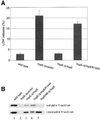
Fig. 6. YopE GAP activity is required to inhibit pore formation. HeLa cells were infected with a wild-type strain (YP126), a YopE mutant harboring an empty vector (YP6/vector), a YopE mutant comple mented with yopE+ (YP6/pYopE), a YopE mutant complemented with yopER144A (YP6/pYopER144A), or a YopEHB mutant comple mented with yopE+ (YP19/pYopE). (A) LDH released from HeLa cells infected for 3 h was measured as described in the legend to Figure 2. (B) HeLa cells infected for 2 h were lysed in a buffer containing 1% Triton X-100. The lysates were separated by centrifugation into soluble and insoluble fractions. Samples of protein equivalent to 1 × 10⁴ HeLa cells (soluble fraction) or 3 × 10⁵ HeLa cells (insoluble fraction) were separated by SDS–PAGE and analyzed by immunoblotting with anti-YopE antibody.
None
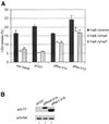
Fig. 7. Constitutively active forms of RhoA and Rac1 rescue pore-forming activity in the presence of YopE or YopT. HeLa cells were not transfected (non transf.) or were transfected with an empty vector (pCGT), pCGTRhoA-V14 (pRhoA-V14) or pCGTRac1-V12 (pRac1-V12). (A) LDH released from HeLa cells infected with YP6/vector, YP6/pYopE or YP6/pYopT for 3 h was determined as described in the legend to Figure 2. (B) Expression of RhoA-V14 and Rac1-V12 in HeLa cells was evaluated 24 h post transfection by immunoblotting. Transfected cells were lysed in detergent and samples of soluble protein equivalent to 1 × 10⁴ HeLa cells were analyzed by immunoblotting with a monoclonal antibody that recognizes the T7 epitope tag appended to RhoA-V14 and Rac1-V12. Duplicate samples of protein were analyzed by immunoblotting with a monoclonal antibody specific for the Raf kinase, to control for loading.
None
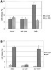
Fig. 8. Actin polymerization inhibitors prevent pore formation in HeLa cells infected with YopE mutant Y.pseudotuberculosis. HeLa cells were exposed to the different actin modifying agents dissolved in DMSO or to DMSO only for 2 h prior to and during infection. Cells were either left uninfected (mock) or infected with a wild-type strain (YP126) or a YopE mutant (YP6) for 3 h. Culture supernatants were assayed for LDH as described in the legend to Figure 2. (A) Cells were treated with 3.9 µM cytochalasin (+CD) or DMSO only (–CD). (B) Cells were treated with 5 µM latrunculin B (Lat), 100 nM Jasplakinolide (Jas) or DMSO only.
None
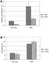
Fig. 9. Wortmannin inhibits bacterial internalization but not LDH release. HeLa cells were exposed to 100 nM wortmannin in DMSO (+Wort.) or to DMSO alone (–Wort.) for 2 h prior to and during infections. Cells were infected with a wild-type strain (YP126) or a YopE mutant (YP6). (A) Bacterial internalization (%) at 1 h post infection was measured by a gentamicin protection assay as described in Materials and methods. (B) LDH release at 3 h post infection was assayed as described in the legend to Figure 2.
None
Fig. 10. Formation of F-actin halos in HeLa cells infected with YopEHJ mutant Y.pseudotuberculosis. Hela cells grown on coverslips were infected with a YopEHJ mutant (YP27) (A and C) or a YopEHJB mutant (YP29) (B and D). At 60 (A and B) or 90 (C and D) min post infection, the cells were fixed, permeabilized, and stained with rhodamine–phalloidin to label F-actin. F-actin was detected by epifluorescence microscopy using a 100× objective. Images were captured with a digital camera. Arrows in (A) and (C) point to F-actin halos. ‘Clumps’ of concentrated F-actin observed in HeLa cells infected with the YopEHJB mutant are morphologically distinct from the F-actin halos observed in cells infected with the YopEHJ mutant.
None
Fig. 11. Bacteria co-localized with F-actin halos. HeLa cells grown on coverslips were exposed to 100 nM wortmannin in DMSO for 2 h prior to and during infection. The HeLa cells were infected with a YopEHJ mutant (YP27) for 60 min. The infected cells were fixed, permeabilized, and bacteria were labeled by sequential incubation with rabbit anti-Yersinia primary antibody and FITC-conjugated goat anti-rabbit IgG secondary antibody. F-actin was labeled with rhodamine–phalloidin. Cells were observed by epifluorescence microscopy using a 100× objective and filters to detect rhodamine (A) or FITC (B) fluorescence. Images were captured with a digital camera. A superimposition of (A) and (B) is presented in (C). Arrows point to F-actin halos in (A) and (C).

Similar articles

Cited by

References

    1. Araki N., Johnson,M.T. and Swanson,J.A. (1996) A role for phosphoinositide 3-kinase in the completion of macropinocytosis and phagocytosis by macrophages. J. Cell Biol., 135, 1249–1260. - PMC - PubMed
    1. Black D.S. and Bliska,J.B. (1997) Identification of p130Cas as a substrate of Yersinia YopH (Yop51), a bacterial protein tyrosine phosphatase that translocates into mammalian cells and targets focal adhesions. EMBO J., 16, 2730–2744. - PMC - PubMed
    1. Black D.S. and Bliska,J.B. (2000) The RhoGAP activity of the Yersinia pseudotuberculosis cytotoxin YopE is required for antiphagocytic function and virulence. Mol. Microbiol., 37, 515–527. - PubMed
    1. Bliska J.B. (2000) Yop effectors of Yersinia spp. and actin rearrangements. Trends Microbiol., 8, 205–208. - PubMed
    1. Bliska J.B. and Black,D.S. (1995) Inhibition of the Fc receptor-mediated oxidative burst in macrophages by the Yersinia pseudotuberculosis tyrosine phosphatase. Infect. Immun., 63, 681–685. - PMC - PubMed

Publication types

MeSH terms