. 2001 Sep 25;98(20):11450-5.
doi: 10.1073/pnas.201415498. Epub 2001 Sep 11.

Delivery of the Cre recombinase by a self-deleting lentiviral vector: efficient gene targeting in vivo

Affiliations

Delivery of the Cre recombinase by a self-deleting lentiviral vector: efficient gene targeting in vivo

A Pfeifer et al. Proc Natl Acad Sci U S A. .

Abstract

The Cre recombinase (Cre) from bacteriophage P1 is an important tool for genetic engineering in mammalian cells. We constructed lentiviral vectors that efficiently deliver Cre in vitro and in vivo. Surprisingly, we found a significant reduction in proliferation and an accumulation in the G(2)/M phase of Cre-expressing cells. To minimize the toxic effect of Cre, we designed a lentiviral vector that integrates into the host genome, expresses Cre in the target cell, and is subsequently deleted from the genome in a Cre-dependent manner. Thus, the activity of Cre terminates its own expression (self-deleting). We showed efficient modification of target genes in vitro and in the brain after transduction with the self-deleting vectors. In contrast to sustained Cre expression, transient expression of Cre from the self-deleting vector induced significantly less cytotoxicity. Such a self-deleting Cre vector is a promising tool for the induction of conditional gene modifications with minimal Cre toxicity in vivo.


Figures

Figure 1
Figure 1
Lentiviral vectors and LV-Cre-mediated recombination in vitro and in vivo. (A) Schematic representation of the lentiviral vectors used in this study. (Top) The Cre lentiviral vector (LV-Cre) contains a Cre expression cassette with an nls. Expression of Cre is driven by the CMV promoter. (Middle) LV-CIG contains an internal ribosomal entry site (I) and a GFP cassette downstream of Cre. (Bottom) LV-Lac carries the lacZ gene instead of Cre. All vectors contain the Rous sarcoma virus promoter in the 5′ long-terminal repeat (LTR) (for efficient virus production in the context of a Tat-free packaging system), and self-inactivating mutations in the 3′ LTR (brown triangle). To enhance transgene expression in the target cells, a central ppt of HIV-1 pol (ppt) and a posttranscriptional element (W) from the woodchuck hepatitis virus were included. (B) Structure of the Cre-responsive lacZ reporter gene unit of CV-1 cells. Before Cre-mediated recombination, transcription from the CMV-β-actin promoter (thin black arrow) stops at the polyadenylation site of the neomycin resistance (neo) cassette. After recombination of the loxP sites (bold black arrows), this cassette is excised and the lacZ reporter gene is transcribed. Wavy line, chromosomal DNA. (C and D) X-gal stain of the CV-1 cells 72 h after infection with LV-Cre (moi ≈ 2) (C) and of uninfected CV-1 cells (D).
Figure 2
Figure 2
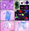
Lentiviral transfer of Cre into tissues and cells of R26R reporter mice. (A) Expression of β-gal 3 weeks after intraparenchymal injection of 6 × 10⁷ IU into the liver. (Scale bar = 200 μm.) (B) Analysis of β-gal expression in colonies formed by progenitor bone marrow cells (Left; scale bar = 400 μm) and in in vitro differentiated progenitor cells (Center and Right). Identification of differentiated progenitors by staining for the myeloid marker CD11b (Center Lower, red) and for the megakaryocyte marker CD41 (Right Lower, red); 4′,6-diamidino-2-phenylindole is shown in blue. Original magnification ×300 (Center) and ×1000 (Right). (C--F) Analysis of Cre expression and recombination in the brain. (C) X-gal stain of coronal section 1 week after injection of 6 × 10⁷ IU LV-Cre into the striatum. (Scale bar = 1 mm.) (D) Identification of transduced cells in the cortex of R26R mice by staining for β-gal (green), the astrocyte marker GFAP (blue), and neuronal marker NeuN (red) (arrowhead, β-gal- and GFAP-positive cell; arrow, β-gal- and NeuN-positive cell). (Scale bar = 30 μm.) (E and F) X-gal stain of coronal section 3 weeks after injection of 6 × 10⁷ IU LV-Cre into the striatum (E) and hippocampus (F). Black arrows indicate tissue damage. (Scale bar = 1 mm.)
Figure 3
Figure 3
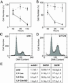
Effect of Cre expression on cell number and cell cycle. (A and B) Quantification of the number of cells attached to the substrate 48, 96, and 140 h after infection with LV-Lac (circles) or LV-Cre (squares). The number of CV-1 (A) and COS (B) cells expressed as percentage of the uninfected control cells (mean ± SEM, n = 4 experiments). (CE) Analysis of propidium iodine incorporation into cellular DNA by flow cytometry. Representative cell-cycle profiles of uninfected (cntr) COS cells (C) and COS cells infected with LV-Cre (D). (E) Statistical analysis of cell-cycle profiles (n = 3 experiments).
Figure 4
Figure 4
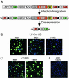
Development of LV-Cre-SD for transient Cre expression. (A) Strategy for regulating Cre expression by using LV-Cre-SD (Top). Self-deletion is achieved by incorporation of a single loxP site (bold black arrow) into the U3 region of the 3′ LTR. Because of the duplication of the U3 region during reverse transcription, the integrated provirus contains two U3 regions with loxP sites (Middle). The internal CMV promoter drives expression of Cre, which in turn will catalyze the recombination of the loxP sites in the U3 regions, resulting in the excision of almost the complete provirus including the Cre cassette (Bottom). In this way, Cre terminates its own expression. (BD) Immunofluorescence analysis of Cre expression (green nuclei) in CV-1 cells transduced with LV-Cre-SD (B), LV-Cre (C), or uninfected (cntr) cells (D) at the indicated times after infection. Representative merged images (green channel, anti-Cre; blue color, 4′,6-diamidino-2-phenylindole). (Scale bar = 90 μm.)
Figure 5
Figure 5
Effect of the conventional and the LV-Cre-SD on cell number. CV-1 (A) and COS (B) cells were counted 140 h after infection with LV-Lac (black columns), LV-Cre (blue), or LV-Cre-SD (red). The number of cells is depicted as percentage of the uninfected (cntr) control cells (mean ± SEM, n = 3 experiments). (C) Analysis of reporter gene (β-gal) activation by the LV-Cre-SD. CV-1 cells were infected with two particles per cell of LV-Cre (Middle) or LV-Cre-SD (Bottom). At 3 days (Left) or 28 days (Right) after infection, cells were stained with X-Gal. (D) Quantification of β-gal+ cells 3 days or 28 days after infection with LV-Cre (blue) or LV-Cre-SD (red) (mean ± SEM, n = 3 experiments).
Figure 6
Figure 6
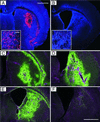
Transient Cre expression and reporter gene activation by the LV-Cre-SD in vivo. (A and B) Immunofluorescence analysis of Cre expression. The striatum of R26R mice was injected with 6 × 10⁷ IU of LV-Cre-SD. At 1 (A) or 5 weeks (B) later, mice were killed and analyzed for Cre expression (red). GFAP was used as background stain (blue). (A Inset and B Inset) Cre-positive cells (red) in the striatum of LV-Cre-SD-injected mice. (Scale bar = 62.5 μm.) (CF) Immunohistochemical analysis of β-gal expression. At 1 (C and D) or 5 (E and F) weeks after injection of 6 × 10⁷ IU of LV-Cre-SD (C and E) or LV-Cre (D and F) into the striatum of R26R mice, coronal sections of fixed brains were stained for β-gal expression (green). The nuclear DNA was stained with 4′,6-diamidino-2-phenylindole (magenta). Coronal sections of the same mice are shown in A and C as well as B and E. (Scale bar for AF = 1 mm.)

Similar articles

Cited by

References

    1. Sternberg N, Hamilton D, Hoess R. J Mol Biol. 1981;150:487–507. - PubMed
    1. Sternberg N, Hamilton D. J Mol Biol. 1981;150:467–486. - PubMed
    1. Sauer B, Henderson N. Proc Natl Acad Sci USA. 1988;85:5166–5170. - PMC - PubMed
    1. Gu H, Marth J D, Orban P C, Mossmann H, Rajewsky K. Science. 1994;265:103–106. - PubMed
    1. Sauer B. Methods Enzymol. 1993;225:890–900. - PubMed

Publication types

LinkOut - more resources