Partitioning of Thy-1, GM1, and cross-linked phospholipid analogs into lipid rafts reconstituted in supported model membrane monolayers
- PMID: 11535814
- PMCID: PMC58519
- DOI: 10.1073/pnas.191168698
Partitioning of Thy-1, GM1, and cross-linked phospholipid analogs into lipid rafts reconstituted in supported model membrane monolayers
Abstract
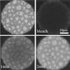
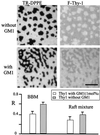
As shown earlier, raft-like domains resembling those thought to be present in natural cell membranes can be formed in supported planar lipid monolayers. These liquid-ordered domains coexist with a liquid-disordered phase and form in monolayers prepared both from synthetic lipid mixtures and lipid extracts of the brush border membrane of mouse kidney cells. The domains are detergent-resistant and are highly enriched in the glycosphingolipid GM1. In this work, the properties of these raft-like domains are further explored and compared with properties thought to be central to raft function in plasma membranes. First, it is shown that domain formation and disruption critically depends on the cholesterol density and can be controlled reversibly by treating the monolayers with the cholesterol-sequestering reagent methyl-beta-cyclodextrin. Second, the glycosylphosphatidylinositol-anchored cell-surface protein Thy-1 significantly partitions into the raft-like domains. The extent of this partitioning is reduced when the monolayers contain GM1, indicating that different molecules can compete for domain occupation. Third, the partitioning of a saturated phospholipid analog into the raft phase is dramatically increased (15% to 65%) after cross-linking with antibodies, whereas the distribution of a doubly unsaturated phospholipid analog is not significantly affected by cross-linking (approximately 10%). This result demonstrates that cross-linking, a process known to be important for certain cell-signaling processes, can selectively translocate molecules to liquid-ordered domains.
Figures


Comment in
-
Seeing is believing: visualization of rafts in model membranes.Proc Natl Acad Sci U S A. 2001 Sep 11;98(19):10517-8. doi: 10.1073/pnas.191386898. Proc Natl Acad Sci U S A. 2001. PMID: 11553797 Free PMC article. No abstract available.
Similar articles
-
Lipid rafts reconstituted in model membranes.Biophys J. 2001 Mar;80(3):1417-28. doi: 10.1016/S0006-3495(01)76114-0. Biophys J. 2001. PMID: 11222302 Free PMC article.
-
Seeing is believing: visualization of rafts in model membranes.Proc Natl Acad Sci U S A. 2001 Sep 11;98(19):10517-8. doi: 10.1073/pnas.191386898. Proc Natl Acad Sci U S A. 2001. PMID: 11553797 Free PMC article. No abstract available.
-
The size of lipid rafts: an atomic force microscopy study of ganglioside GM1 domains in sphingomyelin/DOPC/cholesterol membranes.Biophys J. 2002 May;82(5):2526-35. doi: 10.1016/S0006-3495(02)75596-3. Biophys J. 2002. PMID: 11964241 Free PMC article.
-
Sphingomyelin and cholesterol: from membrane biophysics and rafts to potential medical applications.Subcell Biochem. 2004;37:167-215. doi: 10.1007/978-1-4757-5806-1_5. Subcell Biochem. 2004. PMID: 15376621 Review.
-
Dynamics of raft molecules in the cell and artificial membranes: approaches by pulse EPR spin labeling and single molecule optical microscopy.Biochim Biophys Acta. 2003 Mar 10;1610(2):231-43. doi: 10.1016/s0005-2736(03)00021-x. Biochim Biophys Acta. 2003. PMID: 12648777 Review.
Cited by
-
Structural determinants for partitioning of lipids and proteins between coexisting fluid phases in giant plasma membrane vesicles.Biochim Biophys Acta. 2008 Jan;1778(1):20-32. doi: 10.1016/j.bbamem.2007.08.028. Epub 2007 Sep 12. Biochim Biophys Acta. 2008. PMID: 17936718 Free PMC article.
-
Cholesterol-dependent nanomechanical stability of phase-segregated multicomponent lipid bilayers.Biophys J. 2010 Jul 21;99(2):507-16. doi: 10.1016/j.bpj.2010.04.044. Biophys J. 2010. PMID: 20643069 Free PMC article.
-
Phase diagrams of lipid mixtures relevant to the study of membrane rafts.Biochim Biophys Acta. 2008 Nov-Dec;1781(11-12):665-84. doi: 10.1016/j.bbalip.2008.09.002. Epub 2008 Oct 7. Biochim Biophys Acta. 2008. PMID: 18952002 Free PMC article. Review.
-
Receptor-independent, direct membrane binding leads to cell-surface lipid sorting and Syk kinase activation in dendritic cells.Immunity. 2008 Nov 14;29(5):807-18. doi: 10.1016/j.immuni.2008.09.013. Immunity. 2008. PMID: 18993083 Free PMC article.
-
Probing Gag-Env dynamics at HIV-1 assembly sites using live-cell microscopy.J Virol. 2024 Sep 17;98(9):e0064924. doi: 10.1128/jvi.00649-24. Epub 2024 Aug 13. J Virol. 2024. PMID: 39136462 Free PMC article.
References
-
- Brown D A, Rose J. Cell. 1992;68:533–544. - PubMed
-
- Brown D A, London E. Biochem Biophys Res Commun. 1997;240:1–7. - PubMed
-
- Fridriksson E K, Shipkova P A, Sheets E D, Holowka D, Baird B, McLafferty F W. Biochemistry. 1999;38:8056–8063. - PubMed
-
- Simons K, Ikonen E. Nature (London) 1997;387:569–572. - PubMed
Publication types
MeSH terms
Substances
Grants and funding
LinkOut - more resources
Full Text Sources
Other Literature Sources
Research Materials
Miscellaneous

