. 2001 Sep 3;20(17):4973-86.
doi: 10.1093/emboj/20.17.4973.

An essential role for ARF6-regulated membrane traffic in adherens junction turnover and epithelial cell migration

Affiliations

An essential role for ARF6-regulated membrane traffic in adherens junction turnover and epithelial cell migration

F Palacios et al. EMBO J. .

Abstract

We describe a novel role for the ARF6 GTPase in the regulation of adherens junction (AJ) turnover in MDCK epithelial cells. Expression of a GTPase-defective ARF6 mutant, ARF6(Q67L), led to a loss of AJs and ruffling of the lateral plasma membrane via mechanisms that were mutually exclusive. ARF6-GTP-induced AJ disassembly did not require actin remodeling, but was dependent on the internalization of E-cadherin into the cytoplasm via vesicle transport. ARF6 activation was accompanied by increased migratory potential, and treatment of cells with hepatocyte growth factor (HGF) induced the activation of endogenous ARF6. The effect of ARF6(Q67L) on AJs was specific since ARF6 activation did not perturb tight junction assembly or cell polarity. In contrast, dominant-negative ARF6, ARF6(T27N), localized to AJs and its expression blocked cell migration and HGF-induced internalization of cadherin-based junctional components into the cytoplasm. Finally, we show that ARF6 exerts its role downstream of v-Src activation during the disassembly of AJs. These findings document an essential role for ARF6- regulated membrane traffic in AJ disassembly and epithelial cell migration.


Figures
None
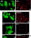
Fig. 1. Selective loss of AJs by expression of constitutively activated ARF6. MDCK cells grown on coverslips were infected with retrovirus encoding ARF6(Q67L) and GFP. Cells were labeled for actin (A and B), E-cadherin (C, D, G and H) and ZO-1 (E and F). Cells were viewed by confocal microscopy and optical sections at the plane of the cell junctions are shown (A–F). Vertical (x/z) sections of E-cadherin-labeled cells from a separate field are also shown (G and H). Infected cells transiently expressing ARF6(Q67L) exhibited diffuse green GFP staining (A, C, E and G). Infected cells exhibit a loss of actin and E-cadherin label at the cell junctions, while the AJs of untransfected cells (arrows) are intact. Also, tight junctions are intact on ARF6(Q67L)-expressing cells.
None
Fig. 2. Distribution of ARF6(Q67L) in MDCK cells. (A) Cells transiently expressing HA-tagged ARF6(Q67L) were labeled for E-cadherin (red) and HA (green). Coincident staining appears yellow in the merged image. (B) MDCK cells stably expressing HA-tagged ARF6(Q67L) were labeled for HA (left), E-cadherin (middle) and β-catenin (right). ARF6(Q67L), E-cadherin and β-catenin localize predominantly to the perinuclear cytoplasm and at the cell periphery in areas of membrane ruffling. (C) Cells transiently expressing HA-tagged ARF6(Q67L) were labeled for HA and ZO-1. Cells were viewed by confocal microscopy and optical sections at two different confocal planes are shown. ARF6(Q67L)-expressing cells have intact tight junctions.
None
Fig. 3. ARF6(Q67L:Q37E:S38I), an effector domain ARF6 mutant incapable of membrane ruffling, can induce AJ disassembly. (A) Cells transiently expressing HA-tagged ARF6(Q67L:Q37E:S38I) were labeled for E-cadherin (red) and HA (green). Coincident staining appears yellow in the merged image. Transfected cells exhibited a loss of E-cadherin label at the cell junctions with a redistribution of the protein to the perinuclear vesicles in the cytoplasm. (B) Cells were labeled for HA (red) and for transferrin receptor (Tfn-R; red). Coincident staining appears yellow in the merged image. A stacked image of optical sections along the z-axis is shown (left). A magnified image across a single confocal plane at the perinuclear region is shown on the right.
None
Fig. 4. ARF6–GTP-generated endosomal vesicles are devoid of PIP2. (A) HA-tagged ARF6(Q67L)-expressing cells were transfected with plasmid encoding PH–GFP, and 24 h post-transfection cells were fixed and labeled with anti-HA monoclonal antibody to determine ARF6 distribution (red). Images were merged and coincident staining appears yellow. Merged images of horizontal x/y sections along the z-axis are shown [1 (top)–6 (bottom)]. An overlapping distribution of PH–GFP (i.e. PIP2) and ARF6(Q67L) was detected in surface ruffles, but not in ARF6(Q67L)-positive perinuclear vesicles. (B) ARF6(Q67L)-expressing cells were transfected with plasmid encoding PH–GFP and labeled for E-cadherin. Coincident staining for E-cadherin (red) and PH–GFP (green) appears yellow. Merged images of a single optical section at the perinuclear plane are shown. (C) Cells infected with retrovirus alone were transfected with plasmid encoding PH–GFP, and 24 h post-transfection, cells were fixed and viewed under a confocal microscope. PH–GFP label was mainly diffuse in the cytoplasm and some label was seen at the cell surface.
None
Fig. 5. ARF6 activation does not perturb cell polarity. MDCK cells infected with retrovirus encoding HA-tagged ARF6(Q67L) for transient protein expression were grown on transwell filters, fixed, double labeled as indicated below, and processed by immunofluorescence microscopy. Cells were viewed using a confocal imaging system. Cells were double labeled with anti-HA rabbit polyclonal antibody (green) and with mouse monoclonal antibodies (red) to either Tfn-R (A), gp135 (B and D) or ZO-1 (C). Merged images of vertical (x/z) sections are shown (A–C). A horizontal x/y section at a single confocal plane at the apical microvilli is also shown (D). Coincident red and green staining appears yellow.
None
Fig. 6. Subcellular distribution of ARF6(T27N) and wild-type ARF6. (A) Cells expressing HA-tagged ARF6(T27N) were double labeled with anti-HA rabbit polyclonal antibody (green) and anti-E-cadherin mouse monoclonal antibody (red). An image across a single confocal plane at the cell junctions is shown to emphasize the localization of ARF6(T27N) at the cell junctions. (B) Cells expressing HA-tagged ARF6(T27N) were double labeled for HA (green) and for either E-cadherin (red, shown on the left) or gp135 (red, shown on the right). Coincident red and green staining appears yellow. Merged images taken along the x/z-axis are shown. ARF6(T27N) localizes to the AJs of MDCK cells; a small fraction of label was also seen at the lateral membrane and subapical region. (C) Cells transiently expressing HA-tagged wild-type (wt) ARF6 were labeled for HA (green) and for either E-cadherin (red, left) or gp135 (red, right). Coincident red and green staining appears yellow. Merged images taken along the x/z-axis are shown. Wild-type ARF6 localizes to the AJs, the lateral membrane and subapical membranes.
None
Fig. 7. Effect of the ARF6 GTPase cycle on the detergent-solubility properties of β-catenin. Cells stably expressing either ARF6, ARF6(Q67L) or ARF6(T27N) were lysed in CSK-A buffer containing 0.5% Triton X-100, centrifuged, and Triton-soluble (s) and -insoluble (i) fractions were analyzed for β-catenin distribution using immunoblotting procedures.
None
Fig. 8. Effect of dominant-negative ARF6 on HGF- and Ca2+ depletion-mediated AJ disassembly. (A) Untransfected MDCK cells or cells stably expressing ARF6(T27N) were grown on coverslips and incubated with 5 ng/ml HGF for 4 h. Cells were fixed and labeled for E-cadherin (green), and stained for actin (red). As shown, ARF6(T27N) expression blocks HGF-induced internalization of E-cadherin. (B) HGF induces the subcellular redistribution of endogenous ARF6. Normal MDCK cells or cells stably expressing either ARF6(Q67L) or ARF6(T27N) were lysed in CSK-A buffer, resolved on 14% SDS gels and the Triton-soluble fraction was analyzed for endogenous ARF6 distribution with anti-ARF6 monoclonal antibody using immunoblotting procedures. Band density was measured using the enhanced UltroScan XL Laser Densitometer (Pharmacia). As a loading control, the membrane was also probed for γ-tubulin expression. HGF treatment led to a marked increase in soluble ARF6. In cells expressing ARF6(Q67L), similar amounts of soluble ARF6 were detected in the presence and absence of HGF, while expression of ARF6(T27N) blocked the redistribution of ARF6 into the soluble pool. Exogenous ARF6(Q67L) runs at a higher molecular weight than the endogenous protein (arrow). Exogenous ARF6(T27N) was not detected on the blot since most of the ARF6(T27N) was present in the insoluble fraction (not shown). The data shown are representative of four independent experiments. (C) Normal MDCK cells (left) and those expressing wild-type ARF6 (center) or ARF67(T27N) (right) were incubated with EDTA (2.5 mM) at 37°C for 15 min. Cells were fixed and labeled for E-cadherin (green) and stained for actin (red). Merged images are shown; coincident staining appears yellow. ARF6(T27N) expression blocks the Ca2+ depletion-induced collapse of E-cadherin into the cytoplasm.
None
Fig. 9. The ARF6 GTPase cycle modulates epithelial cell migration. Cells stably expressing either wild-type ARF6, ARF6(Q67L), ARF6 triple mutant [ARF6(TM)] or ARF6(T27N) were grown on transwell filters and their migratory potential was assessed in the presence or absence of HGF as indicated. The number of cells/field that migrated through the filter after 16 h was counted. The mean of six separate fields is shown. The data are representative of four independent experiments.
None
Fig. 10. Dominant-negative ARF6 blocks pp60v-src-induced cell scattering. Compact colonies of MDCK-pp60vsrc cell lines grown at 41°C, as visualized by phase-contrast microscopy, are shown (A). Alterations in colony morphology induced by temperature shift to 35°C, in the absence (B) or presence (C) of herbimycin, are shown. (D) Phase-contrast image of MDCK-pp60vsrc cell lines expressing ARF6(T27N) at permissive temperature (35°C). Both herbimycin and dominant-negative ARF6 block Src-induced cell scattering. Cells in the latter colonies are less organized and appear to have ruffled edges (see arrows).
None
Fig. 11. Working model for the regulation of AJ turnover by the ARF6 GTPase cycle. Activation on ARF6 at the AJs (by GEFs) occurs in response to extracellular stimuli and downstream of Src activation. This, in turn, promotes AJ disassembly and ruffling of the lateral membrane, although these processes are mediated by distinct effector molecules. GTP hydrolysis on ARF6 (by GAPS) at perinuclear compartments and the lateral membrane results in redistribution of E-cadherin to and at the lateral membrane to reassemble AJs. The scheme alongside shows that the concerted effect of ARF6–GTP on AJ disassembly and membrane ruffling is required for epithelial cell migration.
None
Fig. 12. Expression of wild-type ARF6, ARF6(Q67L) and ARF6(T27N) in MDCK cells using retroviral expression. Lysates of MDCK cells stably expressing either HA-tagged wild-type ARF6, ARF6(Q67L) or ARF6(T27N) were resolved on SDS gels, transferred to nitrocellulose membrane and probed using immunoblotting procedures for either total ARF6 expression or for exogenous ARF6 using anti-HA monoclonal antibody. Expression of ARF6 in transfected cells was barely a fold above endogenous protein expression. Note that unlike the data in Figure 8B, exogenous protein did not migrate at a higher molecular weight compared with endogenous ARF6 since a lower percentage SDS gel was used.

Similar articles

Cited by

References

    1. Al-Awar O., Radhakrishna,H., Powell,N.N. and Donaldson,J.G. (2000) Separation of membrane trafficking and actin remodeling functions of ARF6 with an effector domain mutant. Mol. Cell. Biol., 20, 5998–6007. - PMC - PubMed
    1. Altschuler Y., Liu,S., Katz,L., Tang,K., Hardy,S., Brodsky,F., Apodaca,G. and Mostov,K. (1999) ADP-ribosylation factor 6 and endocytosis at the apical surface of Madin–Darby canine kidney cells. J. Cell Biol., 147, 7–12. - PMC - PubMed
    1. Behrens J., Vakaet,L., Friis,R., Winterhager,E., Van Roy,F., Mareel,M.M. and Birchmeier,W. (1993) Loss of epithelial differentiation and gain of invasiveness correlates with tyrosine phosphorylation of the E-cadherin/β-catenin complex in cells transformed with a temperature-sensitive v-SRC gene. J. Cell Biol., 120, 757–766. - PMC - PubMed
    1. Berx G., Cleton-Jansen,A.M., Nollet,F., de Leeuw,W.J., van de Vijver,M., Cornelisse,C. and van Roy,F. (1995) E-cadherin is a tumour/invasion suppressor gene mutated in human lobular breast cancers. EMBO J., 14, 6107–6115. - PMC - PubMed
    1. Birchmeier C. and Birchmeier,W. (1993) Molecular aspects of mesenchymal–epithelial interactions. Annu. Rev. Cell Biol., 9, 511–540. - PubMed

Publication types

MeSH terms