. 2001 Sep 3;20(17):4952-63.
doi: 10.1093/emboj/20.17.4952.

A moving DNA replication factory in Caulobacter crescentus

Affiliations

A moving DNA replication factory in Caulobacter crescentus

R B Jensen et al. EMBO J. .

Abstract

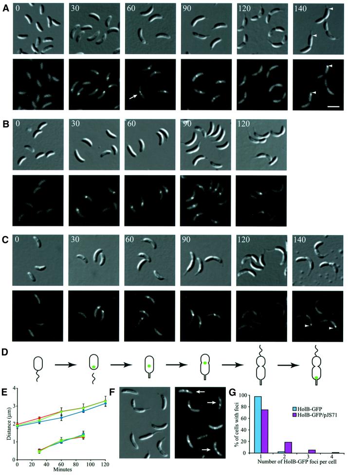
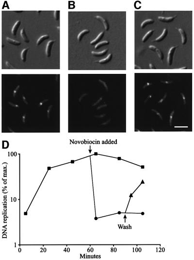
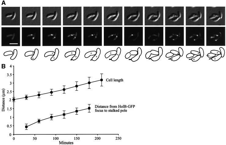
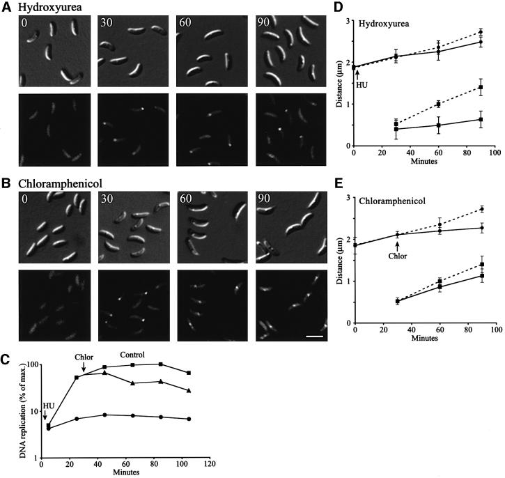
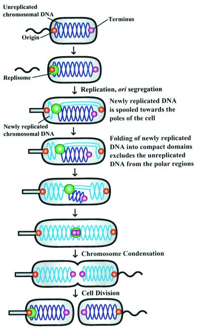
The in vivo intracellular location of components of the Caulobacter replication apparatus was visualized during the cell cycle. Replisome assembly occurs at the chromosomal origin located at the stalked cell pole, coincident with the initiation of DNA replication. The replisome gradually moves to midcell as DNA replication proceeds and disassembles upon completion of DNA replication. Although the newly replicated origin regions of the chromosome are rapidly moved to opposite cell poles by an active process, the replisome appears to be an untethered replication factory that is passively displaced towards the center of the cell by the newly replicated DNA. These results are consistent with a model in which unreplicated DNA is pulled into the replication factory and newly replicated DNA is bidirectionally extruded from the complex, perhaps contributing to chromosome segregation.


Figures

None
Fig. 1. Characterization of the HolB–GFP fusion protein strain. (A) The DNA contents profiles of mixed cultures of the HolB–GFP strain (dashed line) and the parent strain CB15N (solid line) were compared using flow cytometry of chromamycin A3-stained cells. Chromosome equivalents are indicated on the x-axis and relative number of cells on the y-axis. (B) DNA replication during the cell cycle in synchronized populations of HolB–GFP-expressing cells, measured as incorporation of [α-32P]dCTP into chromosomal DNA during a 1 min period. The degree of DNA replication is expressed as a percentage of maximal incorporation of radioactivity into DNA. (C) The abundance of the HolB–GFP fusion protein during the cell cycle analyzed by western blotting using anti-GFP antibodies. Samples were withdrawn at the indicated time points and equal amounts of total protein were loaded in all lanes.
None
Fig. 2. Intracellular localization of replisome proteins. HolB–GFP (A), HolC–YFP (B) or DnaB–YFP (C) expressing swarmer cells were isolated and allowed to progress synchronously through the cell cycle. Samples for microscopy were taken at the indicated time points (in minutes). Top panels show Nomarski DIC microscopy images of the cells and the lower panels show GFP or YFP fluorescence. A white arrow indicates a cell with two closely spaced HolB–GFP foci. White arrowheads indicate the stalked pole of the dividing cells. The white scale bar represents 2 µm. (D) Schematic of cell cycle progression of the strains and the intracellular positions of HolB–GFP, HolC–YFP or DnaB–YFP (green dots). Time-lapse microscopy of HolB–GFP-expressing cells (Figure 4) shows that HolB–GFP specifically assembles at the stalked pole during the swarmer-to-stalked cell transition, as indicated in the schematics. (E) Measurements of the average cell length (circles) and distance from the stalked pole to the middle of the replisome protein foci (squares) at different time points of the cell cycle of the HolB–GFP (blue), HolC–YFP (green) and DnaB–YFP (red) expressing cells. (F) Localization of replisome foci in HolB–GFP-expressing cells containing a freely replicating plasmid (pJS71). Approximately 30–50 copies of the plasmid are present per cell and plasmid replication is initiated by a plasmid-encoded replication-control system. White arrows indicate cells with multiple replisome foci or foci located at positions where they are not observed in plasmid-free cells. (G) Analysis of the number of replisome foci per cell in non-synchronized HolB–GFP-expressing cells in the absence (blue bars) or presence (purple bars) of the pJS71 plasmid. At least 250 cells with clearly visible foci were counted for each strain.
None
Fig. 3. Effect of DNA replication inhibition on the presence of replisome foci. HolB–GFP-expressing cells were synchronized and novobiocin (final concentration 100 µg/ml) was added at the stalked cell stage (60 min into the cell cycle). Some cells were washed and resuspended in fresh growth medium 30 min later. Shown are DIC (top panel) and fluorescence (lower panel) images of (A) untreated stalked cells 60 min into the cell cycle, (B) cells 10 min after novobiocin addition and (C) cells treated with novobiocin for 30 min, washed and incubated in fresh growth medium for 10 min. The scale bar represents 2 µm. (D) Direct measurement of DNA replication using [α-32P]dCTP pulse-labeling in control and novobiocin-treated cells. Squares indicate untreated control cells, circles indicate novobiocin-treated cells and triangles represent novobiocin-treated cells that were washed and resuspended in fresh medium.
None
Fig. 4. Time-lapse microscopy analysis showing gradual movement of HolB–GFP foci from the pole to midcell. HolB–GFP-expressing swarmer cells were placed on a thin layer of agarose containing nutrients, and images of the same cells were acquired every 30 min as the cells progressed synchronously through the cell cycle. Cell division occurs at ∼235 min under these growth conditions. (A) DIC images (top panel), fluorescence images (middle panel) and schematics (lower panel) of the same cells progressing through the cell cycle. Grey circles in the schematics indicate the intracellular positions of HolB–GFP. The white scale bar represents 2 µm. A QuickTime movie of this time-lapse sequence is available as Supplementary data at The EMBO Journal Online. (B) The distance from the stalked pole to the middle of the HolB–GFP focus (squares) and the cell length (circles) were measured at different time points in cells from time-lapse sequences. The graph shows averages and the error bars show the standard deviation. Gradual movement of the HolB–GFP focus from near the stalked pole to midcell was observed for all cells analyzed.
None
Fig. 5. Effect of slowing DNA replication or inhibiting protein synthesis on replisome movement. HolB–GFP-expressing swarmer cells were isolated and allowed to progress synchronously through the cell cycle. (A) To slow down DNA replication, HU (final concentration 3 mg/ml) was added at the beginning of the cell cycle. DIC and fluorescence images show cells at the indicated time points (in minutes) during the cell cycle. (B) Chloramphenicol (final concentration 25 µg/ml) was added to the synchronized cells to inhibit protein synthesis. Since chloramphenicol inhibits initiation of new rounds of DNA replication, but not completion of ongoing DNA replication (Winzeler and Shapiro, 1995), the drug was added at 30 min when DNA replication has been initiated in the majority of the cells. DIC and fluorescence images of cells at the indicated time points are shown. The white scale bar represents 2 µm. (C) The degree of DNA replication in control (squares), HU-treated (circles) and chloramphenicol-treated cells (triangles). (D and E) Measurements of the average distances from the stalked pole to the middle of the HolB–GFP focus (squares) or the cell length (circles) in control (broken lines), HU-treated (D, line) or chloramphenicol-treated (E, line) cells at the indicated time points.
None
Fig. 6. Intracellular localization of DNA replicated at particular time points in the cell cycle determined using BrdU pulse-labeling. Synchronized Caulobacter cells were exposed to BrdU for 15 min prior to the indicated times (in minutes). Cells were then fixed and the sites of BrdU incorporation were visualized by indirect immunofluorescence microscopy using anti-BrdU antibodies. (A) DIC images (top panel), immunofluorescence images (middle panel) of the BrdU-labeled cells, and an overlay of these images (lower panel), where the BrdU immunofluorescence signals were pseudocolored red. The scale bar represents 2 µm. Black arrows indicate typical BrdU foci positions in cells at that stage of the cell cycle. White arrows indicate cells where a weak third BrdU focus is observed, probably at the same intracellular position as the replisome. The schematics show cell cycle progression, the intracellular position of the replisome (green dot) and the organization of the chromosome. Red lines indicate the position of DNA labeled with BrdU during the 15 min pulse-labeling, dark blue lines indicate unreplicated DNA and light blue lines indicate DNA replicated earlier in the cell cycle, prior to addition of BrdU. (B) Measurements of the average intracellular positions of the center of the BrdU foci relative to the cell pole (red circles) at the indicated time points. The schematic shows how the distances were measured. Red dots indicate the BrdU foci and a green dot indicates the replisome focus.
None
Fig. 7. Model describing chromosome segregation and replication factory localization in Caulobacter. Swarmer cells do not replicate their DNA (dark blue lines) and replisome components are distributed throughout the cell. The origin of replication (red dot) is located at the flagellated pole of the cell and the terminus (purple dot) is at the opposite end of the cell (Jensen and Shapiro, 1999a). At the swarmer-to-stalked cell transition, the replication factory (green dot) assembles at the origin and DNA replication is initiated. As DNA replication proceeds, the newly replicated DNA strands (light blue lines) are moved towards both poles of the cell and the replication apparatus gradually migrates to midcell. This model proposes that the unreplicated part of the chromosome and the replication factory is passively displaced by the accumulating bulk of replicated DNA near the poles. When DNA replication is completed, the replication apparatus disassembles.

Similar articles

Cited by

References

    1. Chong J.P., Hayashi,M.K., Simon,M.N., Xu,R.M. and Stillman,B. (2000) A double-hexamer archaeal minichromosome maintenance protein is an ATP-dependent DNA helicase. Proc. Natl Acad. Sci. USA, 97, 1530–1535. - PMC - PubMed
    1. Cook P.R. (1999) The organization of replication and transcription. Science, 284, 1790–1795. - PubMed
    1. Davey M.J. and O’Donnell,M. (2000) Mechanisms of DNA replication. Curr. Opin. Chem. Biol., 4, 581–586. - PubMed
    1. Degnen S.T. and Newton,A. (1972) Dependence of cell division on the completion of chromosome replication in Caulobacter. J. Bacteriol., 110, 852–856. - PMC - PubMed
    1. Dingman C.W. (1974) Bidirectional chromosome replication: some topological considerations. J. Theor. Biol., 43, 187–195. - PubMed

Publication types