. 2001 Sep 3;20(17):4730-41.
doi: 10.1093/emboj/20.17.4730.

Ara6, a plant-unique novel type Rab GTPase, functions in the endocytic pathway of Arabidopsis thaliana

Affiliations

Ara6, a plant-unique novel type Rab GTPase, functions in the endocytic pathway of Arabidopsis thaliana

T Ueda et al. EMBO J. .

Abstract

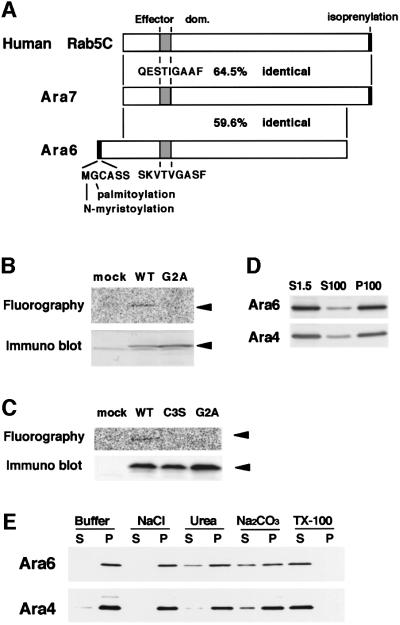
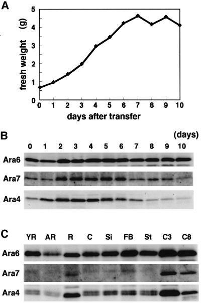
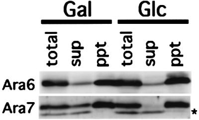
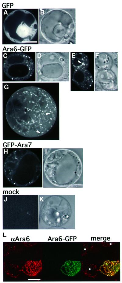
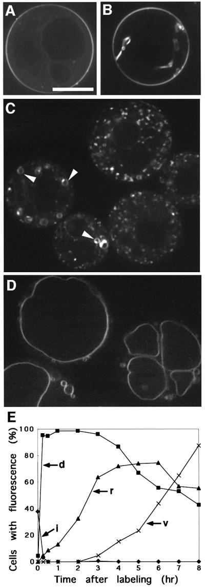
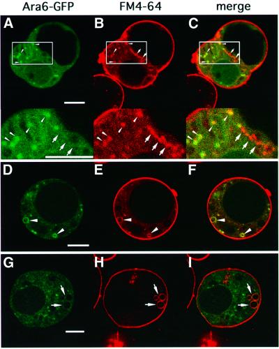
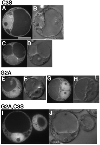
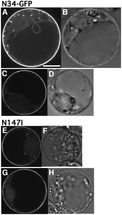
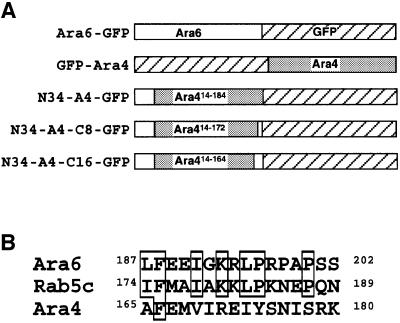
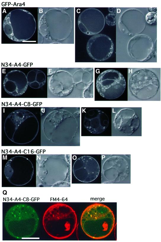
Ara6 of Arabidopsis thaliana is a novel member of the Rab/Ypt GTPase family with unique structural features. It resembles Rab5 GTPases best, but lacks a large part of the C-terminal hypervariable region and the cysteine motif, and instead harbors an extra stretch of amino acid residues containing myristoylation and palmitoylation sites at the N-terminus. Ara6 is tightly associated with membranes and is expressed constitutively. In contrast, the conventional Rab5 ortholog, Ara7, is highly expressed only in actively dividing cells. Examination of green fluorescent protein (GFP)-tagged proteins indicates that both Ara6 and Ara7 are distributed on a subpopulation of endosomes and suggests their roles in endosomal fusion. The endosomal localization of Ara6 requires N-terminal fatty acylation, nucleotide binding and the C-terminal amino acid sequence coordinately. Proteins similar to Ara6 are found only in higher plants and thus represent a novel class of Rab GTPases regulating endocytic function in a plant- specific manner.


Figures

None
Fig. 1. Ara6 is a peripheral membrane protein with a unique structure. (A) Schematic structures of human Rab5c, Ara7 and Ara6. (B) The N-terminus of Ara6 is N-myristoylated in vitro. In vitro transcription and translation of wild-type ARA6 (WT) and ARA6G2A (G2A) were performed in the presence of 14C-labeled myristic acid. The same reaction was carried out without the template (mock). (C) The N-terminus of Ara6 is palmitoylated in vitro. Ara6 (WT), Ara6C3S (C3S) and Ara6G2A (G2A) synthesized in vitro were immuno precipitated and incubated with Arabidopsis lysate in the presence of 14C-labeled palmitoyl-CoA. (D) Membrane binding of Ara6 and Ara4 was examined by western blotting. The total lysate prepared from Arabidopsis cultured cells (S1.5) was ultracentrifuged to separate the cytosolic fraction (S100) from the membrane fraction (P100), and analyzed by immunoblotting with anti-Ara6 polyclonal or anti-Ara4 monoclonal antibody. (E) The P100 fraction was incubated on ice in a buffer containing either salt, urea, alkali (Na2CO3) or Triton X-100. Samples were ultracentrifuged again, then the supernatant and the pellet were analyzed by immunoblotting with anti-Ara6 or anti-Ara4 antibody.
None
Fig. 2. Expression of Ara6, Ara7 and Ara4 in Arabidopsis suspension cultured cells or plants. (A) The Arabidopsis suspension cultured cells were collected every day after transfer to the new medium for 10 days, and fresh weights were plotted. (B) The total protein samples prepared from a series of suspension cultured cells were subjected to immunoblotting analysis with anti-Ara6, anti-Ara7 or anti-Ara4 antibody. (C) The total protein samples were prepared from young rosettes (YR), adult rosettes (AR), roots (R), cotyledons (C), siliques (Si), flower-bud clusters (FB) and stems (St). These samples and total lysates from suspension cultured cells were collected over 3 (C3) or 8 days (C8) after transfer and were subjected to immunoblotting analysis with anti-Ara6, anti-Ara7 or anti-Ara4 antibody.
None
Fig. 3. Recycling of Ara6 is independent of GDI in yeast cells. ARA6 or ARA7 was expressed constitutively in yeast sec19/gdi1 mutant cells harboring AtGDI1 under control of the GAL1 promoter. Transformant cells were cultured in galactose medium at the permissive temperature, shifted up to the restrictive temperature and then transferred to new galactose medium (Gal) or glucose medium (Glc). The total cell lysates (total) were ultracentrifuged to obtain cytosol (sup) and membrane (ppt) fractions, and samples were subjected to immunoblotting analysis with anti-Ara6 or anti-Ara7 antibody. The asterisk indicates a cross-reacting band which is not related to Ara7.
None
Fig. 4. Subcellular localization of Ara6–GFP and GFP–Ara7. ARA6 and ARA7 were tagged with GFP and expressed transiently in the protoplasts of Arabidopsis suspension cultured cells. (A and B) An Arabidopsis cell expressing GFP. (CG) Ara6–GFP was localized on motile dots and ring-like organelles (arrowheads). (H and I) GFP–Ara7 was localized predominantly on the motile dotted organelles. (J and K) Protoplasts not treated with the plasmid showed no fluorescence. (A, C, E, H and J) Confocal images. (B, D, F, I and K) Nomarski images. (G) Projection of serial confocal images taken at 0.25 mm intervals. (B–K) Same scale as (A). Bar, 10 µm. (L) Distribution of Ara6 examined by immunofluorescence analysis. Protoplasts, some of which are expressing ARA6–GFP, were fixed and stained with the anti-Ara6 antibody. Asterisks indicate untransformed cells. Bar, 10 µm.
None
Fig. 5. Endocytic pathway of the Arabidopsis cell visualized with FM4-64. FM4-64 was internalized from the plasma membrane (A and B), via motile dotted organelles (C) and ring-like organelles (C, arrowheads), to the vacuole (D). Bar, 10 µm. (E) Time course of the internalization of FM4-64. Percentages of cells with stained plasma membrane invaginations (i), dotty endosomes (d), ring-like structures (r) and vacuole (v) are plotted.
None
Fig. 6. Ara6–GFP resides on early endosomes. Protoplasts of Arabidopsis cultured cells expressing Ara6–GFP were labeled with FM4-64. (AC) The motile dots with Ara6–GFP (arrowheads) were also stained by FM4-64. Note that some dots labeled by FM4-64 were not marked by Ara6–GFP (arrows). The lower panels are high magnification images of the boxed regions. (DF) Ring-like organelles where Ara6–GFP resides (arrowheads) were also labeled with FM4-64. (GI) Some structures (arrows) were marked by FM4-64 but barely by Ara6–GFP. Bar, 10 µm.
None
Fig. 7. GTP-bound mutant of Ara6 (Ara6Q93L) or Ara7 (Ara7Q69L) tagged by GFP resides on aggregates of ring-like structures, dots and the vacuole membrane. (AF) Protoplasts of Arabidopsis suspension cultured cells expressing Ara6Q93L–GFP. (GL) Protoplasts of Arabidopsis suspension cultured cells expressing Ara7Q69L–GFP. (A, C, E, G, I and K) Confocal images. (B, D, F, H, J and L) Nomarski images. (B–L) Same scale as (A). Bar, 10 µm. (M) The protoplast expressing Ara6Q93L was labeled with FM4-64 and viewed after 30 min. Bar, 10 µm.
None
Fig. 8. N-terminal fatty acylation is essential for the correct localization of Ara6. (AD) Protoplasts of Arabidopsis cells expressing Ara6C3S–GFP, which retain Gly2 to be N-myristoylated but which cannot be the substrate for palmitoylation. (EH) Protoplasts of Arabidopsis cells expressing Ara6G2A–GFP, which cannot be myristoylated. (I and J) Protoplasts of Arabidopsis cells expressing Ara6G2A,C3S–GFP, a double mutant of fatty acylation sites. (A, C, E, G and I) Confocal images. (B, D, F, H and J) Nomarski image. Bar, 10 µm.
None
Fig. 9. (AD) The N-terminal region of Ara6 is not sufficient for endosomal localization. A fusion of the N-terminal 34 amino acids of Ara6 with GFP (N34–GFP) was expressed transiently in Arabidopsis suspension cultured cells. (EH) Nucleotide binding is required for the endosomal localization of Ara6. The nucleotide-free mutant of Ara6 tagged by GFP, Ara6N147I–GFP, was expressed transiently in Arabidopsis suspension cultured cells. (A, C, E and G) Confocal images. (B, D, F and H) Nomarski images. Bar, 10 µm.
None
Fig. 10. (A) Schematic structures of chimeric proteins constructed to determine the minimal region required for endosome localization of Ara6. (B) Sequence comparison among the C-terminal 16 amino acids of Ara6, the corresponding region of human Rab5c and that of Ara4. Conserved amino acids are boxed.
None
Fig. 11. Subcellular localization of chimeric proteins expressed transiently in the protoplasts of Arabidopsis suspension cultured cells. (ADArabidopsis cells expressing GFP–Ara4. (EHArabidopsis cells expressing N34-A4–GFP. (ILArabidopsis cells expressing N34-A4-C8–GFP. (MPArabidopsis cells expressing N34-A4-C16–GFP. (A, C, E, G, I, K, M and O) Confocal images. (B, D, F, H, J, L, N and P) Nomarski images. (B–P) Same scale as (A). Bar, 10 µm. (Q) The protoplast expressing N34-A4-C8–GFP was labeled with FM4-64. Bar, 10 µm.

Similar articles

Cited by

References

    1. Anai T., Hasegawa,K., Watanabe,Y., Uchimiya,H., Ishizaki,R. and Matsui,M. (1991) Isolation and analysis of cDNAs encoding small GTP-binding proteins of Arabidopsis thaliana. Gene, 108, 259–264. - PubMed
    1. Anai T., Aspuria,E.T., Fujii,N., Ueda,T., Matsui,M., Hasegawa,K. and Uchimiya,H. (1995) Immunological analysis of a small GTP-binding protein in higher plant cells. J. Plant Physiol., 147, 48–52.
    1. Anuntalabhochai S., Terryn,N., Van Montagu,M. and Inze,D. (1991) Molecular characterization of an Arabidopsis thaliana cDNA encoding a small GTP-binding protein, Rha1. Plant J., 1, 167–174. - PubMed
    1. Araki S., Kaibuchi,K., Sasaki,T., Hata,Y. and Takai,Y. (1991) Role of the C-terminal region of smg p25A in its interaction with membranes and the GDP/GTP exchange protein. Mol. Cell. Biol., 11, 1438–1447. - PMC - PubMed
    1. Bolte S., Schiene,K. and Dietz,K.J. (2000) Characterization of a small GTP-binding protein of the rab5 family in Mesembryanthemum crystallinum with increased level of expression during early salt stress. Plant Mol. Biol., 42, 923–936. - PubMed

Publication types

MeSH terms

Associated data