

Three-dimensional comparison of ultrastructural characteristics at depressing and facilitating synapses onto cerebellar Purkinje cells
- PMID: 11517256
- PMCID: PMC6763067
- DOI: 10.1523/JNEUROSCI.21-17-06666.2001
Three-dimensional comparison of ultrastructural characteristics at depressing and facilitating synapses onto cerebellar Purkinje cells
Abstract
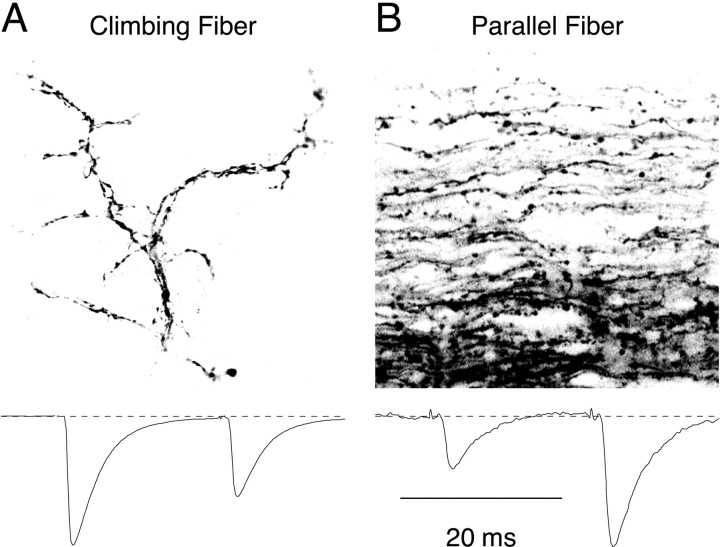
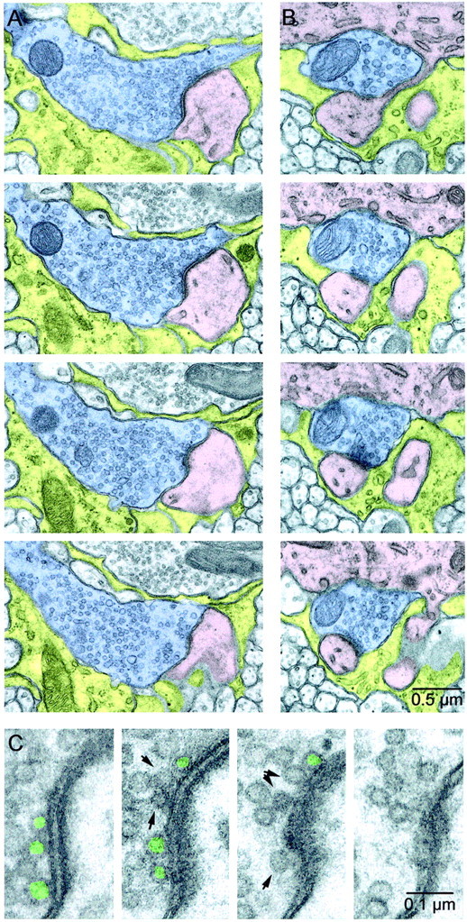
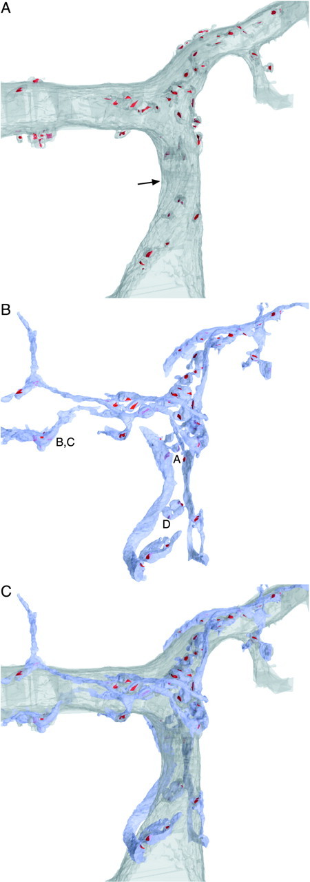
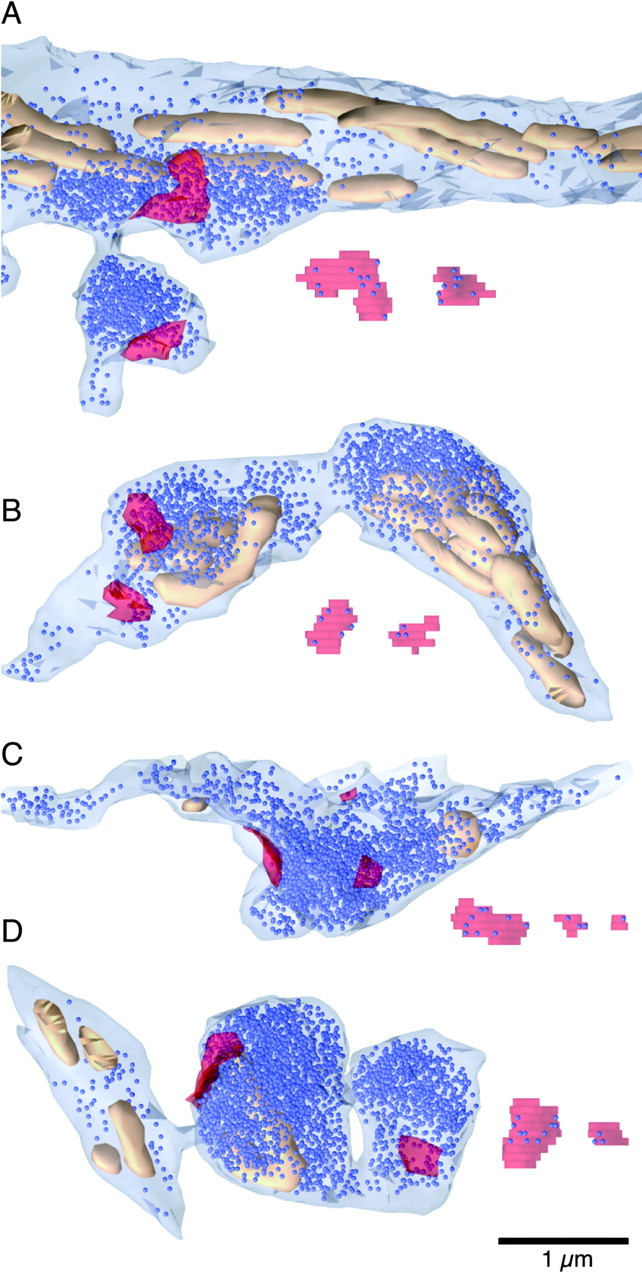
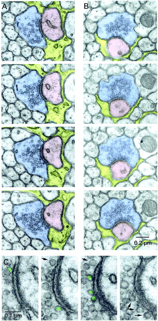
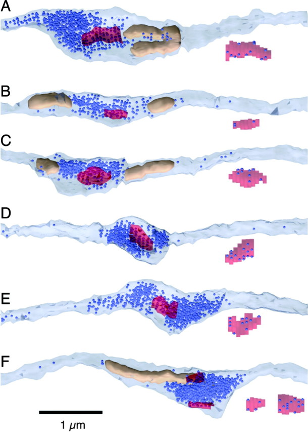
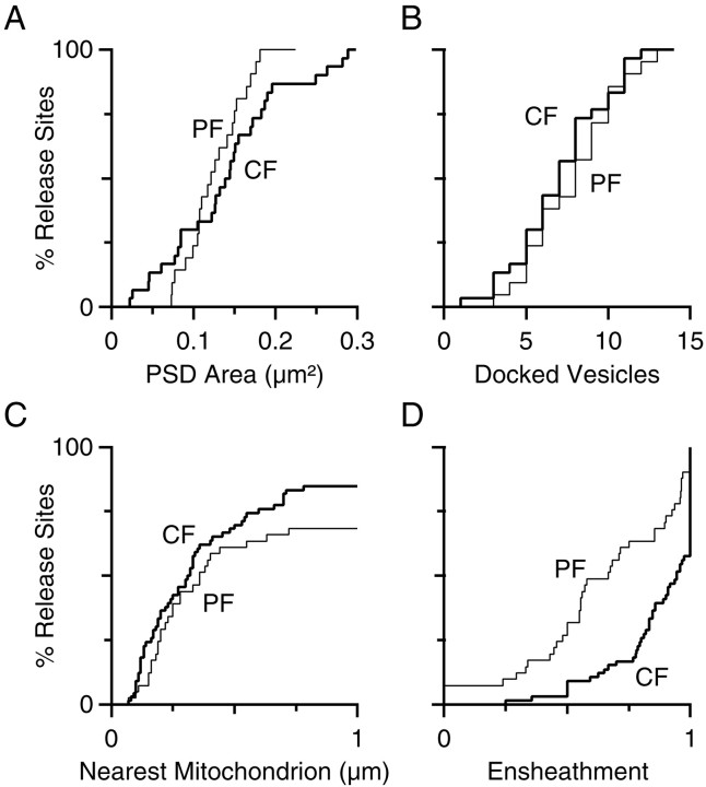
Cerebellar Purkinje cells receive two distinctive types of excitatory inputs. Climbing fiber (CF) synapses have a high probability of release and show paired-pulse depression (PPD), whereas parallel fiber (PF) synapses facilitate and have a low probability of release. We examined both types of synapses using serial electron microscopic reconstructions in 15-d-old rats to look for anatomical correlates of these differences. PF and CF synapses were distinguishable by their overall ultrastructural organization. There were differences between PF and CF synapses in how many release sites were within 1 microm of a mitochondrion (67 vs 84%) and in the degree of astrocytic ensheathment (67 vs 94%). However, the postsynaptic density sizes for both types of synapses were similar (0.13-0.14 microm(2)). For both types of synapses, we counted the number of docked vesicles per release site to test whether this number determines the probability of release and synaptic plasticity. PF and CF synapses had the same number of anatomically docked vesicles (7-8). The number of docked vesicles at the CF does not support a simple model of PPD in which release of a single vesicle during the first pulse depletes the anatomically docked vesicle pool at a synapse. Alternatively, only a fraction of anatomically docked vesicles may be release ready, or PPD could result from multivesicular release at each site. Similarities in the number of docked vesicles for PF and CF synapses indicate that differences in probability of release are unrelated to the number of anatomically docked vesicles at these synapses.
Figures
Similar articles
-
Brain-derived neurotrophic factor modulates cerebellar plasticity and synaptic ultrastructure.J Neurosci. 2002 Feb 15;22(4):1316-27. doi: 10.1523/JNEUROSCI.22-04-01316.2002. J Neurosci. 2002. PMID: 11850459 Free PMC article.
-
GluD2 Endows Parallel Fiber-Purkinje Cell Synapses with a High Regenerative Capacity.J Neurosci. 2016 Apr 27;36(17):4846-58. doi: 10.1523/JNEUROSCI.0161-16.2016. J Neurosci. 2016. PMID: 27122040 Free PMC article.
-
Variation in the number, location and size of synaptic vesicles provides an anatomical basis for the nonuniform probability of release at hippocampal CA1 synapses.Neuropharmacology. 1995 Nov;34(11):1387-95. doi: 10.1016/0028-3908(95)00142-s. Neuropharmacology. 1995. PMID: 8606788
-
Development and fine structure of murine Purkinje cells in dissociated cerebellar cultures: neuronal polarity.Anat Embryol (Berl). 1998 Jan;197(1):9-29. doi: 10.1007/s004290050117. Anat Embryol (Berl). 1998. PMID: 9462856 Review.
-
The morphology of excitatory central synapses: from structure to function.Cell Tissue Res. 2006 Nov;326(2):221-37. doi: 10.1007/s00441-006-0288-z. Epub 2006 Aug 24. Cell Tissue Res. 2006. PMID: 16932936 Review.
Cited by
-
Glycolytic Enzymes Localize to Synapses under Energy Stress to Support Synaptic Function.Neuron. 2016 Apr 20;90(2):278-91. doi: 10.1016/j.neuron.2016.03.011. Epub 2016 Apr 7. Neuron. 2016. PMID: 27068791 Free PMC article.
-
Astrocytic glutamate transport regulates a Drosophila CNS synapse that lacks astrocyte ensheathment.J Comp Neurol. 2016 Jul 1;524(10):1979-98. doi: 10.1002/cne.24016. Epub 2016 Apr 25. J Comp Neurol. 2016. PMID: 27073064 Free PMC article.
-
Intrinsic kinetics determine the time course of neuronal synaptic transporter currents.Proc Natl Acad Sci U S A. 2006 Jan 24;103(4):1083-7. doi: 10.1073/pnas.0510476103. Epub 2006 Jan 17. Proc Natl Acad Sci U S A. 2006. PMID: 16418298 Free PMC article.
-
Differences in transmission properties and susceptibility to long-term depression reveal functional specialization of ascending axon and parallel fiber synapses to Purkinje cells.J Neurosci. 2005 Mar 23;25(12):3246-57. doi: 10.1523/JNEUROSCI.0073-05.2005. J Neurosci. 2005. PMID: 15788782 Free PMC article.
-
Brief bursts of parallel fiber activity trigger calcium signals in bergmann glia.J Neurosci. 2006 Jun 28;26(26):6958-67. doi: 10.1523/JNEUROSCI.0613-06.2006. J Neurosci. 2006. PMID: 16807325 Free PMC article.
References
-
- Brewer PA, Lynch K. Stimulation-associated changes in frog neuromuscular junctions. A quantitative ultrastructural comparison of rapid-frozen and chemically fixed nerve terminals. Neuroscience. 1986;17:881–895. - PubMed
-
- Crepel F, Mariani J, Delhaye-Bouchaud N. Evidence for a multiple innervation of Purkinje cells by climbing fibers in the immature rat cerebellum. J Neurobiol. 1976;7:567–578. - PubMed
Publication types
MeSH terms
Grants and funding
LinkOut - more resources
Full Text Sources