. 2001 Sep;21(18):6122-31.
doi: 10.1128/MCB.21.18.6122-6131.2001.

Transcriptional hyperactivity of human progesterone receptors is coupled to their ligand-dependent down-regulation by mitogen-activated protein kinase-dependent phosphorylation of serine 294

Affiliations

Transcriptional hyperactivity of human progesterone receptors is coupled to their ligand-dependent down-regulation by mitogen-activated protein kinase-dependent phosphorylation of serine 294

T Shen et al. Mol Cell Biol. 2001 Sep.

Abstract

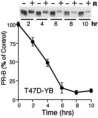
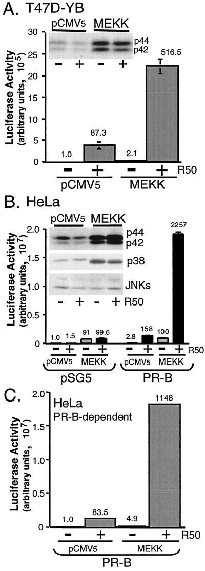
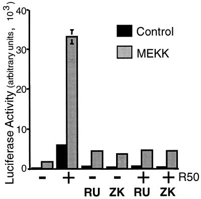
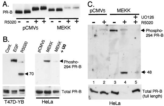
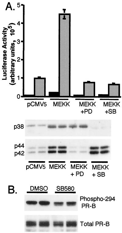
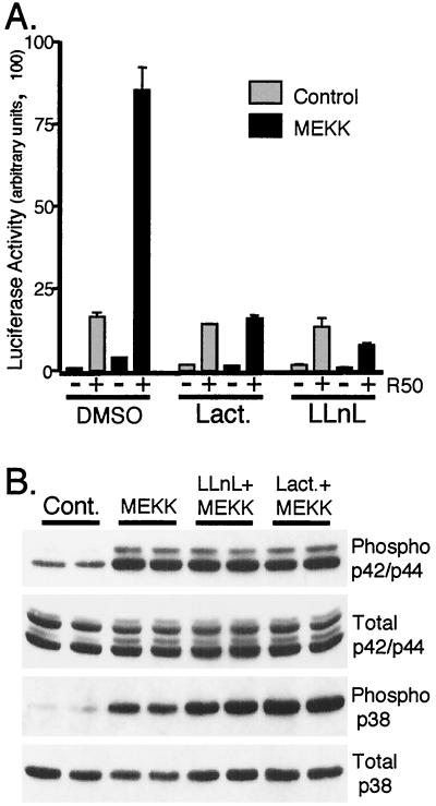
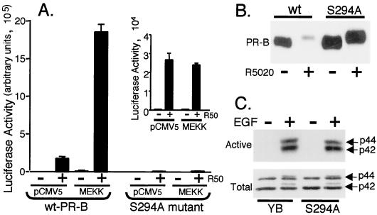
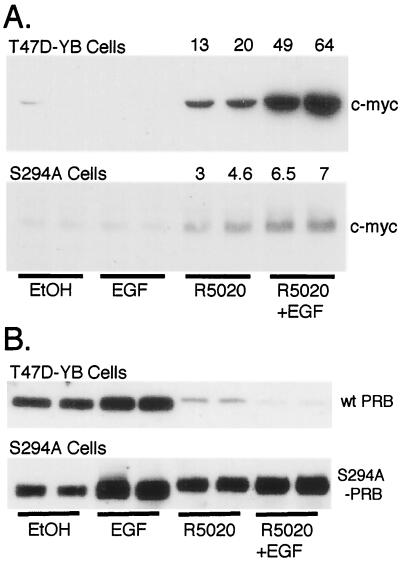
Breast cancers often exhibit elevated expression of tyrosine kinase growth factor receptors; these pathways influence breast cancer cell growth in part by targeting steroid hormone receptors, including progesterone receptors (PR). To mimic activation of molecules downstream of growth factor-initiated signaling pathways, we overexpressed mitogen-activated protein kinase (MAPK; also known as extracellular signal-regulated kinase) kinase kinase 1 (MEKK1) in T47D human breast cancer cells expressing the B isoform of PR. MEKK1 is a strong activator of p42 and p44 MAPKs. MEKK1 expression increased progestin-mediated transcription 8- to 10-fold above normal PR-driven transcription levels. This was dependent on the presence of a progesterone response element and functional PR. PR protein levels were unchanged by MEKK1 alone but were extensively down-regulated by MEKK1 plus the progestin R5020. MEKK1 expression resulted in phosphorylation of PR on Ser294, a MAPK consensus site known to mediate ligand-dependent PR degradation. MEK inhibitors blocked phosphorylation of Ser294 and attenuated PR transcriptional hyperactivity in response to MEKK1 plus R5020; stabilization of PR by inhibition of the 26S proteasome produced similar results. T47D cells stably expressing mutant S294A PR, in which serine 294 is replaced by alanine, fail to undergo ligand-dependent down-regulation and are resistant to MEKK1-plus-R5020-induced transcriptional synergy but respond to progestins alone. Similarly, c-myc protein levels are synergistically increased by epidermal growth factor and R5020 in cells expressing wild-type PR, but not S294A PR. Thus, highly stable mutant PR are functional in response to progestins but are incapable of cross talk with MAPK-driven pathways. These studies demonstrate a paradoxical coupling between steroid receptor down-regulation and transcriptional hyperactivity. They also suggest a link between phosphorylation of PR by MAPKs in response to peptide growth factor signaling and steroid hormone control of breast cancer cell growth.


Figures

FIG. 1
FIG. 1
Ligand-dependent PR down-regulation. T47D-YB cells were treated without or with R5020 (10 nM) for 2 to 10 h; protein levels were measured with PR-specific antibodies as described in Materials and Methods. A total of 100 μg of protein was loaded per lane for Western immunoblotting (inset); band density was plotted as a percentage of untreated control for each time point; graphed data represent the mean and standard error of the mean (error bars) from three independent experiments.
FIG. 2
FIG. 2
MEKK1 increases PR transcriptional activity in the presence of progestins. (A) Triplicate cultures of T47D-YB cells were transiently transfected with a PRE-luciferase reporter construct and either pCMV5 control vector or MEKK1. Cells were placed in serum-free medium for 18 h and then treated without or with R5020 (R50; 10 nM) for 24 h, and luciferase activity in cell lysates was determined as described (see Materials and Methods). Numbers above bars indicate relative fold inductions above untreated vector controls. In a separate experiment, fold inductions for control vector, vector plus R5020, MEKK1, and MEKK1 plus R5020 were 1.0, 100, 1.4, and 380, respectively. (Inset) p42 and p44 MAPK activity in cell lysates was measured from the same experimental set using phospho-specific MAPK antibodies that recognize only activated p42 and p44 MAPKs; total MAPK protein levels remained constant (not shown). (B) MEKK1 increases PR transcriptional activity in the presence of progestins in HeLa cells. Triplicate cultures of HeLa cells were transiently transfected with a PRE-luciferase reporter construct and either pSG5 control vector or human PR-B and/or pCMV5 control vector or MEKK1. Cells were placed in serum-free medium for 18 h and then treated without (gray bars) or with (black bars) R5020 (R50; 10 nM) for 24 h, and luciferase activity in cell lysates was determined as described (see Materials and Methods). Numbers above the bars indicate relative fold inductions above untreated vector controls. (Inset) MAPK activities were measured as described in the legend to panel A with phospho-specific MAPK antibodies that recognize only activated p42 and p44 MAPKs, p38 MAPK, and JNKs. (C) PR-B-dependent transcriptional activity in HeLa cells. Luciferase activity (B) was corrected for background by subtraction of the activity in vector control (pSG5) containing cell lysates from that in PR-B-containing cell lysates for each condition to yield PR-B-dependent transcriptional activities. Numbers above the bars indicate relative fold inductions above untreated vector control.
FIG. 3
FIG. 3
PR antagonists block MEKK1- and R5020-induced transcription. Triplicate cultures of HeLa cells were transiently transfected with a PRE-luciferase reporter construct and human PR-B and either pCMV5 control vector (black bars) or MEKK1 (gray bars). Cells were placed in serum-free medium for 18 h and then treated without or with R5020 (10 nM), RU486 (100 nM), ZK98299 (100 nM), or R5020 plus each antagonist for 24 h. Luciferase activity in cell lysates was determined as described (see Materials and Methods).
FIG. 4
FIG. 4
MEKK1 expression increases ligand-induced PR turnover and Ser294 phosphorylation. (A) MEKK1 augments PR protein turnover in the presence of progestins. Duplicate cultures of HeLa cells were transiently transfected with PR-B and either pCMV5 control vector or MEKK1 as described in Materials and Methods. Cells were treated without or with R5020 (10 nM) for 24 h, and PR protein levels in whole-cell lysates were determined with PR monoclonal antibodies. (B) Regulation of Ser294 phosphorylation in T47D-YB and HeLa cells. T47D-YB cells were treated with EGF (30 ng/ml) for 5 min or with R5020 (10 nM) for 1 h. HeLa cells were transfected with pCMV5 control vector or MEKK1 and treated without or with the MEK inhibitor (U0126) (20 μM) for 12 h. Phospho-Ser294 PR levels in cell lysates were measured with specific monoclonal antibodies to PR phospho-Ser294; a lower-molecular-weight phospho-Ser294-containing PR fragment of approximately 70 kDa in molecular mass was present in lysates from R5020-treated cells only (arrow). Total full-length PR levels remained constant (bottom); PR-B monoclonal antibodies failed to recognize PR fragments (not shown). (C) Regulation of Ser294 by MEKK1 plus R5020. HeLa cells were transfected with pCMV5 control vector or MEKK1 and pretreated without or with the MEK inhibitor, U0126 (20 μM), for 30 min prior to R5020 (10 μM) treatment for 12 h. Phosphorylated PR levels in cell lysates were measured with phospho-Ser294-specific monoclonal antibodies; a lower-molecular-weight phospho-Ser294-containing PR fragment of approximately 48 kDa was present in lysates from R5020-treated HeLa cells in the presence of MEKK1 (arrow). Total full-length PR levels were measured in the same lysates (lower panel); PR-B monoclonal antibodies failed to recognize PR fragments (not shown).
FIG. 5
FIG. 5
Inhibition of MEKK1-induced transcription by MEK inhibitors. (A) Triplicate cultures of HeLa cells were transiently transfected with a PRE-luciferase reporter construct and PR-B and either pCMV5 control vector or MEKK1. Cells were pretreated for 30 min without or with MEK inhibitors specific for p42 and p44 MAPKs (PD98059; 50 μM) or p38 MAPK (SB203580; 20 μM) and then treated without (black bars) or with (gray bars) R5020 (10 nM) for 12 h. Luciferase activity in cell lysates was determined as described above (see Materials and Methods). MAPK activities were measured in duplicate samples from the same experimental set with phospho-specific MAPK antibodies that recognize only activated p42 and p44 MAPKs or activated p38 MAPK. (B) Phosphorylation of PR Ser294 in the presence of MEKK1 and the p38 MAPK inhibitor (SB580). Duplicate cultures of HeLa cells were transfected with MEKK1 and treated with either dimethyl sulfoxide (vehicle) or the p38 MAPK inhibitor (SB203580; 20 μM) for 12 h. Phospho-Ser294 PR levels in cell lysates were measured with monoclonal antibodies specific to PR phospho-Ser294. Total PR levels in the same lysates were measured (bottom).
FIG. 6
FIG. 6
Inhibition of the 26S proteasome blocks MEKK1-induced transcription without effecting MAPK activities. (A) Triplicate cultures of HeLa cells were transiently transfected with a PRE-luciferase reporter construct and PR-B and either pCMV5 control vector (gray bars) or MEKK1 (black bars) as described in Materials and Methods. Cells were pretreated for 30 min without or with specific inhibitors of the 26S proteasome (10 μM lactacystin; 25 μM LLnL) and then treated without or with R5020 (R50; 10 nM) for 16 h. Luciferase activity in cell lysates was determined as described above (see Materials and Methods). Proteasome inhibitors block ligand-dependent PR degradation under these conditions in several cell lines (21). (B) Duplicate cultures of HeLa cells were transiently transfected with either pCMV5 control vector (Cont.) or MEKK1 and treated as described in the legend to panel A, and activated (phospho) and total p42-p44 and p38 MAPKs were measured in cell lysates with specific antibodies. Proteasome inhibitors did not alter the ability of MEKK1 to activate MAPKs.
FIG. 7
FIG. 7
Mutation of PR Ser294 to alanine blocks MEKK1-induced transcription. (A) Triplicate cultures of T47D-YB cells stably expressing wild-type PR or PR-negative T47D cells stably expressing S294A mutant PR (S294A) were transiently transfected with a PRE-luciferase reporter construct and either pCMV5 control vector or MEKK1. Cells were treated without or with R5020 (R50; 10 nM) for 24 h, and luciferase activity in cell lysates was determined as described in Materials and Methods. (Inset) Expanded scale showing R5020-induced transcription in S294A mutant PR-containing cells. Both cell lines exhibited similar transfection efficiencies (not shown). (B) PR protein turnover in T47D cells stably expressing either wild-type or S294A mutant PR-B. Cells were treated without or with R5020 (10 nM) for 24 h, and PR-B protein levels in whole-cell lysates were measured using PR monoclonal antibodies. (C) MAPK activity in T47D cells stably expressing either wild-type or S294A mutant PR-B. Cells were placed in serum-free medium 24 prior to treatment without or with EGF (30 ng/ml) for 5 min, and activated p42 and p44 MAPKs (top) or total MAPKs (bottom) in the same whole-cell lysates were measured with specific antibodies for phosphorylated or total MAPK, respectively.
FIG. 8
FIG. 8
Regulation of c-myc and PR protein expression in T47D cells stably expressing wild-type or mutant S294A PR-B. (A) Duplicate cultures of T47D-YB cells stably expressing wild-type PR or PR-negative T47D cells stably expressing S294A mutant PR were placed in serum-free medium for 24 h prior to treatment without or with EtOH vehicle control, EGF (30 ng/ml), R5020 (10 nM), or EGF plus R5020 for 12 h; levels of c-myc protein in whole-cell lysates were determined with specific antibodies. Numbers above the lanes indicate fold increases over EtOH-treated controls as measured by densitometric analysis of immunoblots. (B) Duplicate cultures of T47D cells stably expressing either wild-type or mutant S294A PR-B were treated as described in the legend to panel A, except that treatment continued for 6 h and PR-B protein levels in whole-cell lysates were measured using PR monoclonal antibodies.

Similar articles

Cited by

References

    1. Aronica S M, Katzenellenbogen B S. Stimulation of estrogen receptor-mediated transcription and alteration in the phosphorylation state of the rat uterine estrogen receptor by estrogen, cyclic adenosine monophosphate, and insulin-like growth factor-I. Mol Endocrinol. 1993;7:743–752. - PubMed
    1. Bagchi M K, Tsai S Y, Tsai M J, O'Malley B W. Ligand and DNA-dependent phosphorylation of human progesterone receptor in vitro. Proc Natl Acad Sci USA. 1992;89:2664–2668. - PMC - PubMed
    1. Bamberger A M, Bamberger C M, Gellersen B, Schulte H M. Modulation of AP-1 activity by the human progesterone receptor in endometrial adenocarcinoma cells. Proc Natl Acad Sci USA. 1996;93:6169–6174. - PMC - PubMed
    1. Beck C A, Weigel N L, Edwards D P. Effects of hormone and cellular modulators of protein phosphorylation on transcriptional activity, DNA binding, and phosphorylation of human progesterone receptors. Mol Endocrinol. 1992;6:607–620. - PubMed
    1. Bunone G, Briand P A, Miksicek R J, Picard D. Activation of the unliganded estrogen receptor by EGF involves the MAP kinase pathway and direct phosphorylation. EMBO J. 1996;15:2174–2183. - PMC - PubMed

Publication types

LinkOut - more resources