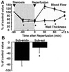
Gene program for cardiac cell survival induced by transient ischemia in conscious pigs
- PMID: 11481491
- PMCID: PMC55421
- DOI: 10.1073/pnas.171297498
Gene program for cardiac cell survival induced by transient ischemia in conscious pigs
Abstract
Therapy for ischemic heart disease has been directed traditionally at limiting cell necrosis. We determined by genome profiling whether ischemic myocardium can trigger a genetic program promoting cardiac cell survival, which would be a novel and potentially equally important mechanism of salvage. Although cardiac genomics is usually performed in rodents, we used a swine model of ischemia/reperfusion followed by ventricular dysfunction (stunning), which more closely resembles clinical conditions. Gene expression profiles were compared by subtractive hybridization between ischemic and normal tissue of the same hearts. About one-third (23/74) of the nuclear-encoded genes that were up-regulated in ischemic myocardium participate in survival mechanisms (inhibition of apoptosis, cytoprotection, cell growth, and stimulation of translation). The specificity of this response was confirmed by Northern blot and quantitative PCR. Unexpectedly, this program also included genes not previously described in cardiomyocytes. Up-regulation of survival genes was more profound in subendocardium over subepicardium, reflecting that this response in stunned myocardium was proportional to the severity of the ischemic insult. Thus, in a swine model that recapitulates human heart disease, nonlethal ischemia activates a genomic program of cell survival that relates to the time course of myocardial stunning and differs transmurally in relation to ischemic stress, which induced the stunning. Understanding the genes up-regulated during myocardial stunning, including those not previously described in the heart, and developing strategies that activate this program may open new avenues for therapy in ischemic heart disease.
Figures


Similar articles
-
Characterization of pDJA1, a cardiac-specific chaperone found by genomic profiling of the post-ischemic swine heart.Cardiovasc Res. 2003 Apr 1;58(1):126-35. doi: 10.1016/s0008-6363(02)00845-3. Cardiovasc Res. 2003. PMID: 12667953
-
Cardioprotection in stunned and hibernating myocardium.Heart Fail Rev. 2007 Dec;12(3-4):307-17. doi: 10.1007/s10741-007-9040-3. Heart Fail Rev. 2007. PMID: 17541819 Review.
-
Cardiac cell survival and reversibility of myocardial ischemia.Arch Mal Coeur Vaiss. 2006 Dec;99(12):1236-43. Arch Mal Coeur Vaiss. 2006. PMID: 18942527 Review.
-
Program of cell survival underlying human and experimental hibernating myocardium.Circ Res. 2004 Aug 20;95(4):433-40. doi: 10.1161/01.RES.0000138301.42713.18. Epub 2004 Jul 8. Circ Res. 2004. PMID: 15242971
-
[Cardiac dysfunction and endogenous cytokines in global ischemia and reperfusion injury].Hokkaido Igaku Zasshi. 1993 Nov;68(6):813-26. Hokkaido Igaku Zasshi. 1993. PMID: 8112707 Japanese.
Cited by
-
Multifunctional protein: cardiac ankyrin repeat protein.J Zhejiang Univ Sci B. 2016 May;17(5):333-41. doi: 10.1631/jzus.B1500247. J Zhejiang Univ Sci B. 2016. PMID: 27143260 Free PMC article. Review.
-
Increased expression of H11 kinase stimulates glycogen synthesis in the heart.Mol Cell Biochem. 2004 Oct;265(1-2):71-8. doi: 10.1023/b:mcbi.0000044311.58653.54. Mol Cell Biochem. 2004. PMID: 15543936
-
Hold me tight: Role of the heat shock protein family of chaperones in cardiac disease.Circulation. 2010 Oct 26;122(17):1740-51. doi: 10.1161/CIRCULATIONAHA.110.942250. Circulation. 2010. PMID: 20975010 Free PMC article. Review. No abstract available.
-
Effect of acute and chronic zofenopril administration on cardiac gene expression.Mol Cell Biochem. 2011 Jun;352(1-2):301-7. doi: 10.1007/s11010-011-0766-9. Epub 2011 Mar 11. Mol Cell Biochem. 2011. PMID: 21394524
-
The BAG3-dependent and -independent roles of cardiac small heat shock proteins.JCI Insight. 2019 Feb 21;4(4):e126464. doi: 10.1172/jci.insight.126464. eCollection 2019 Feb 21. JCI Insight. 2019. PMID: 30830872 Free PMC article. Review.
References
Publication types
MeSH terms
Substances
Grants and funding
- R37 HL033107/HL/NHLBI NIH HHS/United States
- HL65183/HL/NHLBI NIH HHS/United States
- HL33065/HL/NHLBI NIH HHS/United States
- HL65182/HL/NHLBI NIH HHS/United States
- R01 HL033107/HL/NHLBI NIH HHS/United States
- HL69020/HL/NHLBI NIH HHS/United States
- P01 HL 59139/HL/NHLBI NIH HHS/United States
- HL 33107/HL/NHLBI NIH HHS/United States
- P01 HL069020/HL/NHLBI NIH HHS/United States
- R01 HL065182/HL/NHLBI NIH HHS/United States
- P01 HL059139/HL/NHLBI NIH HHS/United States
- AG 14121/AG/NIA NIH HHS/United States
- R01 AG014121/AG/NIA NIH HHS/United States
LinkOut - more resources
Full Text Sources
Other Literature Sources

