. 2001 Jul 23;154(2):293-307.
doi: 10.1083/jcb.200104083.

Residual Cajal bodies in coilin knockout mice fail to recruit Sm snRNPs and SMN, the spinal muscular atrophy gene product

Affiliations

Residual Cajal bodies in coilin knockout mice fail to recruit Sm snRNPs and SMN, the spinal muscular atrophy gene product

K E Tucker et al. J Cell Biol. .

Abstract

Cajal bodies (CBs) are nuclear suborganelles involved in the biogenesis of small nuclear ribonucleoproteins (snRNPs). In addition to snRNPs, they are highly enriched in basal transcription and cell cycle factors, the nucleolar proteins fibrillarin (Fb) and Nopp140 (Nopp), the survival motor neuron (SMN) protein complex, and the CB marker protein, p80 coilin. We report the generation of knockout mice lacking the COOH-terminal 487 amino acids of coilin. Northern and Western blot analyses demonstrate that we have successfully removed the full-length coilin protein from the knockout animals. Some homozygous mutant animals are viable, but their numbers are reduced significantly when crossed to inbred backgrounds. Analysis of tissues and cell lines from mutant animals reveals the presence of extranucleolar foci that contain Fb and Nopp but not other typical nucleolar markers. These so-called "residual" CBs neither condense Sm proteins nor recruit members of the SMN protein complex. Transient expression of wild-type mouse coilin in knockout cells results in formation of CBs and restores these missing epitopes. Our data demonstrate that full-length coilin is essential for proper formation and/or maintenance of CBs and that recruitment of snRNP and SMN complex proteins to these nuclear subdomains requires sequences within the coilin COOH terminus.


Figures

Figure 1.
Figure 1.
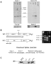
Disruption of the murine coil locus. (A) Maps of the targeting vector, wild-type, and targeted alleles. Open boxes indicate exons, and filled boxes represent selectable markers. Thick horizontal lines indicate intronic sequences. Targeting vector sequences are represented by thin horizontal lines. Large arrows indicate the start and direction of transcription for each gene. The shaded box corresponds to the probe used for confirmation of targeting. PCR primers for use in screening are indicated by small arrows (a, b, and c). NEO, neomycin resistance cassette gene; TK, thymidine kinase cassette gene B, BamHI; E, EcoRI. (B) Southern blot (top) and PCR (bottom) genotyping of progeny. Genomic DNAs for Southern blot analysis were obtained from +/+, +/−, or −/− MEFs. Control DNAs were from 129Sv/J mice (129), NIH 3T3 cells (3T3), or the targeted embryonic stem cell line 3G8 (ES). The positions of the wild-type (WT) and knockout (KO) alleles are shown along with a marker (M). Primers shown in A were used for PCR. (C) Schematic of the coilin protein. Highly conserved regions are indicated as black boxes. Also depicted are NLSs (light gray boxes), acidic patches (hatched boxes), and the putative nucleolar localization signal (dark gray box). A region rich in arginine and glycine dipeptides (RG box) is also indicated. The portion of the protein corresponding to the region deleted in the knockout is indicated by the bar.
Figure 1.
Figure 1.
Disruption of the murine coil locus. (A) Maps of the targeting vector, wild-type, and targeted alleles. Open boxes indicate exons, and filled boxes represent selectable markers. Thick horizontal lines indicate intronic sequences. Targeting vector sequences are represented by thin horizontal lines. Large arrows indicate the start and direction of transcription for each gene. The shaded box corresponds to the probe used for confirmation of targeting. PCR primers for use in screening are indicated by small arrows (a, b, and c). NEO, neomycin resistance cassette gene; TK, thymidine kinase cassette gene B, BamHI; E, EcoRI. (B) Southern blot (top) and PCR (bottom) genotyping of progeny. Genomic DNAs for Southern blot analysis were obtained from +/+, +/−, or −/− MEFs. Control DNAs were from 129Sv/J mice (129), NIH 3T3 cells (3T3), or the targeted embryonic stem cell line 3G8 (ES). The positions of the wild-type (WT) and knockout (KO) alleles are shown along with a marker (M). Primers shown in A were used for PCR. (C) Schematic of the coilin protein. Highly conserved regions are indicated as black boxes. Also depicted are NLSs (light gray boxes), acidic patches (hatched boxes), and the putative nucleolar localization signal (dark gray box). A region rich in arginine and glycine dipeptides (RG box) is also indicated. The portion of the protein corresponding to the region deleted in the knockout is indicated by the bar.
Figure 2.
Figure 2.
Analysis of coilin expression in mutant animals. (A) Northern blots of polyadenylated brain RNAs. The wild-type coilin transcript is ∼2.6 kb. The indicated probes were PCR amplified from mp80-21 as described in Materials and methods. (B) Wild-type and mutant coilin alleles were amplified from polyadenylated RNA followed by digestion with HindIII. Open boxes represent mRNA from the indicated alleles; the length of each ORF is shown below. The small arrows represent the primers used for PCR. Sequence of the cryptic splice junction for the knockout allele transcript is shown below. (C) Western blot analysis of extracts from embryonic tissues with anticoilin antibody R288, which recognizes epitopes in the coilin COOH terminus. The wild-type protein has a molecular weight of 80 kD. The knockout lane was overloaded intentionally to demonstrate the absence of wild-type coilin.
Figure 3.
Figure 3.
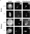
Knockout mice display “residual” CBs. (A) Frozen tissues were sectioned and stained with antibodies against coilin and Fb. In wild-type tissues, coilin is colocalized with Fb in CBs (small arrows) and sometimes is visible in nucleolar caps (see wild-type brain panels). Coilin staining is absent from knockout tissues, but residual CBs (large arrows) are evident as extranucleolar Fb foci. For better visualization, insets display the boxed CBs or residual CBs at higher magnification. (B) Confocal imaging of sensory neurons from dorsal root ganglion of a knockout animal costained for coilin/Gemin2, Nopp140/SMN, or Nopp140/U2B′′. Note the residual CBs denoted by white arrows in the Nopp140 channel and the absence of SMN or U2B′′ within these structures. (C) Sensory ganglion neurons from wild-type and knockout animals were stained with silver to visualize nucleoli and CBs (black arrows). Note the absence of extranucleolar silver deposition in the knockout cells.
Figure 3.
Figure 3.
Knockout mice display “residual” CBs. (A) Frozen tissues were sectioned and stained with antibodies against coilin and Fb. In wild-type tissues, coilin is colocalized with Fb in CBs (small arrows) and sometimes is visible in nucleolar caps (see wild-type brain panels). Coilin staining is absent from knockout tissues, but residual CBs (large arrows) are evident as extranucleolar Fb foci. For better visualization, insets display the boxed CBs or residual CBs at higher magnification. (B) Confocal imaging of sensory neurons from dorsal root ganglion of a knockout animal costained for coilin/Gemin2, Nopp140/SMN, or Nopp140/U2B′′. Note the residual CBs denoted by white arrows in the Nopp140 channel and the absence of SMN or U2B′′ within these structures. (C) Sensory ganglion neurons from wild-type and knockout animals were stained with silver to visualize nucleoli and CBs (black arrows). Note the absence of extranucleolar silver deposition in the knockout cells.
Figure 4.
Figure 4.
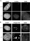
Knockout MEFs display prominent gems. Wild-type and knockout MEFs were costained for SMN and either coilin (A) or Nopp140 (B). SMN foci in wild-type MEFs were nearly always colocalized with coilin or Nopp140 in CBs (A and B, small arrows). In contrast, SMN foci present in knockout MEFs (arrowheads) were never found colocalized with Nopp140, a marker of residual CBs (large arrows). See Table II for additional details.
Figure 5.
Figure 5.
Knockout MEFs display residual CBs. Cells were costained with antibodies against Fb and NOH61 (a nucleolus-only marker) to allow discrimination of small nucleoli (arrowheads) from CBs (small arrows) and residual CBs (large arrow).
Figure 6.
Figure 6.
Residual CBs do not recruit Sm proteins. Triple labeling with antibodies against Nopp140 (red), the Sm proteins (magenta), and a nucleolus specific oligo (anti-28S or anti-MRP, green) demonstrates that Sm proteins are present in the CBs (small arrows) of wild-type MEFs but are absent from the residual CBs (large arrows) found in knockout MEFs.
Figure 7.
Figure 7.
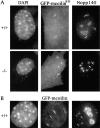
Residual CBs do not accumulate mutant coilin constructs. (A) The knockout allele protein (GFP–mcoilinKO) accumulates in CBs of wild-type MEFs but does not form foci in knockout cells. Cells were costained with anti-Nopp140 to identify nucleoli, CBs (small arrows), and residual CBs (large arrows). Costaining with anticoilin COOH-terminal antibodies in wild-type cells gave identical results (unpublished data). (B) Full-length mouse coilin localizes in two different types of foci when transiently transfected into wild-type or knockout (unpublished data) MEFs. Small arrows denote CBs that costain with Nopp140 (and other CB markers; see Fig. 8), whereas arrowheads mark positions of coilin-only foci.
Figure 8.
Figure 8.
Coilin recruits SMN and Sm proteins to CBs. (A) Cells transiently transfected with GFP–mcoilin (green) were stained for Nopp140 (magenta) and SMN (red). In both wild-type and knockout MEFs, GFP/Nopp140 foci (arrows) also contained SMN. (B) Cells were transfected with GFP–mcoilin (green) and stained for Nopp140 (magenta) and the Sm proteins (red). Regardless of genotype, transfected cells displayed CBs (arrows) that also contained Sm.
Figure 8.
Figure 8.
Coilin recruits SMN and Sm proteins to CBs. (A) Cells transiently transfected with GFP–mcoilin (green) were stained for Nopp140 (magenta) and SMN (red). In both wild-type and knockout MEFs, GFP/Nopp140 foci (arrows) also contained SMN. (B) Cells were transfected with GFP–mcoilin (green) and stained for Nopp140 (magenta) and the Sm proteins (red). Regardless of genotype, transfected cells displayed CBs (arrows) that also contained Sm.

Similar articles

Cited by

References

    1. Alliegro, M., and M. Alliegro. 1998. Protein heterogeneity in the coiled body compartment. Exp. Cell Res. 239:60–68. - PubMed
    1. Almeida, F., R. Saffrich, W. Ansorge, and M. Carmo-Fonseca. 1998. Microinjection of anti-coilin antibodies affects the structure of coiled bodies. J. Cell Biol. 142:899–912. - PMC - PubMed
    1. Andrade, L.E.C., E.K.L. Chan, I. Raska, C.L. Peebles, G. Roos, and E.M. Tan. 1991. Human autoantibody to a novel protein of the nuclear coiled body: immunological characterization and cDNA cloning of p80 coilin. J. Exp. Med. 173:1407–1419. - PMC - PubMed
    1. Andrade, L.E.C., E.M. Tan, and E.K.L. Chan. 1993. Immunocytochemical analysis of the coiled body in the cell cycle and during cell proliferation. Proc. Natl. Acad. Sci. USA. 90:1947–1951. - PMC - PubMed
    1. Bauer, D.W., and J.G. Gall. 1997. Coiled bodies without coilin. Mol. Biol. Cell. 8:73–82. - PMC - PubMed

Publication types

MeSH terms