. 2001 Aug;21(16):5321-31.
doi: 10.1128/MCB.21.16.5321-5331.2001.

Mice with very low expression of the vesicular monoamine transporter 2 gene survive into adulthood: potential mouse model for parkinsonism

Affiliations

Mice with very low expression of the vesicular monoamine transporter 2 gene survive into adulthood: potential mouse model for parkinsonism

K A Mooslehner et al. Mol Cell Biol. 2001 Aug.

Abstract

We have created a transgenic mouse with a hypomorphic allele of the vesicular monoamine transporter 2 (Vmat2) gene by gene targeting. These mice (KA1) have profound changes in monoamine metabolism and function and survive into adulthood. Specifically, these animals express very low levels of VMAT2, an endogenous protein which sequesters monoamines intracellularly into vesicles, a process that, in addition to being important in normal transmission, may also act to keep intracellular levels of the monoamine neurotransmitters below potentially toxic thresholds. Homozygous mice show large reductions in brain tissue monoamines, motor impairments, enhanced sensitivity to dopamine agonism, and changes in the chemical neuroanatomy of the striatum that are consistent with alterations in the balance of the striatonigral (direct) and striatopallidal (indirect) pathways. The VMAT2-deficient KA1 mice are also more vulnerable to the neurotoxic effects of 1-methyl-4-phenyl-1,2,3,6-tetrahydropyridine in terms of nigral dopamine cell death. We suggest that the mice may be of value in examining, long term, the insidious damaging consequences of abnormal intracellular handling of monoamines. On the basis of our current findings, the mice are likely to prove of immediate interest to aspects of the symptomatology of parkinsonism. They may also, however, be of use in probing other aspects of monoaminergic function and dysfunction in the brain, the latter making important contributions to the pathogenesis of schizophrenia and addiction.


Figures
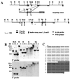
FIG. 1
FIG. 1
Targeted insertion into the third intron of the Vmat2 gene. (A) The Vmat2 gene in the 129 wild-type mouse genome and mutant sequences after targeted insertion of the vector, with prominent restriction endonuclease sites shown (B, BamHI; H, HindIII; K, KpnI; S, SacI; X, XbaI; and Xh, XhoI). Neomycin resistance sequences (neo) and sequences recognized by the Vmat2 3′ and 5′ hybridization probes are indicated. Homologous sequences in the mouse genome and of the targeting vector and the neomycin resistance (neo) and the herpes simplex virus thymidine kinase (HSV tk) sequences are indicated. (B) Southern blot analysis of hybridization of radiolabeled Vmat2 5′ and 3′ hybridization probes with genomic DNA extracted from the tail tips of wild-type (lane 1), heterozygous (lane 3), and homozygous (lanes 2 and 4) mice digested with the restriction endonucleases HindIII and XbaI (H+X), BamHI (B), KpnI (K), or Xhol (Xh). (C) Sequence of the PCR product generated with a forward primer specific for the 5′ flanking genomic sequences (SF1) and a reverse primer specific for the 5′ end of the transgene (SR1) (see also PCR primer in panel A). (D) Vmat2 RNA expression in the major monoaminergic cell body groups in the brain of homozygous KA1 mice. In situ hybridization was carried out using end-labeled radioactive oligonucleotides complementary to the first and second exon of the Vmat2 gene (see also panel A). The representative dark-field photomicrographs show the expression of Vmat2 mRNA in the substantia nigra (SN) and ventral tegmental (VTA), the dorsal raphe nucleus (RAPHE), and in the locus coruleus (LC) in a wild-type mouse. No signal was detected in the homozygous KA1 mutant.
FIG. 1
FIG. 1
Targeted insertion into the third intron of the Vmat2 gene. (A) The Vmat2 gene in the 129 wild-type mouse genome and mutant sequences after targeted insertion of the vector, with prominent restriction endonuclease sites shown (B, BamHI; H, HindIII; K, KpnI; S, SacI; X, XbaI; and Xh, XhoI). Neomycin resistance sequences (neo) and sequences recognized by the Vmat2 3′ and 5′ hybridization probes are indicated. Homologous sequences in the mouse genome and of the targeting vector and the neomycin resistance (neo) and the herpes simplex virus thymidine kinase (HSV tk) sequences are indicated. (B) Southern blot analysis of hybridization of radiolabeled Vmat2 5′ and 3′ hybridization probes with genomic DNA extracted from the tail tips of wild-type (lane 1), heterozygous (lane 3), and homozygous (lanes 2 and 4) mice digested with the restriction endonucleases HindIII and XbaI (H+X), BamHI (B), KpnI (K), or Xhol (Xh). (C) Sequence of the PCR product generated with a forward primer specific for the 5′ flanking genomic sequences (SF1) and a reverse primer specific for the 5′ end of the transgene (SR1) (see also PCR primer in panel A). (D) Vmat2 RNA expression in the major monoaminergic cell body groups in the brain of homozygous KA1 mice. In situ hybridization was carried out using end-labeled radioactive oligonucleotides complementary to the first and second exon of the Vmat2 gene (see also panel A). The representative dark-field photomicrographs show the expression of Vmat2 mRNA in the substantia nigra (SN) and ventral tegmental (VTA), the dorsal raphe nucleus (RAPHE), and in the locus coruleus (LC) in a wild-type mouse. No signal was detected in the homozygous KA1 mutant.
FIG. 2
FIG. 2
VMAT2 and TH immunoreactivity in striatal and midbrain regions of KA1 homozygous mice. (A) Striatal sections from wild-type and homozygous KA1 mice were stained for VMAT2 immunoreactivity (VMAT2) using a specific antiserum. VMAT2 immunoreactivity is present in the caudate putamen (CPu) and the nucleus accumbens (Nacc) of wild-type mice but absent in those structures in homozygous mice. In striatal sections from wild-type and homozygous KA1 mice stained for TH immunoreactivity using a specific antiserum, the level of TH immunoreactivity in the caudate putamen and the nucleus accumbens was similar in both wild-type and homozygous mice. (B) VMAT2 immunostaining is absent in the substantia nigra (SN) and the ventral tegmental area (VTA) of the homozygous KA1 mouse and present in the wild-type mouse, whereas TH immunoreactivity is also present in the dopamine cell body region of homozygous mice. The apparent lack of differences in TH immunoreactivity between wild-type and homozygous mice, seen on gross visual inspection, was confirmed by quantitative analysis of four wild-type and five homozygous animals.
FIG. 3
FIG. 3
RT-PCR and Western blot analysis of homozygous KA1 insertional mutants and homozygous GB1/1 knockout mice. (A) Ethidium-stained agarose electrophoresis of RT-PCR products. Total midbrain RNA from homozygous (hom) and wild-type (wt) GB1/1 and KA1 neonates was isolated and reverse transcribed. Expression of Vmat2 mRNA was detected using specific primers for exon 2 and exon 12 of the mouse Vmat2 gene (33). These primers amplified Vmat2 cDNA made from homozygous KA1 mice, but no amplification product was generated with cDNA made from homozygous GB1/1 neonates. The quality of all cDNA preparations was examined by performing control RT-PCRs with primers to hypoxanthine phosphoribosyltransferase (Hprt) (22). (B) Schematic representation of the genomic organization of the Vmat2 wild-type allele, the KA1 insertion allele, and the GB1/1 knockout allele. The wild-type allele is transcriptionally active. The KA1 insertion interferes severely with transcription, and only a small amount of Vmat2 message is generated. In the GB1/1 knockout line, the first and second exon of the Vmat2 gene are deleted, completely abolishing the generation of normal Vmat2 message. (C) Western blot of striatal membrane preparations from homozygous and wild-type KA1 mice probed with a polyclonal antibody against the VMAT2 protein. Both lanes contain comparable amounts of protein, as determined by the Bradford method. Quantitative analysis of the blot using SeeScan indicated a decrease in signal of more than 95% in homozygotes below that of the wild-type signal. The position of the VMAT2-specific band is indicated by the arrowhead. The higher molecular weight band is nonspecific, being found in extracts from controls and mutants.
FIG. 4
FIG. 4
Quantification of monoamines and metabolites in different brain regions of wild-type, heterozygous, and homozygous mice. Dopamine, noradrenaline, and serotonin, as well as their main metabolites, DOPAC, HVA, and 5-HIAA, were measured in perchloric acid extracts prepared from brain areas of 13 wild-type, 17 heterozygous, and 6 homozygous mice using HPLC with electrochemical detection (12). In comparison to wild-type controls, KA1 homozygous mice showed widespread, major reductions in the levels of all three monoamines. The results are represented as the mean ± SEM. ∗, P < 0.05; ∗∗, P < 0.001, compared with the wild-type group (Student t test). In contrast, metabolite levels of all three monoamines were, in general, much less affected in the VMAT2-deficient KA1 mice, consistent with higher rates of monoamine turnover in these animals.
FIG. 5
FIG. 5
Motor functioning and dopamine supersensitivity in KA1 mice. (A) Homozygous (light gray bars) and heterozygous (dark gray bars) KA1 mice could not perform as well as their wild-type (black bars) littermates in a beam walking task, but they showed a normal preference for a novel environment. The results, for each beam diameter and shape, are the mean latencies to cross the beam ± SEM of six crossings done by 15 males of each genotype in the fourth (i.e., the final) test session. Data were analyzed by the Tukey-Kramer multiple comparison test, which indicated significant group differences at each level of difficulty (∗, P < 0.05; ∗∗, P < 0.001, compared with the wild-type group [Student t test]). The novelty place preference, assessed using the Tukey-Kramer multiple comparisons test, did not show any significant group differences in the time spent on the novel site. There were, however, significant group differences in the number of visits and rears in the novel environment. The results are represented as mean ± SEM for 15 mice of each genotype. ∗, P < 0.05; ∗∗, P < 0.001, compared with the wild-type group (Student t test). (B) Increased responsiveness to systemically administered d-amphetamine (3 mg/kg) given i.p., on stereotyped behaviors in wild-type, heterozygous, and homozygous animals. Stereotypy ratings, based on the scoring system of Mittleman et al. (23), were taken blind for 1 h postinjection. The lower the rating, the lower the observed rate of stereotypy, and vice versa. The results are the mean scores ± SEM, registered in 12 5-min bins, for 11 wild-type, 8 heterozygous, and 9 homozygous mice. Analysis of variance indicated a significant main effect of group (P < 0.01), consistent with an increased response to d-amphetamine in the homozygous mice. (C) An altered chemical neuroanatomy in the striatum of male VMAT2-deficient KA1 mice expressing substance P and enkephalin mRNA is shown in striatal sections from wild-type mice and homozygous KA1 mutants, hybridized with oligonucleotides complementary to the substance P and enkephalin messages. The relative down-regulation of substance P mRNA expression and the relative up-regulation of enkephalin mRNA expression in the striatum of KA1 homozygous mice was quantified by silver grain counting on mRNA-positive cells. The difference between the two groups was compared by the Student t test and showed a significant difference in the number of silver grains per square millimeter of cell area between wild-type and homozygous mice for both substance P (P < 0.001) and enkephalin (P < 0.05).
FIG. 6
FIG. 6
MPTP treatment of wild-type, heterozygous, and homozygous KA1 mice. (A) TH-immunopositive cells in sections from the substantia nigra pars compacta of saline-treated and MPTP-treated wild-type, heterozygous, and homozygous KA1 mice. (B) Number of TH-immunopositive cells in the substantia nigra pars compacta of saline-treated and MPTP-treated wild-type, heterozygous, and homozygous KA1 mice. TH-immunopositive cells were counted from 30 serial sections from 4 wild-type, 3 heterozygous, and 3 homozygous MPTP-treated and 4 saline-treated (2 homozygous and 2 wild-type) mice. The cell loss was significantly increased in MPTP-treated heterozygous and homozygous KA1 mice compared to the MPTP-treated wild-type littermates. The difference between the three groups was compared by the Student t test and showed a significant difference in the number of TH-positive cells between wild-type and heterozygous MPTP-treated mice (P < 0.01) and between wild-type and homozygous MPTP-treated mice (P = 0.001).

Similar articles

Cited by

References

    1. Bergman H, Feingold A, Nini A, Raz A, Slovin H, Abeles M, Vaadia E. Physiological aspects of information processing in the basal ganglia of normal and parkinsonian primates. Trends Neurosci. 1998;21:32–38. - PubMed
    1. Bergman H, Wichmann T, DeLong M R. Reversal of experimental parkinsonism by lesions of the subthalamic nucleus. Science. 1990;249:1436–1438. - PubMed
    1. Björklund A, Lindvall O. Dopamine in dendrites of substantia nigra neurons: suggestions for a role in dendritic terminals. Brain Res. 1975;83:531–537. - PubMed
    1. Chase T N, Oh J D, Blanchet P J. Neostriatal mechanisms in Parkinson's disease. Neurology. 1998;51:30–35. - PubMed
    1. Dahlström A, Fuxe K. Evidence for the existence of monoamine-containing neurons in the central nervous system. 1. Demonstration of monoamines in the cell bodies of brain stem neurons. Acta Physiol Scand Suppl. 1964;62:232. - PubMed

Publication types

MeSH terms

Substances

LinkOut - more resources