. 2001 Jun 25;153(7):1441-52.
doi: 10.1083/jcb.153.7.1441.

Effects of purified recombinant neural and muscle agrin on skeletal muscle fibers in vivo

Affiliations

Effects of purified recombinant neural and muscle agrin on skeletal muscle fibers in vivo

G Bezakova et al. J Cell Biol. .

Abstract

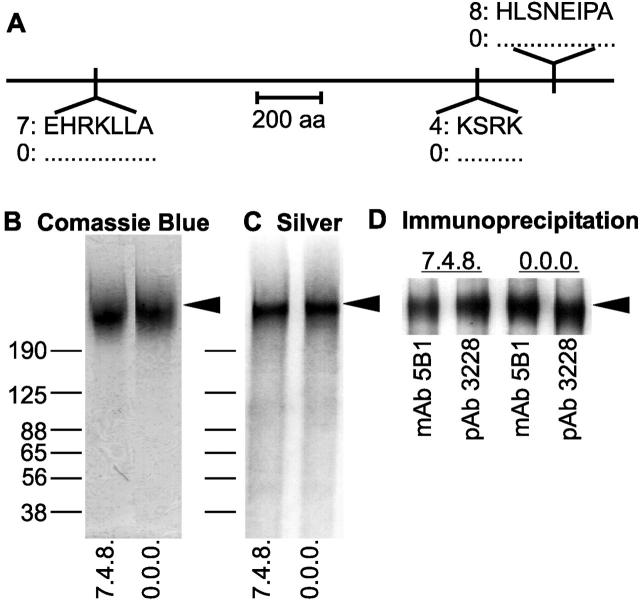
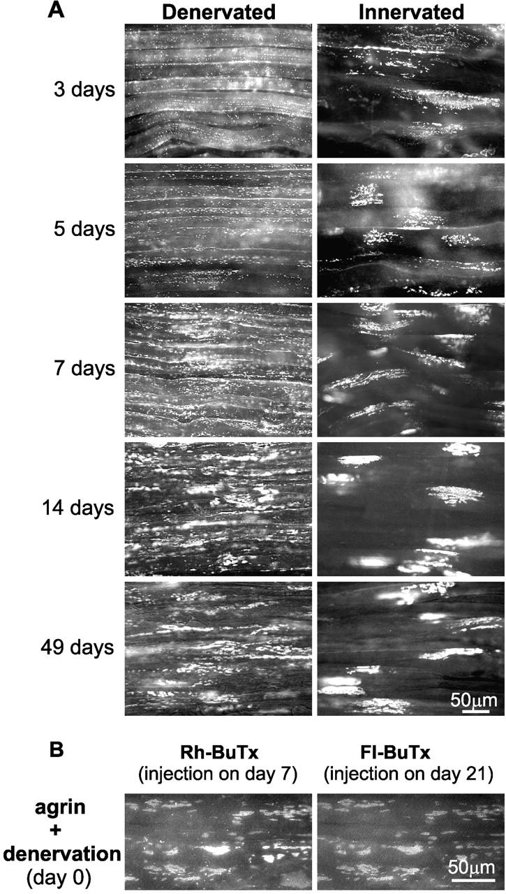
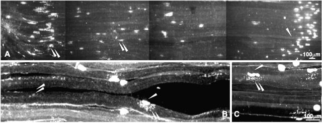
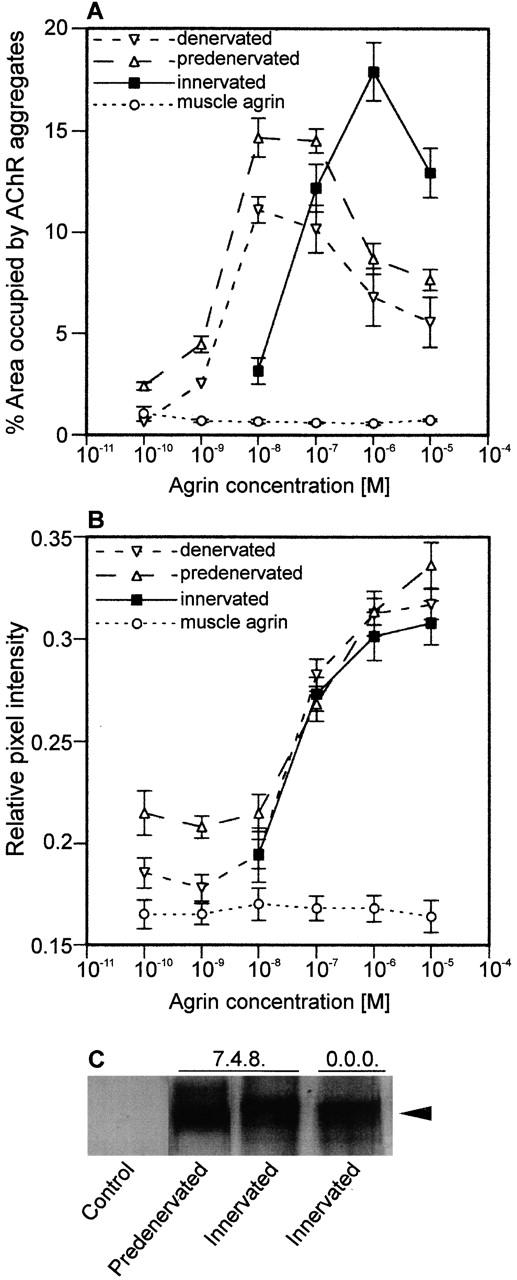
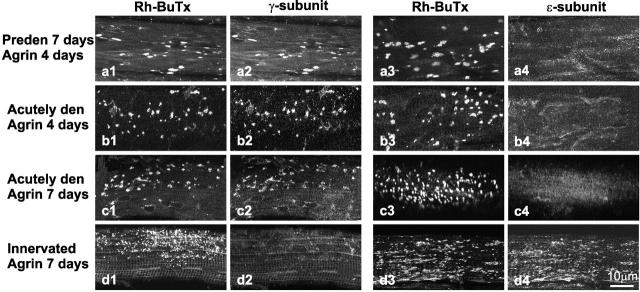
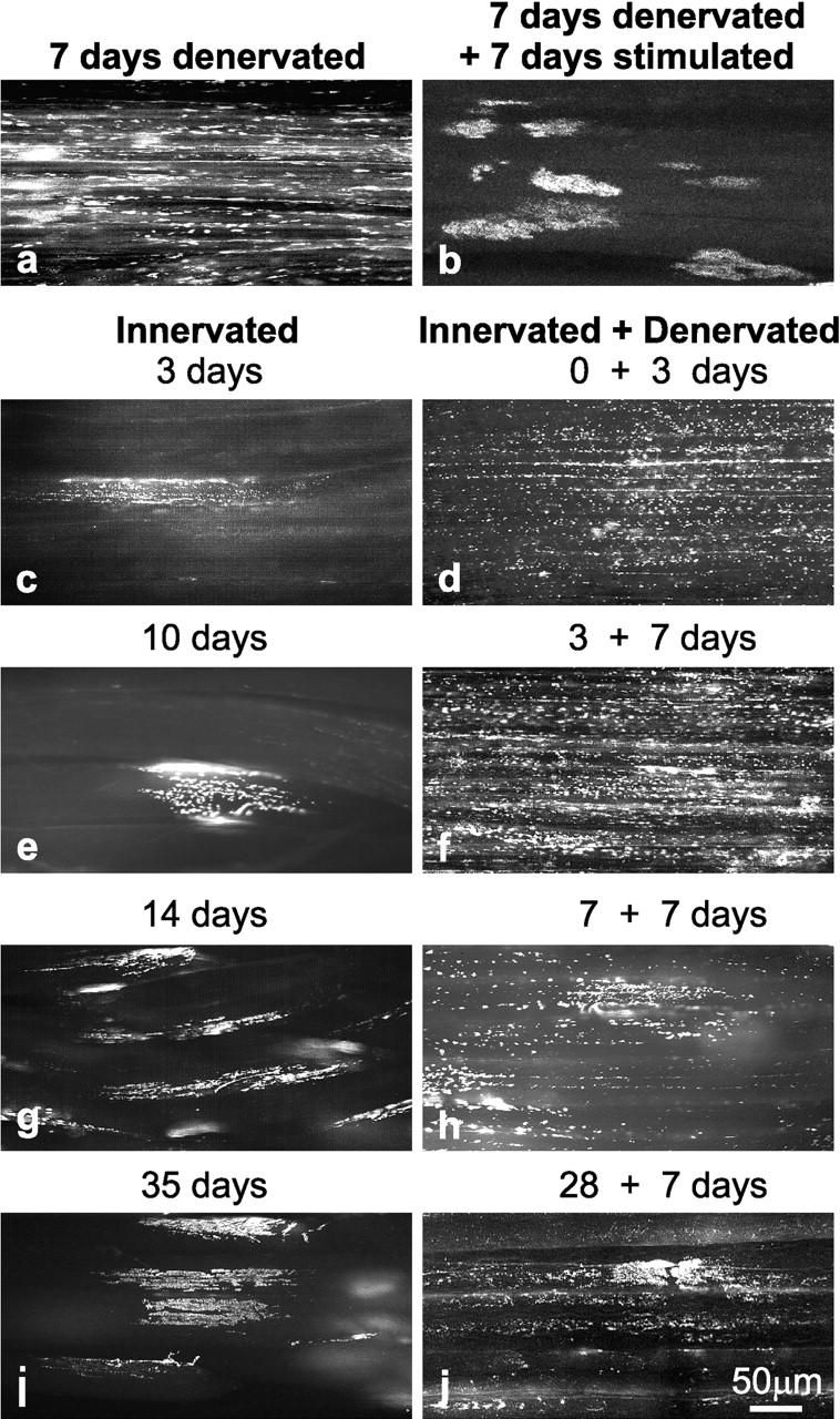
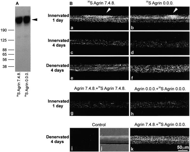
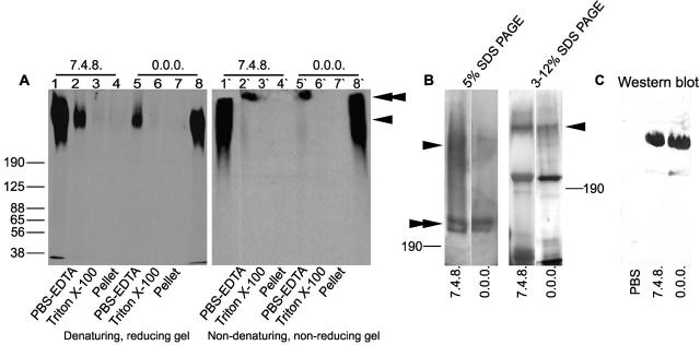
Aggregation of acetylcholine receptors (AChRs) in muscle fibers by nerve-derived agrin plays a key role in the formation of neuromuscular junctions. So far, the effects of agrin on muscle fibers have been studied in culture systems, transgenic animals, and in animals injected with agrin--cDNA constructs. We have applied purified recombinant chick neural and muscle agrin to rat soleus muscle in vivo and obtained the following results. Both neural and muscle agrin bind uniformly to the surface of innervated and denervated muscle fibers along their entire length. Neural agrin causes a dose-dependent appearance of AChR aggregates, which persist > or = 7 wk after a single application. Muscle agrin does not cluster AChRs and at 10 times the concentration of neural agrin does not reduce binding or AChR-aggregating activity of neural agrin. Electrical muscle activity affects the stability of agrin binding and the number, size, and spatial distribution of the neural agrin--induced AChR aggregates. Injected agrin is recovered from the muscles together with laminin and both proteins coimmunoprecipitate, indicating that agrin binds to laminin in vivo. Thus, the present approach provides a novel, simple, and efficient method for studying the effects of agrin on muscle under controlled conditions in vivo.


Figures

Figure 1
Figure 1
Characterization of full-length chick neural and muscle agrin purified from stably transfected 293 HEK cells. (A) Location of alternatively spliced sites characteristic for neural (7.4.8.) and nonneural or muscle (0.0.0.) agrin isoforms together with their amino acid sequences. The fractions of purified agrin collected by ion exchange chromatography contained high concentrations of agrin that were easily detectable by Comassie blue staining (B) and were >90% pure as shown by Silver staining (C). Immunoprecipitation (D) using the mAb against agrin (mAb 5B1) or antiagrin antiserum (polyclonal antibody 3228) confirmed that the purified protein is chick agrin. Proteins were separated by 3–12% SDS-PAGE. Molecular masses of standard proteins are indicated in kD in B. Arrows point to the smeared bands of purified neural and muscle agrin with the apparent molecular of 400–600 kD.
Figure 2
Figure 2
Ectopic AChR aggregates induced by purified recombinant chick neural agrin. (A) A single injection of 1 μM agrin into acutely denervated (left) or innervated (right) SOL muscles caused the appearance of numerous AChR aggregates at the indicated times after injection. Note differences in number, size, and distribution of aggregates labeled with Rh-BuTx in denervated and innervated muscles. In innervated muscles, the aggregates appeared predominantly near myotendinous junctions (see also Fig. 3). (B) SOL muscle was injected with agrin and denervated on day 0. On day 7, it was injected with Rh-BuTx, removed, and labeled with Fl-BuTx on day 21. Presence of Rh-BuTx–labeled aggregates on day 21 shows their metabolic stability; colocalization of Rh-BuTx and Fl-BuTx indicates the structural stability of aggregates.
Figure 3
Figure 3
Distribution of AChR aggregates induced by injection of 1 μM neural agrin into innervated (A and C) or denervated (B) muscles. In innervated muscles, most of the aggregates appeared near myotendinous junction (to the left in A). In denervated muscles, AChR aggregates were essentially absent near the original NMJs (B) but abundant elsewhere (see also Fig. 2). Small punctate aggregates appeared in the area immediately adjacent to the original NMJs in both innervated and denervated muscles (B and C). The large majority of agrin-induced aggregates were distinguished from the original NMJs by their different appearance, location outside the characteristic band of NMJs across the middle of the muscle, and weaker intensity of Rh-BuTx staining. The few punctate aggregates close to the NMJs were distinguished by their appearance since nothing like them was observed near innervated or denervated NMJs without injecting neural agrin. Double arrow points to agrin-induced ectopic AChR aggregates; single arrow points to original NMJ.
Figure 4
Figure 4
AChR-aggregating activity of neural agrin is dose dependent. 7-d denervated (predenervated), acutely denervated, or innervated SOL muscles were bathed in the solution of neural or muscle agrin at concentrations as indicated in vivo for 2 h. The muscles were dissected out after 4 (predenervated and acutely denervated) or 7 (innervated) d and labeled with Rh-BuTx. Only neural agrin induced AChR aggregates whose appearance was dependent on the dose applied.
Figure 5
Figure 5
Dose-response curves from experiments illustrated in Fig. 4. The area of AChR aggregates as percentage of a given area of muscle fiber surface (A) and the relative fluorescence of the Rh-BuTx that labeled the aggregates (B) are plotted against concentration of applied agrin (see Materials and Methods). Plots represent mean values ± SEM of 20 collections of aggregates in each of three experiments for each concentration. Amount of neural and muscle agrin bound to the muscle was determined by immunoprecipitation 12 h after application (C).
Figure 6
Figure 6
Presence of γ- and ε- AChR subunits in AChR aggregates induced by neural agrin. SOL muscles that were 7-d denervated (a), acutely denervated (b and c), or innervated (d) were bathed in PBS containing 10 μM neural agrin in vivo for 2 h. 4 (a and b) and 7 (c and d) d later, the muscles were excised and examined for AChR aggregates on surface fibers after labeling with Rh-BuTx and antibodies against γ- and ε-subunits, as indicated. Note that AChR aggregates on denervated fibers contained γ-subunits (a2, b2, and c2) and no detectable ε-subunits (a4, b4, and c4), whereas innervated fibers contained ε-subunits (d4) but little or no detectable γ-subunits (d2).
Figure 7
Figure 7
Electrical muscle activity affects number, size, and distribution of neural agrin–induced AChR aggregates. 1 μM neural agrin was injected into SOL muscles that were immediately denervated (a, b, and d) or kept innervated (c and e–j). Muscles denervated for 7 d (a) were then electrically stimulated for additional 7 d (b). Some innervated muscles were denervated 0 (d), 3 (f), 7 (h), or 28 (j) d after the injection. At the indicated days after these treatments (3–35 d), the muscles were excised and treated with Rh-BuTx to label AChR aggregates as shown. Note the changes in number, size, and distribution of AChR aggregates that were caused by electrical stimulation of denervated muscles (compare a with b) and the similarity between aggregates in denervated stimulated (b) and innervated (c, e, g, and i) muscles. Also, note the appearance of a declining number of additional small AChR aggregates after denervating the innervated muscles ≤28 d after the injection of neural agrin (d, f, h, and j).
Figure 8
Figure 8
Binding of neural and muscle agrin to the surface of SOL muscle fibers. Radioactive (35S) neural (isoform 7.4.8) and muscle (isoform 0.0.0) chick agrin were purified from stably transfected 293 HEK cells (see Materials and Methods) and separated by SDS-PAGE 3–12% gradient gel electrophoresis (A). 1 μM 35S-labeled neural or muscle agrin was injected into SOL muscles, which were removed 1 or 4 d later for isolation of single fibers and subsequent autoradiography (B). Arrows in a and b, respectively, point to high density of bound neural and muscle agrin at a site in the middle of the fiber that probably corresponds to original NMJ. Note similar densities of neural and muscle agrin 4 d after injection in innervated (compare c with d) and denervated (compare e with f) fibers and lower densities in innervated (c and d) compared with denervated (e and f) fibers. Injection of 10 μM unlabeled agrin 6 h before injection of 1 μM radioactive agrin markedly reduced binding of the corresponding radioactive agrin (g and h). Fibers teased from muscles injected with PBS displayed essentially no grains (i and j, autoradiography and phase contrast, respectively). Injection of 10 μM unlabeled neural agrin did not reduce the binding of radioactive muscle agrin injected at 1 μM concentration 6 h later (k). The images show representative distribution of agrin after different treatments. Three muscles for each condition, and 10 randomly teased fibers from each muscle were examined.
Figure 9
Figure 9
Agrin binds to laminin. SOL muscles were injected with 1 μM radioactively labeled neural or muscle agrin. 1 d later the muscles were excised, sequentially extracted, and analyzed by 3–12% SDS-PAGE electrophoresis under denaturing and reducing conditions (A, lanes 1–8) or nondenaturing and nonreducing conditions (A, lanes 1′–8′) followed by autoradiography. Lanes 1, 8, 1′, and 8′ represent the migration of purified radioactive neural and muscle agrin, respectively. Injected recombinant agrin was extracted from the muscle by PBS containing EDTA (lanes 2, 5, 2′, and 5′). The migration of extracted agrin was dependent on the conditions used to run the gel, indicating its binding to other protein(s). (B) SOL extracts were also immunoprecipitated with mAb 5B1 against agrin. Silver staining revealed the presence of bands having molecular mass ∼400 and ∼200 kD. Western blots of the immunoprecipitated complex and use of polyclonal antibody against α2-laminin confirmed the presence of α2-laminin (C). The molecular masses of standard proteins are indicated in kD.

Comment in

Similar articles

Cited by

References

    1. Bezakova G., Lømo T. Muscle activity and muscle agrin regulate the organization of cytoskeletal proteins and attached acetylcholine esterase (AChR) aggregates in skeletal muscle fibers. J. Cell Biol. 2001;153:1453–1463. - PMC - PubMed
    1. Bork P., Downing A.K., Kieffer B., Campbell I.D. Structure and distribution of modules in extracellular proteins. Q. Rev. Biophys. 1996;29:119–167. - PubMed
    1. Brenner H.R., Lømo T., Williamson R. Control of end-plate channel properties by neurotrophic effects and by muscle activity in rat. J. Physiol. 1987;388:367–381. - PMC - PubMed
    1. Burkin D.J., Gu M., Hodges B.L., Campanelli J.T., Kaufman S.J. A functional role for specific spliced variants of the α7β1 integrin in acetylcholine receptor clustering. J. Cell Biol. 1998;143:1067–1075. - PMC - PubMed
    1. Chiu A.Y., Sanes J.R. Development of basal lamina in synaptic and extrasynaptic portions of embryonic rat muscle. Dev. Biol. 1984;103:456–467. - PubMed

Publication types

MeSH terms