. 2001 Jul;21(14):4818-28.
doi: 10.1128/MCB.21.14.4818-4828.2001.

The cell cycle-regulatory CDC25A phosphatase inhibits apoptosis signal-regulating kinase 1

Affiliations

The cell cycle-regulatory CDC25A phosphatase inhibits apoptosis signal-regulating kinase 1

X Zou et al. Mol Cell Biol. 2001 Jul.

Abstract

CDC25A phosphatase promotes cell cycle progression by activating G(1) cyclin-dependent kinases and has been postulated to be an oncogene because of its ability to cooperate with RAS to transform rodent fibroblasts. In this study, we have identified apoptosis signal-regulating kinase 1 (ASK1) as a CDC25A-interacting protein by yeast two-hybrid screening. ASK1 activates the p38 mitogen-activated protein kinase (MAPK) and c-Jun NH(2)-terminal protein kinase-stress-activated protein kinase (JNK/SAPK) pathways upon various cellular stresses. Coimmunoprecipitation studies demonstrated that CDC25A physically associates with ASK1 in mammalian cells, and immunocytochemistry with confocal laser-scanning microscopy showed that these two proteins colocalize in the cytoplasm. The carboxyl terminus of CDC25A binds to a domain of ASK1 adjacent to its kinase domain and inhibits the kinase activity of ASK1, independent of and without effect on the phosphatase activity of CDC25A. This inhibitory action of CDC25A on ASK1 activity involves diminished homo-oligomerization of ASK1. Increased cellular expression of wild-type or phosphatase-inactive CDC25A from inducible transgenes suppresses oxidant-dependent activation of ASK1, p38, and JNK1 and reduces specific sensitivity to cell death triggered by oxidative stress, but not other apoptotic stimuli. Thus, increased expression of CDC25A, frequently observed in human cancers, could contribute to reduced cellular responsiveness to oxidative stress under mitogenic or oncogenic conditions, while it promotes cell cycle progression. These observations propose a mechanism of oncogenic transformation by the dual function of CDC25A on cell cycle progression and stress responses.


Figures
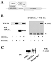
FIG. 1
FIG. 1
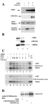
ASK1 is a CDC25A-interacting protein. (A) Yeast two-hybrid screening for CDC25A-interacting proteins. AD, activation domain; BD, binding domain; UAS, upstream activation sequence; minimum, TATA box minimum promoter; X, protein encoded in the library. (B) Complex formation of CDC25A and ASK1 in transfected COS cells. COS-7 cells were transfected with pcDNA3 expression vectors encoding the proteins shown in the panel. At 48 h posttransfection, extracts were prepared and analyzed by Western blotting (WB) with antibodies as shown (left panels). To detect complexes, extracts were immunoprecipitated (IP) with anti-CDC25A monoclonal antibody, followed by Western blotting with anti-HA monoclonal antibody (right panel). The arrow indicates HA-tagged ASK1 detected in CDC25A immunoprecipitation. Ig, IgG heavy chain in immunoprecipitation cross-reacted with the secondary antibody. (C) CDC25A-ASK1 complex in untransfected ovarian carcinoma cells. Extracts of exponentially growing OVCAR-8 cells were immunoprecipitated with anti-ASK1 polyclonal antibody, anti-CDC25A monoclonal antibody, or control mouse IgG, followed by Western blotting with the anti-ASK1 antibody.
FIG. 2
FIG. 2
Subcellular localization of CDC25A and ASK1. Human embryonic kidney 293 cells, transfected with pcDNA3 expression vectors for CDC25A and HA-tagged ASK1, were stained with anti-CDC25A monoclonal antibody and anti-HA polyclonal antibody (upper panels). Human ovarian carcinoma OVCAR-8 cells, without transfection, were stained with anti-CDC25A monoclonal antibody and anti-ASK1 polyclonal antibody (lower panels). Signals were visualized by incubation with fluorescein-conjugated antimouse IgG antibody and Texas red-conjugated anti-rabbit IgG antibody, followed by analysis with a confocal laser-scanning microscope. These results are representative of three independent sets of experiments. Bar, 5 μm.
FIG. 3
FIG. 3
CDC25A inhibits ASK1 in a manner independent of its phosphatase activity. (A) Decreased ASK1-dependent MKK phosphorylation by cotransfection with CDC25A. COS-7 cells were transfected with pcDNA3 vectors encoding the proteins shown in the top panel. Cell extracts were prepared at 48 h after transfection and analyzed by immunoblotting with antibodies for ASK1, CDC25A, MKK3, and phosphorylated active MKK3/6. (B) CDC25A is both an inhibitor and a substrate of ASK1. ASK1 was immunoprecipitated from Sf9 cells infected with a human ASK1-encoding recombinant baculovirus and incubated at 30°C for 30 min with purified GST (lane 1) or GST-CDC25A (lane 2) in the presence of [γ-32P]ATP and purified GST-MKK6 (kinase-inactive KM mutant form). Immunoprecipitates with normal IgG were used as a negative control (lane 3). Phosphorylation of the proteins was analyzed by SDS-polyacrylamide gel electrophoresis (PAGE) and autoradiography. (C) Inhibition of ASK1 by CDC25A and CDC25A(D383N/C430S). Extracts of Sf9 cells infected with the ASK1-encoding baculovirus were preincubated for 30 min at 30°C with purified GST-CDC25A, GST-CDC25A(D383N/C430S), or GST at the indicated concentration, before the addition of [γ-32P]ATP and GST- MKK6(KM). After incubation for 30 more min, phosphorylation of the proteins was analyzed by SDS-PAGE and autoradiography. Extracts of uninfected Sf9 cells were used as negative controls (lanes 1 and 9). wt, wild type. (D) The CDC25A(D383N/C430S) double mutant is phosphatase inactive. The activity of CDC25A was assessed by its ability to activate cyclin E-CDK2 in vitro. Sf9 cells were coinfected with baculoviruses for the expression of human cyclin E and CDK2(R169L), a mutant CDK2 known to accumulate inhibitory phosphorylation at Tyr 15. Cyclin E-CDK2 complexes were immunoprecipitated with anti-CDK2 antibody and incubated at 30°C for 30 min with GST-CDC25A(D383N/C430S) or wild-type GST-CDC25A. CDK2 kinase activity then was measured with histone H1 as a substrate, and phosphorylation of histone H1 was analyzed by SDS-PAGE and autoradiography.
FIG. 4
FIG. 4
Increased expression of CDC25A diminishes homo-oligomerization of ASK1. 293 cells were cotransfected with pcDNA3 expression vectors encoding the proteins shown in the panel. After 36 h, cell lysates were prepared and subjected to Western blotting (WB) or immunoprecipitation (IP) followed by Western blotting, with antibodies against the epitopes shown in the panel. To confirm the specificity of immunoprecipitation, normal mouse IgG-conjugated protein A-Sepharose was used in lane 1, instead of Sepharose conjugated with anti-Flag monoclonal antibody (lanes 2 and 3).
FIG. 5
FIG. 5
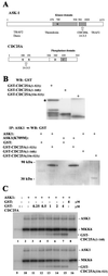
The carboxyl terminus of CDC25A is sufficient to bind and inhibit ASK1 in vitro. (A) Domain structures of ASK1 and CDC25A. The numbers shown represent amino acid residues. Names of proteins that bind to these proteins are listed below the putative binding domains. (B) The carboxyl terminus of CDC25A binds to ASK1 in vitro. The full-length and truncated fragments of CDC25A were purified from bacteria and analyzed by Western blotting (WB) with anti-GST antibody (upper panel). The asterisk indicates full-length GST-CDC25A(1–523), and other bands in the lane are degraded products in the preparation. ASK1 was immunoprecipitated (IP) from Sf9 cells infected with an ASK1-encoding baculovirus and then incubated at 30°C for 45 min with the GST-CDC25A fusion proteins. After extensive wash of the protein A-Sepharose beads, complexes were analyzed by Western blotting with anti-GST antibody (lower panel). (C) The carboxyl terminus of CDC25A can inhibit ASK1 in vitro. Extracts of Sf9 cells infected with the ASK1 baculovirus were incubated at 30°C for 30 min with purified GST or GST-tagged truncated CDC25A mutants at the indicated concentrations, before the addition of [γ-32P]ATP and GST-MKK6(KM). After incubation for 30 more min, phosphorylation of the proteins was analyzed by SDS-polyacrylamide gel electrophoresis and autoradiography. Extracts of uninfected Sf9 cells were used as negative controls (lanes 1 and 9).
FIG. 6
FIG. 6
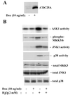
Increased expression of CDC25A suppresses activation of the stress kinase cascades upon oxidative stress. (A) CDC25A expression after a 4-h induction with doxycycline (Dox; 10 ng/ml) in 293 cells carrying a tetracycline-inducible CDC25A transgene. Expression of CDC25A was measured by immunoblotting. (B) Induced CDC25A expression diminishes the activation of ASK1, JNK1, and p38 by H2O2 treatment. Cells were treated with doxycycline for 4 h, and then exposed to H2O2 (2 mM) for 20 min. For the activities of the stress-responsive kinases, cell extracts were immunoprecipitated with antibodies against ASK1, JNK1, or p38, and the activities were assayed as described in Materials and Methods. Phosphorylation of MKK3/6 was examined by Western blotting with an antibody specific for phosphorylated active forms. Total expression of MKK3, JNK1, and p38 was determined by Western blotting.
FIG. 7
FIG. 7
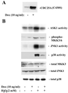
Ectopic expression of phosphatase-inactive CDC25A(C430S) suppresses activation of the stress kinase cascades upon oxidative stress. (A) The expression of CDC25A(C430S) after a 4-h induction with doxycycline (Dox; 10 ng/ml) in 293 cells carrying a tetracycline-inducible transgene. The sum of expression of CDC25A(C430S) and endogenous CDC25A was measured by immunoblotting. (B) Ectopic CDC25A(C430S) expression diminishes the activation of ASK1, JNK1 and p38 by H2O2 treatment. Cells were treated with doxycycline for 4 h and then exposed to H2O2 (2 mM) for 20 min. For the activities of the stress-responsive kinases, cell extracts were immunoprecipitated with antibodies against ASK1, JNK1, or p38, and the activities were assayed as described in Materials and Methods. Phosphorylation of MKK3/6 was examined by Western blotting with an antibody specific for phosphorylated active forms. Total expression of MKK3, JNK1, and p38 was determined by Western blotting.
FIG. 8
FIG. 8
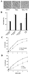
Ectopic expression of wild-type or phosphatase-inactive CDC25A reduces the sensitivity of cells to oxidant-induced death specifically. (A) The morphology of cells after a 3-h exposure to H2O2 (2 mM) in 293 cells carrying a tetracycline-inducible CDC25A transgene (tet-CDC25A-293 cells). The Dox group was preincubated for 3 h with doxycycline (10 ng/ml) to increase cellular expression of CDC25A (Fig. 6A). Cells undergoing apoptosis exhibit round and condensed bodies with decreased adherence to the culture dish. (B) The extent of cell death was evaluated in cultures of tet-CDC25A-293 cells after treatment for 9 h with actinomycin (100 ng/ml), staurosporine (500 nM), cycloheximide (3 μg/ml), and H2O2 (1 mM). Death was assessed as the loss of mitochondrial membrane potential (diminished TMRE staining) as well as diminution of cell size (forward-angle light scatter). The open and solid columns represent cultures with and without a 3-h preincubation with doxycycline. Data from three independent experiments are presented as means ± standard errors. (C) Percentages of dead nonadherent cells in cultures of tet-CDC25A-293 cells after a 12-h exposure to H2O2 at the concentrations shown. The Dox group was preincubated for 3 h with doxycycline (10 ng/ml). Data from three independent experiments are shown as means ± standard errors. (D) Percentages of dead nonadherent cells in cultures of tet-CDC25A(C430S)-293 cells after a 12-h exposure to H2O2 at the concentrations shown. The Dox group was preincubated for 3 h with doxycycline (10 ng/ml). See Fig. 7A for induced expression of the phosphatase-inactive CDC25A(C430S). Data from three independent experiments are shown as means ± standard errors.

Similar articles

Cited by

References

    1. Adler V, Yin Z, Tew K D, Ronai Z. Role of redox potential and reactive oxygen species in stress signaling. Oncogene. 1999;18:6104–6111. - PubMed
    1. Behrens A, Sibilia M, Wagner E F. Amino-terminal phosphorylation of c-Jun regulates stress-induced apoptosis and cellular proliferation. Nat Genet. 1999;21:326–329. - PubMed
    1. Biguet C, Wakasugi N, Mishal Z, Holmgren A, Chouaib S, Tursz T, Wakasugi H. Thioredoxin increases the proliferation of human B-cell lines through a protein kinase C-dependent mechanism. J Biol Chem. 1994;269:28865–28870. - PubMed
    1. Blomberg I, Hoffmann I. Ectopic expression of Cdc25A accelerates the G1/S transition and leads to premature activation of cyclin E- and cyclin A-dependent kinases. Mol Cell Biol. 1999;19:6183–6194. - PMC - PubMed
    1. Bunz F, Dutriaux A, Lengauer C, Waldman T, Zhou S, Brown J P, Sedivy J M, Kinzler K W, Vogelstein B. Requirement for p53 and p21 to sustain G2 arrest after DNA damage. Science. 1998;282:1497–1501. - PubMed

Publication types