. 2001 Jul;75(14):6460-71.
doi: 10.1128/JVI.75.14.6460-6471.2001.

Repression of vaccinia virus Holliday junction resolvase inhibits processing of viral DNA into unit-length genomes

Affiliations

Repression of vaccinia virus Holliday junction resolvase inhibits processing of viral DNA into unit-length genomes

A D Garcia et al. J Virol. 2001 Jul.

Abstract

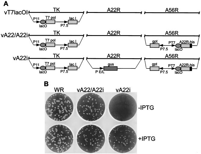
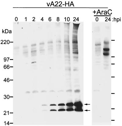
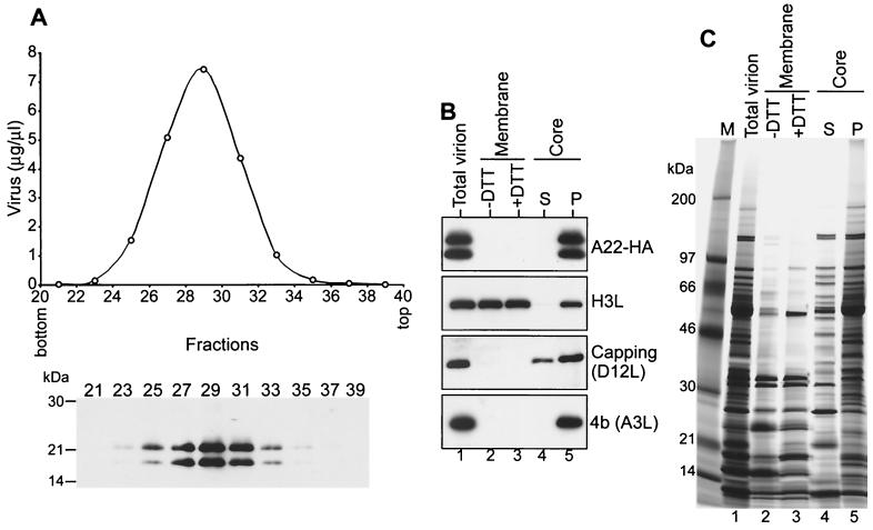
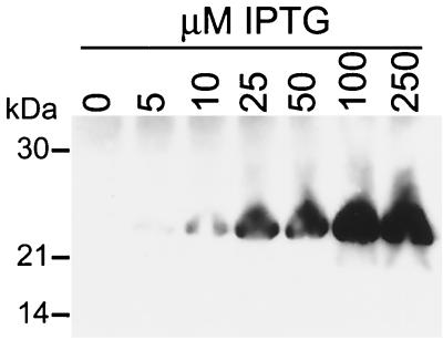
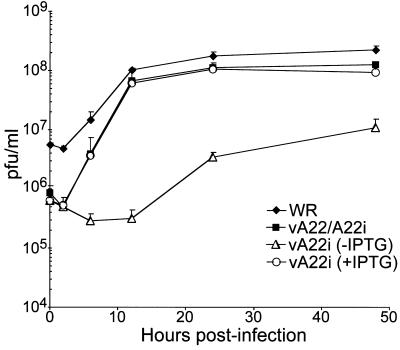
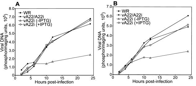
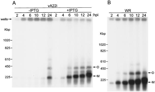
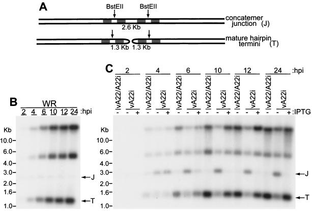
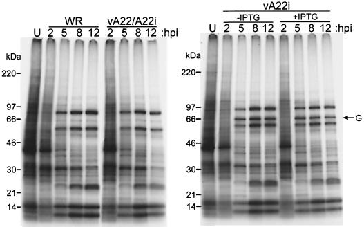
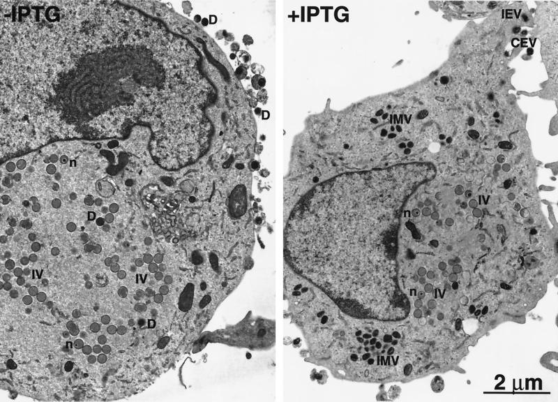
The vaccinia virus A22R gene encodes a protein that is homologous to the bacterial enzyme RuvC and specifically cleaves and resolves four-way DNA Holliday junctions into linear duplex products. To investigate the role of the vaccinia virus Holliday junction resolvase during an infection, we constructed two recombinant viruses: vA22-HA, which has a short C-terminal epitope tag appended to the A22R open reading frame, and vA22i, in which the original A22R gene is deleted and replaced by an inducible copy. Polyacrylamide gel electrophoresis and Western blot analysis of extracts and purified virions from cells infected with vA22-HA revealed that the resolvase was expressed after the onset of DNA replication and incorporated into virion cores. vA22i exhibited a conditional replication defect. In the absence of an inducer, (i) viral protein synthesis was unaffected, (ii) late-stage viral DNA replication was reduced, (iii) most of the newly synthesized viral DNA remained in a branched or concatemeric form that caused it to be trapped at the application site during pulsed-field gel electrophoresis, (iv) cleavage of concatemer junctions was inhibited, and (v) virion morphogenesis was arrested at an immature stage. These data indicated multiple roles for the vaccinia virus Holliday junction resolvase in the replication and processing of viral DNA into unit-length genomes.


Figures

FIG. 1
FIG. 1
Construction of a conditional lethal mutant with an inducible A22R gene. (A) Representations of relevant portions of the genomes of vT7lacOI, vA22/A22i, and vA22i corresponding to the J2R (thymidine kinase [TK]), A22R (HJ resolvase), and A56R (HA) genes. Insertions of exogenous genes into these regions are depicted below the diagrams. Abbreviations: PT7 and T7 pol, bacteriophage T7 promoter and RNA polymerase gene, respectively; lac I and lacO, E. coli lac repressor gene and lac operator element, respectively; gus, color marker gene; gpt, mycophenolic acid resistance gene; P11, P7.5, and P E/L, late, early-late, and synthetic early-late VV promoters, respectively. (B) Plaque formation. BS-C-1 cell monolayers were infected with wild-type VV (WR), vA22/A22i, or vA22i in the absence (−) or presence (+) of 50 μM IPTG. After 48 h, monolayers were stained with crystal violet and photographed.
FIG. 2
FIG. 2
Construction of an rVV with an HA epitope-tagged A22R gene. (A) Representations of relevant portions of the genomes of vA22i, vA22-HA/A22i, and vA22-HA. Abbreviations are the same as those used in Fig. 1. (B). Plaque formation. BS-C-1 cell monolayers were infected with wild-type VV or vA22-HA. After 48 h, monolayers were stained with crystal violet and photographed.
FIG. 3
FIG. 3
HJ resolvase synthesis during virus infection. BS-C-1 cells were infected at a multiplicity of 5 in the absence or presence of 44 μM cytosine arabinoside (AraC). At the indicated hours postinfection (hpi), cells were lysed in hypotonic buffer containing 0.2% NP-40 and 10 μg of micrococcal nuclease per ml and analyzed by Western blotting using a horseradish peroxidase-conjugated anti-HA antibody. Proteins were detected by chemiluminescence. The positions and masses of marker proteins are indicated. Arrows point to the A22R-HA doublet.
FIG. 4
FIG. 4
Association of the HJ resolvase with purified virions. (A) Cosedimentation of the A22-HA protein with virus particles. Sucrose gradient fractions were collected and analyzed for particles by light scattering and for the A22-HA protein by Western blotting as described in the legend to Fig. 3. (B) Detergent extraction of purified vA22-HA virions. Virions were incubated with 0.5% NP-40 in the absence or presence of 50 mM DTT, and the released membrane-associated and insoluble core proteins were separated by centrifugation. The viral cores were then resuspended in deoxycholate-containing lysis buffer and separated by centrifugation into supernatant (S) and pellet (P) fractions. The samples were analyzed by SDS-PAGE and Western blotting using antibodies to the HA epitope tag, the H3L protein, the large subunit of capping enzyme (D12L), and major core protein 4b (A3L). (C) The fractions described in panel B were analyzed by SDS-PAGE and silver staining.
FIG. 5
FIG. 5
Inducer-dependent expression of the HJ resolvase. BS-C-1 cells were infected with 5 PFU of vA22i per cell in the absence (0) or presence of 5 to 250 μM IPTG. Cells were harvested after 20 h and analyzed by SDS-PAGE and Western blotting using an antitetrahistidine antibody. Proteins were detected by chemiluminescence. The positions and masses of marker proteins are indicated.
FIG. 6
FIG. 6
Inducer-dependent replication of vA22i. BS-C-1 cells were infected with 5 PFU of WR, vA22/A22i, or vA22i in the absence or presence of 50 μM IPTG. At the indicated times, the cells were harvested and virus titers were determined by plaque assays in the presence of IPTG. Upper bars of standard deviations are shown.
FIG. 7
FIG. 7
Synthesis of viral DNA. BS-C-1 cells were infected with 5 PFU of WR, vA22/A22i, or vA22i in the absence or presence of 50 μM IPTG. At the indicated times, the cells were harvested and lysed in a high-salt solution. (A) Duplicate DNA samples were spotted directly onto a membrane and hybridized to 32P-labeled VV DNA. (B) DNA samples were deproteinized, sonicated, and denatured with alkali at ∼100°C prior to spotting onto a membrane and hybridization. Radioactivity was measured with a PhosphorImager and quantified with ImageQuant software.
FIG. 8
FIG. 8
Analysis of viral DNA by pulsed-field gel electrophoresis. BS-C-1 cells were infected with a 5 PFU of vA22i in the absence or presence of 50 μM IPTG (A) or WR (B). Cells were harvested at the indicated times (hpi, hours postinfection) and embedded in agarose. The DNA was subjected to pulsed-field electrophoresis, transferred onto a nylon membrane, and hybridized with 32P-labeled VV DNA. An autoradiograph is shown with the position of the wells and DNA size markers on the left. The positions of monomer (M) and dimer (D) viral genomes are indicated by arrows on the right.
FIG. 9
FIG. 9
Analysis of concatemer junctions. (A) Diagram of a concatemer junction (J) and mature hairpin termini (T). BstEII cleavage sites are shown. (B and C) BS-C-1 cells were infected with WR (B) or with vA22/A22i or vA22i in the absence (−) or presence (+) of 50 μM IPTG (C). At the indicated times (hpi, hours postinfection), DNA was purified; 2 μg was digested with restriction enzyme BstEII, electrophoresed through an agarose gel, transferred onto a nylon membrane, and hybridized with a 32P-labeled oligonucleotide complementary to the repeated sequence near the ends of the genome. The positions of monomer termini (T) and concatemer junction (J) intermediates are indicated by arrows.
FIG. 10
FIG. 10
Viral protein synthesis under permissive and nonpermissive conditions. BS-C-1 cell monolayers were infected with 5 PFU (per cell) of WR, vA22/A22i, or vA22i in the absence (−) or presence (+) of 50 μM IPTG. At the indicated hour postinfection (hpi), cells were labeled with a mixture of [35S]methionine and [35S]cysteine for 30 min. The labeled proteins were analyzed by SDS-PAGE and autoradiography. Lanes U contain proteins from uninfected cells. G, position of the band presumed to be the gus protein, which is present only in vA22i samples. The positions and masses of marker proteins are indicated.
FIG. 11
FIG. 11
Electron microscopy of cells infected with vA22i. BS-C-1 cells were infected with vA22i at a multiplicity of 10 in the absence (−) or presence (+) of 50 μM IPTG. After 24 h, the cells were harvested, fixed, and embedded in Epon. Ultrathin sections were prepared for electron microscopy. Abbreviations: IV, immature virion; n, nucleoid; D, dense immature virion; IMV, intracellular mature virion; CEV, cell-associated enveloped virion; IEV, intracellular mature enveloped virion.

Similar articles

Cited by

References

    1. Alexander W A, Moss B, Fuerst T R. Regulated expression of foreign genes in vaccinia virus under the control of bacteriophage T7 RNA polymerase and the Escherichia coli lac repressor. J Virol. 1992;66:2934–2942. - PMC - PubMed
    1. Baroudy B M, Venkatesan S, Moss B. Incompletely base-paired flip-flop terminal loops link the two DNA strands of the vaccinia virus genome into one uninterrupted polynucleotide chain. Cell. 1982;28:315–324. - PubMed
    1. Baroudy B M, Venkatesan S, Moss B. Structure and replication of vaccinia virus telomeres. Cold Spring Harbor Symp Quant Biol. 1982;47:723–729. - PubMed
    1. Black L W. DNA packaging and cutting by phage terminases: control in phage T4 by a synaptic mechanism. Bioessays. 1995;17:1025–1030. - PubMed
    1. Carpenter M S, DeLange A M. A temperature-sensitive lesion in the small subunit of the vaccinia virus-encoded mRNA capping enzyme causes a defect in viral telomere resolution. J Virol. 1991;65:4042–4050. - PMC - PubMed

LinkOut - more resources