Review
. 2001 Jun;65(2):187-207 ; first page, table of contents.
doi: 10.1128/MMBR.65.2.187-207.2001.

Pneumococcal virulence factors: structure and function

Affiliations
Review

Pneumococcal virulence factors: structure and function

M J Jedrzejas. Microbiol Mol Biol Rev. 2001 Jun.

Abstract

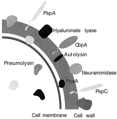
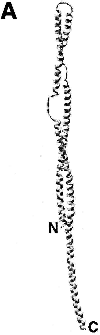
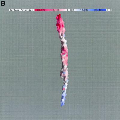
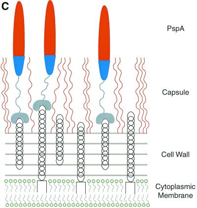
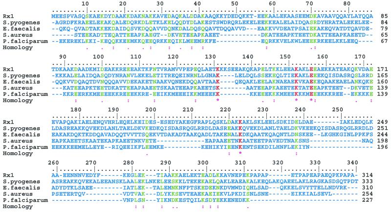
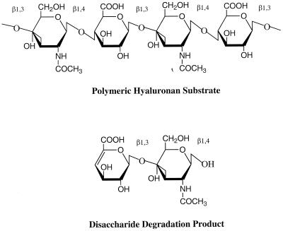
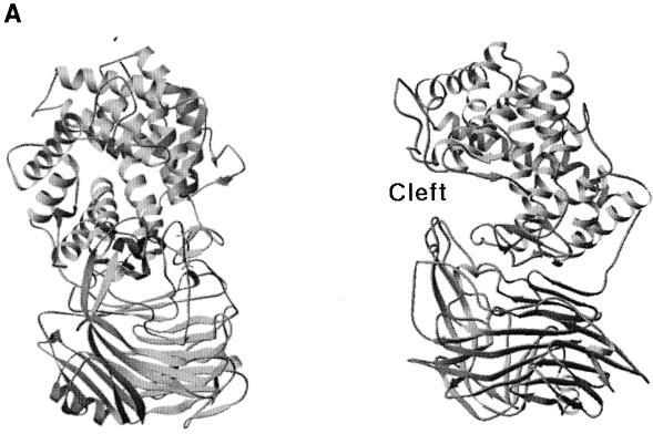
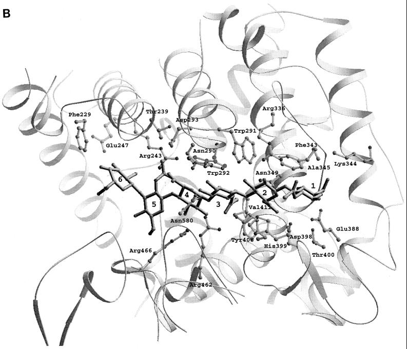
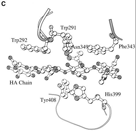
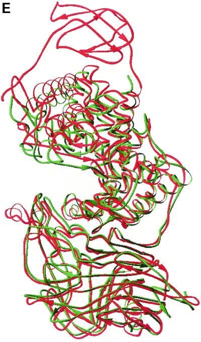
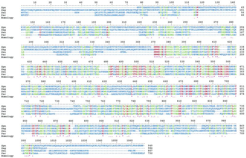
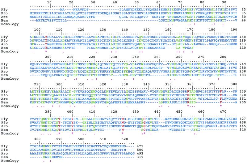
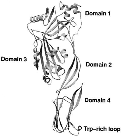
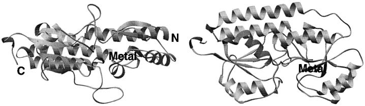
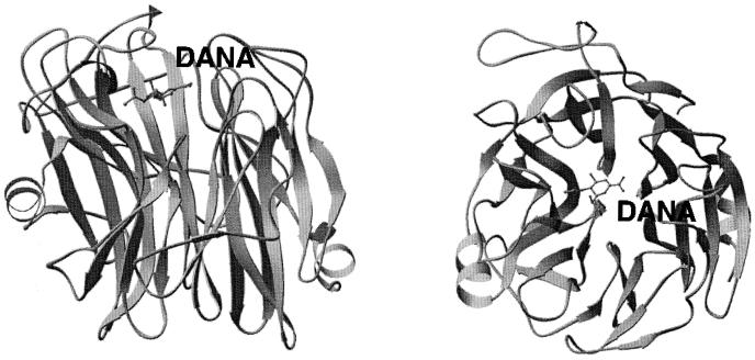
The overall goal for this review is to summarize the current body of knowledge about the structure and function of major known antigens of Streptococcus pneumoniae, a major gram-positive bacterial pathogen of humans. This information is then related to the role of these proteins in pneumococcal pathogenesis and in the development of new vaccines and/or other antimicrobial agents. S. pneumoniae is the most common cause of fatal community-acquired pneumonia in the elderly and is also one of the most common causes of middle ear infections and meningitis in children. The present vaccine for the pneumococcus consists of a mixture of 23 different capsular polysaccharides. While this vaccine is very effective in young adults, who are normally at low risk of serious disease, it is only about 60% effective in the elderly. In children younger than 2 years the vaccine is ineffective and is not recommended due to the inability of this age group to mount an antibody response to the pneumococcal polysaccharides. Antimicrobial drugs such as penicillin have diminished the risk from pneumococcal disease. Several pneumococcal proteins including pneumococcal surface proteins A and C, hyaluronate lyase, pneumolysin, autolysin, pneumococcal surface antigen A, choline binding protein A, and two neuraminidase enzymes are being investigated as potential vaccine or drug targets. Essentially all of these antigens have been or are being investigated on a structural level in addition to being characterized biochemically. Recently, three-dimensional structures for hyaluronate lyase and pneumococcal surface antigen A became available from X-ray crystallography determinations. Also, modeling studies based on biophysical measurements provided more information about the structures of pneumolysin and pneumococcal surface protein A. Structural and biochemical studies of these pneumococcal virulence factors have facilitated the development of novel antibiotics or protein antigen-based vaccines as an alternative to polysaccharide-based vaccines for the treatment of pneumococcal disease.


Figures

FIG. 1
FIG. 1
Schematic diagram of the virulence factors of S. pneumoniae.
FIG. 2
FIG. 2
The heptad repeat pattern of the N-terminal helical part of the deduced amino acid sequence of the Rx1 PspA. The sequence of the first 288 amino acids of the Rx1 PspA is arranged in heptad repeat blocks characteristic of coiled-coil proteins. The residues in the right part of the figure (residues 120 to 123, 173 to 175, and 208 to 212) do not satisfy the requirements for the coiled-coil structure.
FIG. 3
FIG. 3
Structural model of PspA (A), the electrostatic potential distribution on its surface (B), and the schematic arrangement of PspA molecules on the surface of S. pneumoniae (C). (A) The N-terminal part of PspA has an elongated rod-like shape built from antiparallel coiled-coil α-helices. The drawing is based on the model of a PspA molecule containing amino acids 1 to 288 (70). (B) The color coding of the PspA surface corresponds to the magnitude of the electrostatic potential: blue, electropositive; red, electronegative. The model also depicts the highly charged and polar character of PspA. (C) The character of the surface interactions of PspA with teichoic acids and the capsule exposes the highly electronegative end of the molecule outside of the bacterial cell.
FIG. 3
FIG. 3
Structural model of PspA (A), the electrostatic potential distribution on its surface (B), and the schematic arrangement of PspA molecules on the surface of S. pneumoniae (C). (A) The N-terminal part of PspA has an elongated rod-like shape built from antiparallel coiled-coil α-helices. The drawing is based on the model of a PspA molecule containing amino acids 1 to 288 (70). (B) The color coding of the PspA surface corresponds to the magnitude of the electrostatic potential: blue, electropositive; red, electronegative. The model also depicts the highly charged and polar character of PspA. (C) The character of the surface interactions of PspA with teichoic acids and the capsule exposes the highly electronegative end of the molecule outside of the bacterial cell.
FIG. 4
FIG. 4
Multiple alignment of the N-terminal part of Rx1 PspA with sequences from selected microbial genomes. The sequences were aligned using Multalin program (32) and displayed using Multiple Protein Sequence Alignment (MPSA) software (14). The sequence data represented correspond to unfinished genome sequences obtained from www.ncbi.nlm.nih.gov/BLAST/unfinishedgenome.html: S. pyogenes, Streptococcus pyogenes Contig1; E.faecalis, Enterococcus faecalis gef_6217; S.aureus, Staphylococcus aureus 4433; P.falciparum, Plasmodium falciparum Contig34. The color coding for the alignment is as follows: red, single fully conserved residue; green, fully conserved strong or weaker groups; blue, no particularity.
FIG. 5
FIG. 5
Structure of hyaluronan. Alternating units of glucuronic acid (GlcUA) and N-acetylglucosamine (GlcNAc) are the building blocks of the hyaluronan polymer. The main digestive product of the action of bacterial hyaluronate lyase on hyaluronan is the disaccharide unit. The glycosidic linkages are also marked.
FIG. 6
FIG. 6
(A) Overall structure of S. pneumoniae hyaluronate lyase (pdb: 1egu). (B) Structure of the modeled hexasaccharide substrate in the binding/catalytic cleft. (C) Catalytic residues of the enzyme. (D) Proposed mechanism of catalysis. (E) Alignment with the structure of S. agalactiae hyaluronate lyase (pdb: 1f1s). (A) The enzyme is built from an α-helical catalytic domain and a supportive β-sheet domain. The two domains are connected by one flexible peptide linker. Two perpendicular views of the enzyme are shown. (B) The cleft present at one end of the α-domain is where the substrate binds and is being degraded. (C) The residues directly involved in catalysis are Asn-349, His-399, and Tyr-408. (D) These residues degrade hyaluronan through a five-step proton acceptance and donation mechanism. (E) The S. agalactiae enzyme structure shows the presence of an additional supportive β-sheet domain at its N terminus.
FIG. 6
FIG. 6
(A) Overall structure of S. pneumoniae hyaluronate lyase (pdb: 1egu). (B) Structure of the modeled hexasaccharide substrate in the binding/catalytic cleft. (C) Catalytic residues of the enzyme. (D) Proposed mechanism of catalysis. (E) Alignment with the structure of S. agalactiae hyaluronate lyase (pdb: 1f1s). (A) The enzyme is built from an α-helical catalytic domain and a supportive β-sheet domain. The two domains are connected by one flexible peptide linker. Two perpendicular views of the enzyme are shown. (B) The cleft present at one end of the α-domain is where the substrate binds and is being degraded. (C) The residues directly involved in catalysis are Asn-349, His-399, and Tyr-408. (D) These residues degrade hyaluronan through a five-step proton acceptance and donation mechanism. (E) The S. agalactiae enzyme structure shows the presence of an additional supportive β-sheet domain at its N terminus.
FIG. 7
FIG. 7
Sequence alignment of selected bacterial hyaluronate lyases. The sequence data correspond to the following enzymes: Spn, S. pneumoniae; Sag, S. agalactiae; Sau, Staphylococcus aureus; Pac, Propionibacterium acnes. The sequence data were obtained from GenBank. The method used to produce this figure and the color coding used are the same as in Fig. 4.
FIG. 8
FIG. 8
Sequence alignment of CDCs of known three-dimensional structures. The origin of the proteins is as follows: Ply, S. pneumoniae; Pfo, Clostridium perfringens; Aro, Aeromonas hydrophilia; Hem, Staphylococcus aureus. Overall, the molecules have sequences that are only 14% homologous, but the three-dimensional structures have a similar fold and similar general structural arrangement. The sequence data were from GenBank. The method used to construct this figure and the color coding used are the same as in Fig. 4.
FIG. 9
FIG. 9
Structure of perfringolysin, a model structure for pneumolysin. All domains are labeled from 1 to 4, along with the Trp-rich loop in domain 4 (pdb: 1pfo). Domains 3 and 4 were implicated in membrane insertion accompanied by conformational changes leading to an α-helix–to–β-strand secondary structure shift.
FIG. 10
FIG. 10
Three-dimensional structure of PsaA based on the X-ray diffraction data (pdb: 1psz). The metal bound in its binding site is labeled. Two perpendicular views of the protein are shown.
FIG. 11
FIG. 11
Sequence alignment of selected microbial neuraminidase enzymes. The abbreviations are as follows: NanA and NanB, two neuraminidases of Streptococcus pneumoniae; Tcru, Trypanosoma cruzi; Cper, Clostridium perfringens; Styp = Salmonella enterica serovar Typhimurium. The sequence data were from GenBank. The method used to construct this figure and the color coding used are the same as in Fig. 4.
FIG. 12
FIG. 12
Three-dimensional structure of Salmonella enterica serovar Typhimurium LT2 neuraminidase with 2-deoxy-2,3-dehydro-N-acetylneuraminic acid (DANA) bound in its active site (pdb: 3sim). Two perpendicular views of the enzyme are shown.

Similar articles

Cited by

References

    1. Afessa B, Greaves W L, Frederick W R. Pneumococcal bacteremia in adults: a 14-year experience in an inner-city university hospital. Clin Infect Dis. 1995;21:345–351. - PubMed
    1. Alexander J E, Lock R A, Peeters C C, Poolman J T, Andrew P W, Mitchell T J, Hansman D, Paton J C. Immunization of mice with pneumolysin toxoid confers a significant degree of protection against at least nine serotypes of Streptococcus pneumoniae. Infect Immun. 1994;62:5683–5688. - PMC - PubMed
    1. Arnold K E, Leggiadro R J, Breiman R F, et al. Risk factors for carriage of drug-resistant Streptococcus pneumoniae among children in Memphis, Tennessee. J Pediatr. 1996;128:757–764. - PubMed
    1. Aruffo A, Stamenkovic I, Melnick M, Underhill C B, Seed B. CD44 is the principal cell surface receptor for hyaluronan. Cell. 1990;61:1303–1313. - PubMed
    1. Austrian R. Some observations on the pneumococcus and on the current status of pneumococcal disease and its prevention. In: Quie P G, Kass E H, editors. The pneumococcus and the pneumococcal vaccine. Chicago, Ill: University of Chicago Press; 1982. pp. 191–207.

Publication types

MeSH terms