. 2001 May 28;153(5):933-46.
doi: 10.1083/jcb.153.5.933.

Endothelial cell laminin isoforms, laminins 8 and 10, play decisive roles in T cell recruitment across the blood-brain barrier in experimental autoimmune encephalomyelitis

Affiliations

Endothelial cell laminin isoforms, laminins 8 and 10, play decisive roles in T cell recruitment across the blood-brain barrier in experimental autoimmune encephalomyelitis

M Sixt et al. J Cell Biol. .

Abstract

An active involvement of blood-brain barrier endothelial cell basement membranes in development of inflammatory lesions in the central nervous system (CNS) has not been considered to date. Here we investigated the molecular composition and possible function of the extracellular matrix encountered by extravasating T lymphocytes during experimental autoimmune encephalomyelitis (EAE). Endothelial basement membranes contained laminin 8 (alpha4beta1gamma1) and/or 10 (alpha5beta1gamma1) and their expression was influenced by proinflammatory cytokines or angiostatic agents. T cells emigrating into the CNS during EAE encountered two biochemically distinct basement membranes, the endothelial (containing laminins 8 and 10) and the parenchymal (containing laminins 1 and 2) basement membranes. However, inflammatory cuffs occurred exclusively around endothelial basement membranes containing laminin 8, whereas in the presence of laminin 10 no infiltration was detectable. In vitro assays using encephalitogenic T cell lines revealed adhesion to laminins 8 and 10, whereas binding to laminins 1 and 2 could not be induced. Downregulation of integrin alpha6 on cerebral endothelium at sites of T cell infiltration, plus a high turnover of laminin 8 at these sites, suggested two possible roles for laminin 8 in the endothelial basement membrane: one at the level of the endothelial cells resulting in reduced adhesion and, thereby, increased penetrability of the monolayer; and secondly at the level of the T cells providing direct signals to the transmigrating cells.


Figures

Figure 2
Figure 2
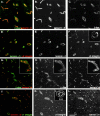
Confocal microscopy analysis of consecutive sections of EAE brains (clinical score +1 to +2) double stained for pan–laminin 1 (PAN-LM1) and CD45 (A), laminins α1 and α4 (B), laminins α1 and α5 (C), laminins α1 and α2 (D–F), or α-smooth muscle actin (SMA) alone (G). Inset in B shows double staining for PECAM-1 and laminin α1 at sites of monoclear infiltration. Superimposed images are shown in A–D; individual images for laminins α1 and α2 are shown in E and F, respectively. Arrowheads in all sections mark the same blood vessel where mononuclear infiltration has occurred, revealing the presence of laminin α4 alone in the endothelial cell basement membrane. The arrows mark vessels where no infiltration has occurred, revealing the presence of laminin α4 and α5 in the endothelial cell basement membrane and α-smooth muscle actin reactivity (G). Note the presence of laminin α1 and α2 in parenchymal basement membrane only (B–F) and the restricted distribution of laminin α1 (D and E). Double staining for laminin β1 and β2 (H) and for laminin γ1 alone (I) revealed the presence of laminin β1 and γ1 chains in association with all blood vessels, whereas laminin β2 is restricted to larger vessels. Inset in H shows laminin β1 staining at a site of mononuclear infiltration, localizing this chain to endothelial and parenchymal basement membranes. The same pattern of results was observed for laminin γ1. Bar, 80 μm.
Figure 1
Figure 1
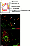
Cell layers and basement membranes which occur in association with blood vessels in the CNS. (A) Schematic representation of blood vessels showing the inner endothelial cell layer and its basement membrane (BM), bordered by epithelial meningeal cells and associated extracellular matrix and outer astroglial basement membrane and astrocyte endfeet. Collectively, the meningeal component and astroglial basement membrane are termed the parenchymal basement membrane. (B) Double staining of EAE brains (clinical scores +1 to +2) for PECAM-1 (red) and pan–laminin 1 antibody (PAN-LM1, green) which recognizes laminin α1, β1, and γ1 chains and thereby many laminin isoforms. Inset shows pan–laminin 1 staining alone. At sites of mononuclear infiltration the internal endothelial cell layer, bordered by its basement membrane, is distinct from the outer parenchymal basement membrane (arrowhead), whereas where no infiltration has occurred these two basement membranes are indistinguishable (arrows). (C) Double staining of EAE brains (clinical scores +3 to +4) with the leukocyte marker, CD45 and pan–laminin 1 antibody, demonstrating infiltrating mononuclear cells accumulating in the perivascular space and penetrating the outer parenchymal basement membrane. Bar, 50 μm.
Figure 3
Figure 3
Confocal microscopy analysis of individual blood vessels in the CNS double stained for laminin α1 and α2 (A–C) or laminin α2 and GFAP (D–F). Superimposed images are shown in A and D; individual images are shown in B, C, E, and F. Note the restricted distribution of laminin α1 which disappears at the precapillary level (A–C). Laminin α2 localizes at the GFAP-positive astrocyte endfeet (D and E). Bars: (A–C) 8 μm; (D–F) 12 μm.
Figure 5
Figure 5
Confocal microscopy analysis for the expression of integrin α6 and β-dystroglycan in mouse brain blood vessels. A–C show double staining for integrin α6 and β-dystroglycan (β-dys) showing mutually exclusive distributions. D–F show double staining for integrin α6 and the parenchymal basement membrane component, laminin α1, localizing integrin α6 on the inner endothelial cell layer; inset in F shows integrin α6 expression at sites of mononuclear infiltration revealing discontinuous staining. G–I show double staining for β-dystroglycan and the endothelial cell basement membrane–specific laminin α4, demonstrating β-dystroglycan expression on astrocyte endfeet; the insets in G–I show a higher magnification of a single vessel. J–L show double staining for integrin β1 and laminin α4 showing overlapping distribution patterns; the inset in K shows integrin β1 expression at sites of mononuclear infiltration, demonstrating concentration on the inner endothelial cell layer, but also on infiltration cells, with weaker expression on the astrocyte endfeet. Bars: (A–C) 80 μm; (D–L) 100 μm.
Figure 4
Figure 4
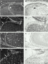
In situ hybridization of consecutive sections of E16 embryonic mouse brains with probes specific for laminin α1 (A and B), α2, (C and D), α4 (E and F), and an endothelial cell–specific probe, flk-1 (G and H). Data shown are for similar areas, encompassing meninges (arrows) and a portion of the cortex of the cerebellum. Similar results were observed for E18 embryos and, in the case of laminin α4, also for newborn and adult tissues. Laminin α1 mRNA was expressed by the leptomeningeal cells of the pia mater that encase the brain and are infolded from the brain surface (arrowheads in A and B); laminin α2 mRNA was restricted to the meninges (C and D), whereas laminin α4 mRNA was detected in endothelial cells within and outside the brain (E and F) in a similar pattern to that observed with the endothelial cell–specific probe, flk-1 (G and H). Bar, 25 μm.
Figure 6
Figure 6
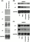
Northern blot analysis for the expression of laminin α1, α2, α4, and α5 mRNA in control and EAE (clinical score 4+/5+) mouse brains (A), and for laminins α4 and α5 mRNA in sEND1 (B) and bEND3 (C) mouse endothelial cell lines. Endothelial cell lines were either untreated, activated with IL-1 or transforming growth factor α (TNFα), or treated with hydroxymethylprogesterone (Progester.). Data in A for laminin α1 and α2 is after 2 wk of exposure, for laminin α5 after 6 d of exposure, and for laminin α4 after 24 h of exposure.
Figure 8
Figure 8
Illustration of cell layers, basement membranes (BM), and their laminin composition of CNS blood vessels with and without an inflammatory cuff. Larger blood vessels consist of an inner endothelial cell layer with a basement membrane (containing laminins α4 and α5), bordered by the meningeal epithelium and its basement membrane (containing laminin α1) and an outer astroglial basement membrane (containing laminin α2) and astrocyte endfeet. The meningeal and astroglial basement membranes are collectively termed the parenchymal basement membrane as they delineate the border to the brain parenchyma. Only at sites of local inflammation are the endothelial and parenchymal basement membranes distinguishable and define the inner and outer limits of the perivascular space where leukocytes accumulate before infiltrating the brain parenchyma. Examination of such sites demonstrates that mononuclear infiltration occurs across endothelial basement membranes containing only the laminin α4 and bordered by a parenchymal basement membrane containing laminin α1 and α2. The basement membrane of microvessels where no epithelial meningeal contribution occurs appear to have a composite basement membrane containing the endothelial cell laminins, laminin α4 and α5, and laminin α2 produced by the astrocytes and deposited at their endfeet.
Figure 7
Figure 7
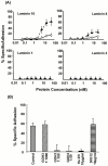
(A) Specific binding of nonactivated (○) and PMA-activated (▴) PLP-specific, encephalitogenic T cell lines to increasing concentrations of the endothelial cell basement membrane laminins 10 and 8, and the parenchymal basement membrane laminins 1 and 2. Saturable T cell binding occurred only to laminins 10 and 8; binding to laminins 1 and 2 could not be induced, even under maximal integrin activation conditions (10 mM Mn2+). (B) Binding to 30 nM laminin 10 was completely inhibited by antiintegrin α6 (GoH3) or antiintegrin β1 (Ha2/5), whereas anti-CD45 antibody (30G12) had no effect. The same pattern of results was observed for laminin 8. Values represent means of at least six experiments ± SD.

Similar articles

Cited by

References

    1. Alcolado R., Weller R.O., Parrish E.P., Garrod D. The cranial arachnoid and piamater in mananatomical and ultrastructural observations. Neuropathol. Appl. Neurobiol. 1988;14:1–17. - PubMed
    1. Aumailley M., Smyth N. The role of laminins in basement membranes. J. Anat. 1998;193:1–21. - PMC - PubMed
    1. Boado R.J., Pardridge W.M. Differential expression of alpha-actin mRNA and immunoreactive protein in brain microvascular pericytes and smooth muscle cells. J. Neurosci. Res. 1994;39:430–435. - PubMed
    1. Breier G., Albrecht U., Sterrer S., Risau W. Expression of vascular endothelial growth factor during embryonic angiogenesis and endothelial cell differentiation. Development. 1992;114:521–532. - PubMed
    1. Chandler S., Miller K.M., Clements J.M., Lury J., Corkill D., Anthony D.C.C., Adams S.E., Gearing A.J.H. Matrix metalloproteinases, tumor necrosis factor and multiple sclerosisan overview. J. Neuroimmunol. 1997;72:155–161. - PubMed

Publication types

MeSH terms