

Review
doi: 10.1136/bmj.322.7296.1222.
Microbubble contrast agents: a new era in ultrasound
Affiliations
- PMID: 11358777
- PMCID: PMC1120332
- DOI: 10.1136/bmj.322.7296.1222
Item in Clipboard
Review
Microbubble contrast agents: a new era in ultrasound
BMJ.
.
No abstract available
Figures
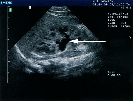
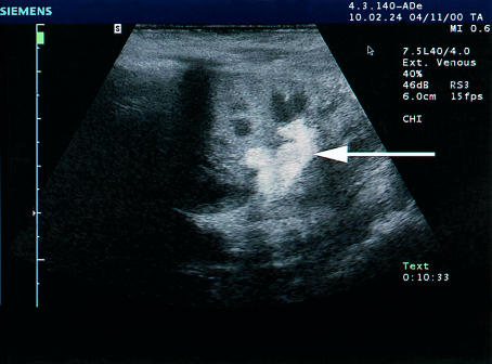
Use of microbubbles to demonstrate vesicoureteric reflux. Top figure shows left kidney with a slightly dilated renal pelvis (arrow). Bottom figure shows the same area after microbubbles have been instilled into the bladder. Bright echoes, representing microbubbles can now be seen in the renal pelvis (arrow), indicating vesicoureteric reflux, which was confirmed on x ray micturating cystourethrogram. (Images courtesy of Dr Thomas Albrecht, Benjamin Franklin University, Berlin, Germany)
Use of microbubbles to demonstrate vesicoureteric reflux. Top figure shows left kidney with a slightly dilated renal pelvis (arrow). Bottom figure shows the same area after microbubbles have been instilled into the bladder. Bright echoes, representing microbubbles can now be seen in the renal pelvis (arrow), indicating vesicoureteric reflux, which was confirmed on x ray micturating cystourethrogram. (Images courtesy of Dr Thomas Albrecht, Benjamin Franklin University, Berlin, Germany)
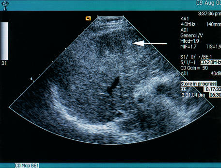
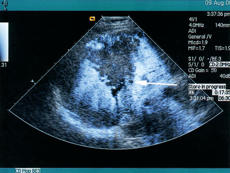
Effect of liver-specific microbubbles on the visualisation of a hepatocellular carcinoma complicating chronic hepatitis B infection. Top figure shows the liver is heterogeneous with an ill defined lesion (arrow). Bottom figure shows the presence of liver-specific microbubbles (Levovist) administered 5 minutes earlier. A defect is clearly seen in the central right lobe of the liver, with several additional defects thought to represent additional satellite foci of hepatocellular carcinoma
Effect of liver-specific microbubbles on the visualisation of a hepatocellular carcinoma complicating chronic hepatitis B infection. Top figure shows the liver is heterogeneous with an ill defined lesion (arrow). Bottom figure shows the presence of liver-specific microbubbles (Levovist) administered 5 minutes earlier. A defect is clearly seen in the central right lobe of the liver, with several additional defects thought to represent additional satellite foci of hepatocellular carcinoma

Echocardiographic image of the left ventricle using real time imaging of perfusion and an intravenous microbubble as contrast agent. The contrast agent fills the ventricular cavity, clearly delineating the endocardial border, and gives colour enhancement in the myocardium, showing perfusion of the apex and septum (arrows)
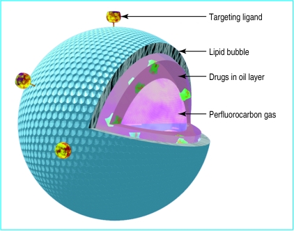
Diagram of a microbubble constructed for drug delivery. Gas-filled microspheres may be designed so that their interior is loaded with drug and gas. A stabilising material, here a lipid, surrounds the perfluorocarbon bubble. Drugs may be incorporated by themselves or, if insoluble in water, in an oil layer. The microsphere may be targeted to specific tissue by incorporating protein ligands on the surface
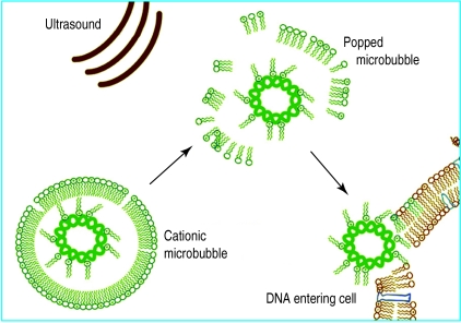
Gene delivery using ultrasound and microbubbles. The presence of gas in the gene-filled microbubble allows ultrasound energy to “pop” the bubble. An energetic wave is then created which allows the genetic material to enter surrounding cells
Similar articles
-
Voiding urosonography: an additional important indication for use of US contrast agents.Radiology. 2011 May;259(2):614-5; author reply 615. doi: 10.1148/radiol.11102479. Radiology. 2011. PMID: 21502399 No abstract available.
-
Early assessment of the therapeutic response to radio frequency ablation for hepatocellular carcinoma: utility of gray scale harmonic ultrasonography with a microbubble contrast agent.J Ultrasound Med. 2003 Nov;22(11):1163-72. doi: 10.7863/jum.2003.22.11.1163. J Ultrasound Med. 2003. PMID: 14620886
-
Contrast-enhanced ultrasound of the liver: technical and lexicon recommendations from the ACR CEUS LI-RADS working group.Abdom Radiol (NY). 2018 Apr;43(4):861-879. doi: 10.1007/s00261-017-1392-0. Abdom Radiol (NY). 2018. PMID: 29151131 Free PMC article.
-
[Guidelines for contrast enhanced ultrasound (CEUS)--update 2008].J Radiol. 2009 Jan;90(1 Pt 2):123-38; quiz 139-40. doi: 10.1016/s0221-0363(09)70090-3. J Radiol. 2009. PMID: 19212280 Review. French.
-
Contrast-Enhanced Ultrasound of Focal Liver Lesions.Semin Roentgenol. 2016 Oct;51(4):334-357. doi: 10.1053/j.ro.2016.05.018. Epub 2016 May 30. Semin Roentgenol. 2016. PMID: 27743569 Review. No abstract available.
Cited by
-
The future of collateral artery research.Curr Cardiol Rev. 2014 Feb;10(1):73-86. doi: 10.2174/1573403x113099990001. Curr Cardiol Rev. 2014. PMID: 23638829 Free PMC article. Review.
-
Therapeutic potential of low-intensity ultrasound (part 1): thermal and sonomechanical effects.J Med Ultrason (2001). 2008 Dec;35(4):153-60. doi: 10.1007/s10396-008-0194-y. Epub 2008 Dec 16. J Med Ultrason (2001). 2008. PMID: 27278986 Review.
-
Ultrasound-Guided Delivery of siRNA and a Chemotherapeutic Drug by Using Microbubble Complexes: In Vitro and In Vivo Evaluations in a Prostate Cancer Model.Korean J Radiol. 2016 Jul-Aug;17(4):497-508. doi: 10.3348/kjr.2016.17.4.497. Epub 2016 Jun 27. Korean J Radiol. 2016. PMID: 27390541 Free PMC article.
-
Role of microbubble ultrasound contrast agents in the non-invasive assessment of chronic hepatitis C-related liver disease.World J Gastroenterol. 2006 Jun 14;12(22):3461-5. doi: 10.3748/wjg.v12.i22.3461. World J Gastroenterol. 2006. PMID: 16773702 Free PMC article. Review.
-
Cavitation threshold of microbubbles in gel tunnels by focused ultrasound.Ultrasound Med Biol. 2007 Oct;33(10):1651-60. doi: 10.1016/j.ultrasmedbio.2007.04.018. Epub 2007 Jun 27. Ultrasound Med Biol. 2007. PMID: 17590501 Free PMC article.
References
-
- Nanda NC, Carstensen C. Echo-enhancing agents: safety. In: Nanda NC, Schlief R, Goldberg BB, editors. Advances in echo imaging using contrast enhancers. Dordrecht: Kluwer; 1997. pp. 115–131.
-
- Albrecht T, Urbank A, Mahler M, Bauer A, Dore CJ, Blomley MJ, et al. Prolongation and optimization of Doppler enhancement with a microbubble US contrast agent by using continuous infusion: preliminary experience. Radiology. 1998;207:339–347. - PubMed
-
- Ries F, Honisch C, Lambertz M, Schlief R. A transpulmonary contrast medium enhances the transcranial Doppler signal in humans. Stroke. 1993;24:1903–1909. - PubMed
-
- Cosgrove D. Why do we need contrast agents for ultrasound? Clin Radiol. 1996;51(suppl 1):1–4. - PubMed
-
- Darge K, Troeger J, Duetting T, Zieger B, Rohrschneider W, Moehring K, et al. Reflux in young patients: comparison of voiding US of the bladder and retrovesical space with echo enhancement versus voiding cystourethrography for diagnosis. Radiology. 1999;210:201–207. - PubMed
Publication types
MeSH terms
Substances
LinkOut - more resources
Full Text Sources
Other Literature Sources