

Large involuntary forces consistent with plateau-like behavior of human motoneurons
- PMID: 11356893
- PMCID: PMC6762712
- DOI: 10.1523/JNEUROSCI.21-11-04059.2001
Large involuntary forces consistent with plateau-like behavior of human motoneurons
Abstract
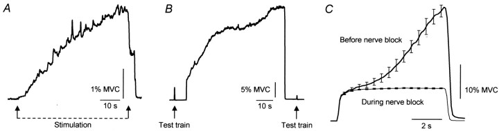
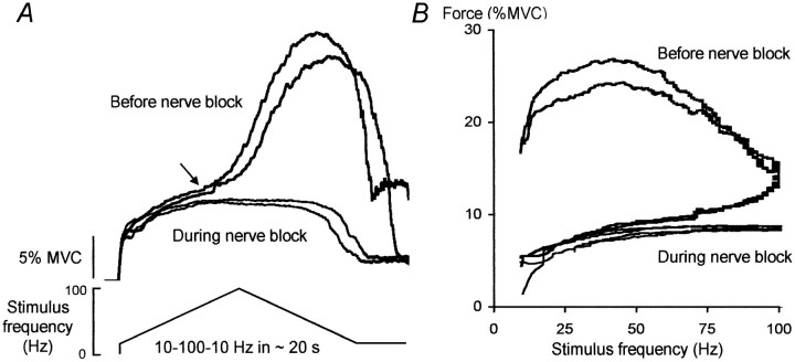
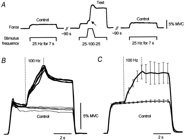
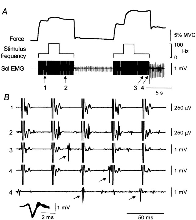
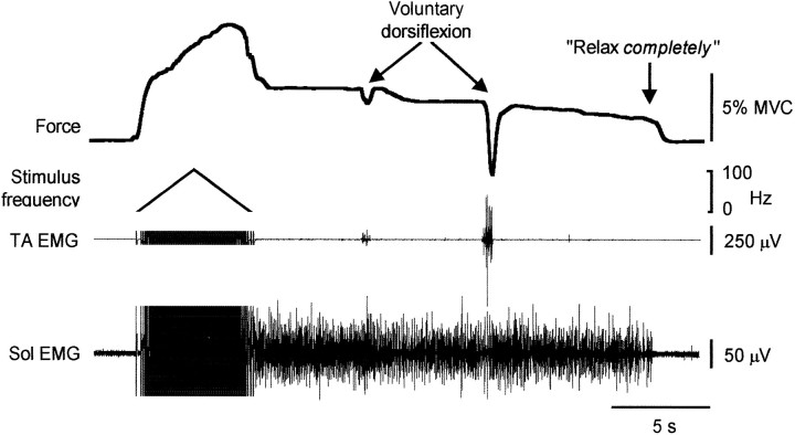
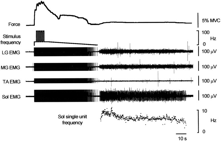
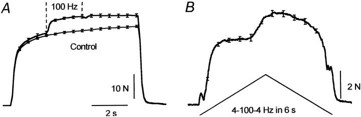
When electrical stimulation is applied over human muscle, the evoked force is generally considered to be of peripheral origin. However, in relaxed humans, stimulation (1 msec pulses, 100 Hz) over the muscles that plantarflex the ankle produced more than five times more force than could be accounted for by peripheral properties. This additional force was superimposed on the direct response to motor axon stimulation, produced up to 40% of the force generated during a maximal voluntary contraction, and was abolished during anesthesia of the tibial nerve proximal to the stimulation site. It therefore must have resulted from the activation of motoneurons within the spinal cord. The additional force could be initiated by stimulation of low-threshold afferents, distorted the classical relationship between force and stimulus frequency, and often outlasted the stimulation. The mean firing rate of 27 soleus motor units recorded during the sustained involuntary activity after the stimulation was 5.8 +/- 0.2 Hz. The additional force increments were not attributable to voluntary intervention because they were present in three sleeping subjects and in two subjects with lesions of the thoracic spinal cord. The phenomenon is consistent with activation of plateau potentials within motoneurons and, if so, the present findings imply that plateau potentials can make a large contribution to forces produced by the human nervous system.
Figures
Similar articles
-
Sustained contractions produced by plateau-like behaviour in human motoneurones.J Physiol. 2002 Jan 1;538(Pt 1):289-301. doi: 10.1113/jphysiol.2001.012825. J Physiol. 2002. PMID: 11773336 Free PMC article.
-
Contractile properties of single motor units in human toe extensors assessed by intraneural motor axon stimulation.J Neurophysiol. 1996 Jun;75(6):2509-19. doi: 10.1152/jn.1996.75.6.2509. J Neurophysiol. 1996. PMID: 8793760 Clinical Trial.
-
Recruitment of triceps surae motor units in the decerebrate cat. II. Heterogeneity among soleus motor units.J Neurophysiol. 1996 May;75(5):2005-16. doi: 10.1152/jn.1996.75.5.2005. J Neurophysiol. 1996. PMID: 8734599
-
Muscle weakness, paralysis, and atrophy after human cervical spinal cord injury.Exp Neurol. 1997 Dec;148(2):414-23. doi: 10.1006/exnr.1997.6690. Exp Neurol. 1997. PMID: 9417821
-
Human spinal cord injury: motor unit properties and behaviour.Acta Physiol (Oxf). 2014 Jan;210(1):5-19. doi: 10.1111/apha.12153. Epub 2013 Sep 13. Acta Physiol (Oxf). 2014. PMID: 23901835 Review.
Cited by
-
Neuromuscular electrical stimulation at submaximal intensity combined with motor imagery increases corticospinal excitability.Eur J Appl Physiol. 2024 Oct 2. doi: 10.1007/s00421-024-05615-y. Online ahead of print. Eur J Appl Physiol. 2024. PMID: 39356322
-
Motoneuron persistent inward current contribution to increased torque responses to wide-pulse high-frequency neuromuscular electrical stimulation.Eur J Appl Physiol. 2024 Nov;124(11):3377-3386. doi: 10.1007/s00421-024-05538-8. Epub 2024 Jun 28. Eur J Appl Physiol. 2024. PMID: 38940932 Free PMC article.
-
Effect of electromyostimulation training on soleus and gastrocnemii H- and T-reflex properties.Eur J Appl Physiol. 2003 Nov;90(5-6):601-7. doi: 10.1007/s00421-003-0914-3. Epub 2003 Aug 16. Eur J Appl Physiol. 2003. PMID: 12923640
-
Evidence for increased activation of persistent inward currents in individuals with chronic hemiparetic stroke.J Neurophysiol. 2008 Dec;100(6):3236-43. doi: 10.1152/jn.90563.2008. Epub 2008 Oct 1. J Neurophysiol. 2008. PMID: 18829849 Free PMC article.
-
Effect of tendon vibration during wide-pulse neuromuscular electrical stimulation (NMES) on muscle force production in people with spinal cord injury (SCI).BMC Neurol. 2018 Feb 13;18(1):17. doi: 10.1186/s12883-018-1020-9. BMC Neurol. 2018. PMID: 29433467 Free PMC article.
References
-
- Ashby P, Zilm D. Characteristics of postsynaptic potentials produced in single human motoneurons by homonymous group 1 volleys. Exp Brain Res. 1982;47:41–48. - PubMed
-
- Baldissera F, Cavallari P, Dworzak F. Cramps: a sign of motoneurone “bistability” in a human patient. Neurosci Lett. 1991;133:303–306. - PubMed
-
- Baldissera F, Cavallari P, Dworzak F. Motor neuron “bistability”. A pathogenetic mechanism for cramps and myokymia. Brain. 1994;117:929–939. - PubMed
-
- Bennett DJ, Hultborn H, Fedirchuk B, Gorassini M. Synaptic activation of plateaus in hindlimb motoneurons of decerebrate cats. J Neurophysiol. 1998a;80:2023–2037. - PubMed
-
- Bennett DJ, Hultborn H, Fedirchuk B, Gorassini M. Short-term plasticity in hindlimb motoneurons of decerebrate cats. J Neurophysiol. 1998b;80:2038–2045. - PubMed
Publication types
MeSH terms
Substances
LinkOut - more resources
Full Text Sources
Other Literature Sources