Regeneration of a lytic central vacuole and of neutral peripheral vacuoles can be visualized by green fluorescent proteins targeted to either type of vacuoles
- PMID: 11351072
- PMCID: PMC102283
- DOI: 10.1104/pp.126.1.78
Regeneration of a lytic central vacuole and of neutral peripheral vacuoles can be visualized by green fluorescent proteins targeted to either type of vacuoles
Abstract
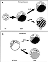
Protein trafficking to two different types of vacuoles was investigated in tobacco (Nicotiana tabacum cv SR1) mesophyll protoplasts using two different vacuolar green fluorescent proteins (GFPs). One GFP is targeted to a pH-neutral vacuole by the C-terminal vacuolar sorting determinant of tobacco chitinase A, whereas the other GFP is targeted to an acidic lytic vacuole by the N-terminal propeptide of barley aleurain, which contains a sequence-specific vacuolar sorting determinant. The trafficking and final accumulation in the central vacuole (CV) or in smaller peripheral vacuoles differed for the two reporter proteins, depending on the cell type. Within 2 d, evacuolated (mini-) protoplasts regenerate a large CV. Expression of the two vacuolar GFPs in miniprotoplasts indicated that the newly formed CV was a lytic vacuole, whereas neutral vacuoles always remained peripheral. Only later, once the regeneration of the CV was completed, the content of peripheral storage vacuoles could be seen to appear in the CV of a third of the cells, apparently by heterotypic fusion.
Figures


Similar articles
-
Specific accumulation of GFP in a non-acidic vacuolar compartment via a C-terminal propeptide-mediated sorting pathway.Plant J. 1998 Aug;15(4):449-57. doi: 10.1046/j.1365-313x.1998.00210.x. Plant J. 1998. PMID: 9753772
-
Two glycosylated vacuolar GFPs are new markers for ER-to-vacuole sorting.Plant Physiol Biochem. 2013 Dec;73:337-43. doi: 10.1016/j.plaphy.2013.10.010. Epub 2013 Oct 23. Plant Physiol Biochem. 2013. PMID: 24184454
-
The vacuolar transport of aleurain-GFP and 2S albumin-GFP fusions is mediated by the same pre-vacuolar compartments in tobacco BY-2 and Arabidopsis suspension cultured cells.Plant J. 2008 Dec;56(5):824-39. doi: 10.1111/j.1365-313X.2008.03645.x. Epub 2008 Sep 4. Plant J. 2008. PMID: 18680561
-
Contribution of chitinase A's C-terminal vacuolar sorting determinant to the study of soluble protein compartmentation.Int J Mol Sci. 2014 Jun 18;15(6):11030-9. doi: 10.3390/ijms150611030. Int J Mol Sci. 2014. PMID: 24945312 Free PMC article. Review.
-
Vacuolar deposition of recombinant proteins in plant vegetative organs as a strategy to increase yields.Bioengineered. 2017 May 4;8(3):203-211. doi: 10.1080/21655979.2016.1222994. Epub 2016 Sep 20. Bioengineered. 2017. PMID: 27644793 Free PMC article. Review.
Cited by
-
What is moving in the secretory pathway of plants?Plant Physiol. 2008 Aug;147(4):1493-503. doi: 10.1104/pp.108.124552. Plant Physiol. 2008. PMID: 18678741 Free PMC article. Review. No abstract available.
-
Dynamic protein trafficking to the cell wall.Plant Signal Behav. 2011 Jul;6(7):1012-5. doi: 10.4161/psb.6.7.15550. Plant Signal Behav. 2011. PMID: 21701253 Free PMC article.
-
In vivo imaging and quantitative monitoring of autophagic flux in tobacco BY-2 cells.Plant Signal Behav. 2013 Jan;8(1):e22510. doi: 10.4161/psb.22510. Epub 2012 Nov 3. Plant Signal Behav. 2013. PMID: 23123450 Free PMC article.
-
SNAREs: cogs and coordinators in signaling and development.Plant Physiol. 2008 Aug;147(4):1504-15. doi: 10.1104/pp.108.121129. Plant Physiol. 2008. PMID: 18678742 Free PMC article. Review. No abstract available.
-
Cotyledon cells of Vigna mungo seedlings use at least two distinct autophagic machineries for degradation of starch granules and cellular components.J Cell Biol. 2001 Sep 3;154(5):973-82. doi: 10.1083/jcb.200105096. Epub 2001 Aug 27. J Cell Biol. 2001. PMID: 11524437 Free PMC article.
References
-
- Beevers L, Raikhel NV. Transport to the vacuole: receptors and trans elements. J Exp Bot. 1998;49:1271–1279.
-
- Buvat R. Some aspects of the origin and evolution of vacuoles-cytophysiological properties and related consequences. Bull Soc Bot. 1982;129:7–17.
-
- Cormack BP, Valdivia RH, Falkow S. FACS-optimized mutants of the green fluorescent protein (GFP) Gene. 1996;173:33–38. - PubMed
Publication types
MeSH terms
Substances
LinkOut - more resources
Full Text Sources
Other Literature Sources

