. 2001 May 15;20(10):2424-34.
doi: 10.1093/emboj/20.10.2424.

Phospholipase D1: a key factor for the exocytotic machinery in neuroendocrine cells

Affiliations

Phospholipase D1: a key factor for the exocytotic machinery in neuroendocrine cells

N Vitale et al. EMBO J. .

Abstract

Phospholipase D (PLD) has been proposed to mediate cytoskeletal remodeling and vesicular trafficking along the secretory pathway. We recently described the activation of an ADP ribosylation factor-regulated PLD at the plasma membrane of chromaffin cells undergoing secretagogue-stimulated exocytosis. We show here that the isoform involved is PLD1b, and, using a real-time assay for individual cells, that PLD activation and exocytosis are closely correlated. Moreover, overexpressed PLD1, but not PLD2, increases stimulated exocytosis in a phosphatidylinositol 4,5-bisphosphate-dependent manner, whereas catalytically inactive PLD1 inhibits it. These results provide the first direct evidence that PLD1 is an important component of the exocytotic machinery in neuroendocrine cells.


Figures
None
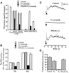
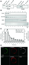
Fig. 1. Chromaffin cells express PLD1b, which localizes to and is activated at the plasma membrane. (A) Cultured chromaffin cells were collected, homogenized and centrifuged at 20 000 g to separate cytosol (C) from the crude membranes (Mb). Note that light membranes, such as the Golgi and endosomes, are not pelleted under these conditions. The fractions were then subjected to protein determination, gel electrophoresis and immunodetection on nitrocellulose sheets using anti-PLD1 and anti-PLD2 antibodies. Lysates from HEK293 cells transfected with pCGN-PLD1a, pCGN-PLD1b and pCGN-PLD2 plasmids were used as positive controls. (B) Fractions 2–11 (40 µg of protein per fraction) collected from a continuous sucrose density gradient layered with the crude chromaffin membrane pellet were immunodetected as above using antibodies against PLD1, SNAP-25 (plasma membrane marker), dopamine-β-hydroxylase (chromaffin granule marker) and β-COP (Golgi membrane marker). (C) Chromaffin cells labeled with [3H]myristic acid were permeabilized with SLO and subsequently incubated in KG medium containing 1% ethanol and 100 µM GTPγS in the presence (closed bars) or absence (open bars) of 20 µM free Ca2+. Cells were then collected and processed for subcellular fractionation on a continuous sucrose density gradient. Fractions were assayed for Na+/K+-ATPase for plasma membranes. Phospholipids were extracted from each fraction and the amount of [3H]phosphatidylethanolamine formed determined by TLC. Most of the Ca2+-stimulated PLD activity is detected in the plasma membrane-enriched fractions, which similarly contained PLD1. Data are the mean values of duplicate determinations. Similar results were obtained in four independent experiments. (D) Immunofluorescence confocal micrographs of cultured chromaffin cells double-labeled with anti-PLD1 or anti-PLD2 antibodies, detected with secondary Cy2-conjugated anti-rabbit antibodies, and anti-SNAP-25 antibodies detected with Cy3-conjugated anti-mouse antibodies. The same optical section recorded sequentially in the Cy2 (PLD) and Cy3 (SNAP-25) channels is presented. Masks representing the regions of co-localization are obtained by selecting the pixels double-labeled with Cy2 and Cy3.
None
Fig. 2. Effects of ceramides on PLD activation and catecholamine secretion from stimulated chromaffin cells. (A) Purified PLD1 was assayed in vitro under basal and stimulated conditions in the presence of the indicated ceramides. ARF-stimulated PLD1 activity was assayed in the absence or presence of either C8-, C2- or dihydro-C2 ceramides (20 or 50 µM). Basal, ARF-stimulated and Rho-stimulated PLD1 were assayed in the absence or presence of 50 µM C2-ceramide. (B) Chromaffin cells labeled with [3H]myristic acid were incubated for 1 h with the indicated ceramides at 50 µM. Cells were then permeabilized with SLO and stimulated with 20 µM free Ca2+ in the presence of 1% ethanol. Phospholipids subsequently were extracted and analyzed by TLC. Data are the mean values of duplicate determinations ± SEM. (C) Chromaffin cells were incubated for 1 h in the presence of the indicated ceramides at 50 µM. Cells were then stimulated by a local application of 100 µM nicotine for 10 s (arrow) and the exocytotic release of catecholamines was estimated by electrochemical detection with a carbon fiber electrode placed adjacent to single chromaffin cells. Typical amperometric responses from nicotine-stimulated chromaffin cells pre-incubated in Locke’s solution (control) or in Locke’s solution containing either C2- or dihydro-C2-ceramide are shown. (D) Amperometric responses were integrated to obtain the total catecholamine secretion expressed in pA/s. Results are expressed relative to the secretory response obtained for the control cells. Data are the means of 30 cells/group from the same experiment ± SEM. Similar results were obtained in three independent experiments performed on different culture preparations.
None
Fig. 3. Overexpression of wild-type or catalytically inactive PLD1 but not PLD2 alters secretion of co-expressed GH from PC12 cells. PC12 cells were transfected with either pCGN (empty vector), pCGN-PLD1, pCGN-PLD1(K898R), pCGN-PLD2 or pCGN-PLD2(K758R) plasmids along with the plasmid encoding GH (4 µg/well of each plasmid). (A) At 48 h post-transfection, the proteins expressed were visualized by immunocytochemistry and confocal microscopy. Approximately 10–20% of cells were transfected successfully with the GH-expressing vector and, of these, >90% became co-transfected with the second plasmid. Neither wild-type nor mutated PLD1 or PLD2 proteins affected the punctuate pattern of fluorescence observed with the anti-GH antibodies, which is consistent with the storage of GH within secretory granules. Note the distinct PLD and GH labelings, revealing that the overexpressed PLDs are not associated with secretory granules. (B) Transfected cells were washed and subsequently incubated for 10 min in calcium-free Locke’s solution (open bars) or stimulated for 10 min with elevated K+ (closed bars). Extracellular fluids were then collected and GH present in solutions and in cells was estimated by radioimmunoassay. GH release is expressed as the percentage of total GH present in the cells before the 10 min incubation period. Data are given as the mean values ± SEM (n = 3). Similar results were obtained in three independent experiments performed with different cell cultures.
None
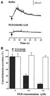
Fig. 4. Catalytically inactive PLD1(K898R) but not catalytically inactive PLD2(K758R) inhibits nicotine-evoked catecholamine secretion from single chromaffin cells. Chromaffin cells were microinjected with buffer, PLD1(K898R) or PLD2(K758R). The concentration of recombinant proteins in the pipet was 0.2 or 1 µM. Cells were stimulated 10–15 min later by a local application of 100 µM nicotine for 5 s (arrow). Catecholamine secretion was recorded with a carbon fiber electrode. (A) Representative amperometric responses. (B) Total catecholamine secretion expressed in pA/s. Results are expressed relative to the secretory response obtained for the control cells. Data are the means of 25 cells/group from the same dish ± SEM. Similar results were obtained in four independent experiments performed on two culture preparations and with different batches of recombinant proteins.
None
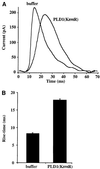
Fig. 5. Catalytically inactive PLD1(K898R) increases the rising phase of individual amperometric spikes. (A) Example spikes representing the average spikes in control buffer-injected chromaffin cells and PLD1(K898R)-injected cells on an expanded time base. (B) Mean values (±SEM) for the rise time of amperometric spikes from control (n = 285 spikes) and PLD1(K898R)-injected (n = 203 spikes) cells.
None
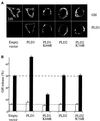
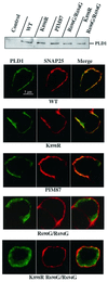
Fig. 6. PtdIns(4,5)P2 interaction targets PLD1 to sites of regulated exocytosis in PC12 cells. PC12 cells cultured on glass coverslips were co-transfected with the GH expression plasmid in combination with wild-type PLD1, PLD1(K898R), PIM87, PLD1(R690G/R694G) or PLD1(K898R/R690G/R694G). At 48 h post-transfection, the proteins expressed were detected by western blots and their intracellular distribution visualized by immunocytochemistry. Confocal fluorescent images were taken through the center of the nucleus. Images obtained in the Cy2 (green; PLD1) and Cy3 (red; SNAP-25) channels were recorded simultaneously in the same optical section by a double exposure procedure. The yellow–orange staining indicates areas of co-localization. Other than PLD1(R690G/R694G) and PLD1(K898R/R690G/R694G), the wild-type and mutated PLD1 proteins co-localized with SNAP-25 at the plasma membrane.
None
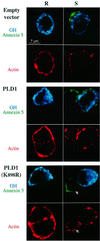
Fig. 7. Overexpression of catalytically inactive PLD1(K898R) blocks vesicular fusion but not the actin cytoskeletal reorganization required for regulated exocytosis. PC12 cells were transfected with either pCGN (empty vector), pCGN-PLD1 or pCGN-PLD1(K898R) in combination with the GH expression plasmid (4 µg/well of each plasmid). At 48 h post-transfection, cells were washed and then incubated for 10 min in either Locke’s solution (resting cells) or elevated K+ (stimulated cells) in the presence of Alexa 488-conjugated annexin 5 to reveal the exocytotic activity (green). Cells subsequently were fixed and stained with rabbit anti-GH and secondary Cy5-labeled anti-rabbit antibodies to identify transfected cells (blue), and with rhodamine-conjugated phalloidin to visualize actin filaments (red). Stimulation with high K+ triggers a partial disruption of the cortical actin ring in control, PLD1- and PLD1(K898R)-expressing PC12 cells. Exocytotic patches at the cell surface were observed only in control and PLD1-expressing cells. Overexpression of the inactive PLD1(K898R) inhibited the exocytotic response without affecting the disassembly of the subplasmalemmal actin. Arrows point to a non-transfected cell undergoing actin depolymerization and displaying exocytotic patches in response to elevated K+, while the adjacent transfected cell expressing GH and PLD1(K898R) exhibits no exocytotic patches. Similar patterns of inhibition mediated by PLD1(K898R) but not wild-type PLD1 were observed in three separate experiments.

Similar articles

Cited by

References

    1. Abousalham A., Liossis,C., O’Brien,L. and Brindley,D.N. (1997) Cell-permeable ceramides prevent the activation of phospholipase D by ADP-ribosylation factor and RhoA. J. Biol. Chem., 272, 1069–1075. - PubMed
    1. Albillos A., Dernick,G., Horstmann,H., Almers,W., Alvarez de Toledo, G. and Lindau,M. (1997) The exocytotic event in chromaffin cells revealed by patch amperometry. Nature, 389, 509–512. - PubMed
    1. Ashery U., Varoqueaux,F., Voets,T., Betz,A., Thakur,P., Koch,H., Neher,E., Brose,N. and Rettig,J. (2000) Munc13-1 acts as a priming factor for large dense-core vesicles in bovine chromaffin cells. EMBO J., 19, 3586–3596. - PMC - PubMed
    1. Bi K., Roth,M.G. and Ktistakis,N.T. (1997) Phosphatidic acid formation by phospholipase D is required for transport from the endoplasmic reticulum to the Golgi complex. Curr. Biol., 7, 301–307. - PubMed
    1. Brown F.D., Thompson,N., Saqib,K., Clark,J.M., Powner,D., Thompson,N.T., Solari,R. and Wakelam,M.J.O. (1998) Phospholipase D1 localises to secretory granules and lysosomes and is plasma-membrane translocated on cellular stimulation. Curr. Biol., 8, 835–838. - PubMed

Publication types

MeSH terms