. 2001 May 15;20(10):2404-12.
doi: 10.1093/emboj/20.10.2404.

Regulation of glutamate dehydrogenase by reversible ADP-ribosylation in mitochondria

Affiliations

Regulation of glutamate dehydrogenase by reversible ADP-ribosylation in mitochondria

A Herrero-Yraola et al. EMBO J. .

Abstract

Mitochondrial ADP-ribosylation leads to modification of two proteins of approximately 26 and 53 kDA: The nature of these proteins and, hence, the physiological consequences of their modification have remained unknown. Here, a 55 kDa protein, glutamate dehydrogenase (GDH), was established as a specific acceptor for enzymatic, cysteine-specific ADP-ribosylation in mitochondria. The modified protein was isolated from the mitochondrial preparation and identified as GDH by N-terminal sequencing and mass spectrometric analyses of tryptic digests. Incubation of human hepatoma cells with [14C]adenine demonstrated the occurrence of the modification in vivo. Purified GDH was ADP-ribosylated in a cysteine residue in the presence of the mitochondrial activity that transferred the ADP-ribose from NAD+ onto the acceptor site. ADP- ribosylation of GDH led to substantial inhibition of its catalytic activity. The stoichiometry between incorporated ADP-ribose and GDH subunits suggests that modification of one subunit per catalytically active homohexamer causes the inactivation of the enzyme. Isolated, ADP-ribosylated GDH was reactivated by an Mg2+-dependent mitochondrial ADP-ribosylcysteine hydrolase. GDH, a highly regulated enzyme, is the first mitochondrial protein identified whose activity may be modulated by ADP-ribosylation.


Figures
None
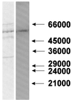
Fig. 1. Separation of the ADP-ribosylated ∼53 kDa protein. Specific modification of solubilized mitochondrial proteins in the presence of 100 µM [32P]NAD+ (0.6 µCi/nmol) was performed. After incubation, the suspension was separated in a non-denaturing polyacrylamide gel. The band containing the majority of the radioactive label was excised from the gel and subjected to SDS–PAGE. The proteins were then blotted onto a PVDF membrane and subsequently revealed by staining with Coomassie Blue (left panel). The right panel shows the auto radiogram of the blot. Numbers (in daltons) on the right indicate the mobility of marker proteins.
None
Fig. 2. Mass spectrometric analysis of a tryptic digest obtained from the ADP-ribosylated ∼53 kDa mitochondrial protein. The incubation and separation of the modified protein were conducted as described in the legend to Figure 1. Following SDS–PAGE, the protein band containing the radioactive label was subjected to ‘in-gel’ digestion with trypsin. The mass spectrum of the resulting peptides was obtained by MALDI-TOF spectrometry. Only masses corresponding to internal peptides of GDH are indicated, assuming a maximum of one missed cleavage site per peptide. The asterisks indicate peaks corresponding to GDH peptide masses, considering more than one missed cleavage site or oxidized methionine residues.
None
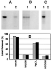
Fig. 3. Cysteine-specific ADP-ribosylation of purified bovine liver GDH and its co-migration with the modified mitochondrial ∼53 kDa protein. (A) Purified bovine liver GDH (10 µg) was ADP-ribosylated in the presence of the mitochondrial preparation (10 µg) and 100 µM [32P]NAD+ (1 µCi) for 30 min (lane 1). For lane 2, 100 µg of the mitochondrial preparation were treated as described for lane 1, except that no GDH was added. (B) The experiment was conducted as described for (A), lane 1, except that instead of [32P]NAD+, [14C]NAD+ labeled in either the adenine (lane 1) or the nicotinamide moiety (lane 2) was used (0.25 µCi each). (C) The sample used for lane 1 was prepared as described for (A), lane 1. For lane 2, purified GDH was subjected to a similar incubation with [32P]NAD+ in the absence of the mitochondrial suspension. Following the incubations, samples were precipitated with acetone and the pellets subjected to SDS–PAGE. The autoradiograms of the gels are shown. (D) Specific [32P]ADP-ribosylation of mitochondrial proteins or purified GDH was conducted as described in Materials and methods. Proteins of several identical samples were separated by SDS–PAGE and subsequently blotted onto PVDF membranes. The blots were washed twice in a solution containing 50 mM MOPS–KOH pH 7.0, 100 mM NaCl and 1 mM EDTA for 15 min. The regions of the ∼53 kDa protein (gray bars) or purified GDH (black bars) were excised and the incorporated ADP-ribose determined by Cerenkov counting. The individual pieces of the blots were then subjected to treatment at 37°C for 2 h with the reagents indicated below. Thereafter, the pieces were washed twice as detailed above and the remaining radioactivity determined. Subsequent staining with Coomassie Blue gave no indication that the amount of protein adsorbed to the blot membranes was affected by any of these treatments. The additions indicated in the figure were 10 mM HgCl2 (in wash buffer without EDTA), 2 M NH2OH (adjusted to pH 7.0) in wash buffer, 0.2 M NaOH plus 1 mM EDTA, or 44% HCOOH. Note that NaOH releases bound ADP-ribose non-specifically from modified amino acids. The data represent the average of two independent experiments.
None
Fig. 4. Inhibition of GDH activity by ADP-ribosylation. Mitochondrial proteins or purified GDH were ADP-ribosylated in the presence of 100 µM NAD+. Samples containing 0.25 µg of GDH (squares) or ∼15 µg of mitochondrial protein (triangles) were withdrawn at the times indicated and their GDH activity determined. The initial specific activity of the GDH (100%) was 186 µmol/min/mg. The uninhibited activity of the solubilized mitochondrial preparation was ∼1.7 µmol/min/mg. In the absence of added NAD+, the GDH activity decreased only by ∼5–8% after 2 h. The inset shows the modification with ADP-ribose of endogenous GDH during a parallel experiment conducted in the presence of [32P]NAD+. Aliquots were withdrawn at the times indicated (in minutes) and precipitated with acetone. The autoradiogram of the SDS–PAGE of the samples is presented. The data represent five independent experiments.
None
Fig. 5. Correlation of inhibition and ADP-ribosylation of GDH. Purified GDH was inhibited by ADP-ribosylation to various extents by incubating the enzyme for different time intervals (0–60 min) in the presence of [32P]NAD+ and mitochondrial preparation (cf. Figure 4). Parallel samples (20 µl) were withdrawn and used for analysis of enzyme activity or subjected to gel filtration by HPLC. Prior to gel filtration, the samples were made 5 mM in unlabeled NAD+ to minimize co-migration with GDH of non-covalently bound radioactive NAD+. The eluting fractions containing GDH were pooled and both their protein content and bound radioactivity determined. From these data, the stoichiometry of ADP-ribose per enzyme hexamer was calculated assuming a subunit molecular mass of 55 561 Da. The data were corrected for the values obtained using mitochondria in the absence of added GDH or purified GDH without mitochondria. The straight line indicates the result of a linear regression analysis of the data (R2 = 0.92). The values represent two independent experiments using two different mitochondrial preparations.
None
Fig. 6. Pyridine nucleotides prevent ADP-ribosylation of GDH. ADP-ribosylation assays were conducted under standard conditions in the presence of 25 µM [32P]NAD+, 10 µg of GDH, 10 µg of the mitochondrial suspension and the nucleotide (1 mM) indicated in the figure. Incubations were stopped after 15 min and the samples processed for SDS–PAGE. The autoradiogram of the gel is shown.
None
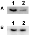
Fig. 7. ADP-ribosylation of GDH in vivo. Hep-G2 cells were grown in the presence of [14C]adenine and extracts immunoprecipitated with purified rabbit anti-GDH IgG. (A) The growth medium contained either 2 mM glutamate (lane 1), 1 mM glutamate and 1 mM glutamine (lane 2), or 2 mM glutamine (lane 3). Immunoprecipitated proteins were separated by 12% SDS–PAGE and their modification revealed by autoradiography. The film was exposed for 25 days. The arrow indicates the position at which GDH migrated. (B) Immunoblot of Hep-G2 cells (lane 1) and bovine liver mitochondria (lane 2) developed with the purified anti-GDH IgG used for the immunoprecipitation experiment shown in (A).
None
Fig. 8. Reversal of ADP-ribosylation and concomitant reactivation of GDH catalyzed by a mitochondrial hydrolase activity. (A and B) Purified GDH was [32P]ADP-ribosylated and then freed of non-covalently bound nucleotide by HPLC as described in the legend to Figure 5. The modified enzyme (10 µg) was then incubated in the presence of 5 mM MgCl2 (lanes 1), 5 mM MgCl2 and 10 µg of the mitochondrial preparation (lanes 2), or 10 µg of the mitochondrial preparation alone (lanes 3). After 30 min at 30°C, the protein was subjected to SDS–PAGE and the remaining modification visualized by autoradiography (A). The liberated nucleotides were analyzed by thin-layer chromatography (B). The autoradiogram is shown. On the right, the migration of standard compounds is indicated (ADPR, ADP-ribose). A control sample (no addition during incubation) was indistinguishable from the sample represented by lanes 1. (C) Purified GDH or endogenous GDH of the mitochondrial preparation was ADP-ribosylated to achieve ∼50% inhibition by incubating mitochondria and purified GDH (squares and circles) or mitochondria alone (triangles) in the presence of 100 µM NAD+ for ∼45 min (cf. Figure 4). The sample represented by the circles was then centrifuged to remove the added mitochondria. Thereafter (time zero in this figure), 5 mM MgCl2 was added to all samples and incubation continued. At the times indicated, aliquots received 10 mM EDTA and their GDH activity was determined. The arrow indicates re-addition of mitochondria (3 µg per µg of GDH, after 30 min of incubation) to the sample previously freed of mitochondria. The values given are related to the activities of purified or endogenous GDH, respectively, prior to the incubation with NAD+. The data represent 2–4 independent experiments using two different mitochondrial preparations.
None
Fig. 9. Specificity of the mitochondrial hydrolase activity for ADP-ribosylcysteine. (A) A 5 µg aliquot of Gαi1 (Calbiochem) was ADP-ribosylated in a cysteine residue by 0.5 µg of activated pertussis toxin (Sigma) with 25 µM [32P]NAD+ for 30 min in a medium consisting of 50 mM MOPS–KOH pH 7.5, 10 mM DTT, 1 mM EDTA, 1 mM ATP and 1 mM GTP. Thereafter, 10 mM MgCl2 was added and incubation continued for 20 min in the absence (lane 1) or presence (lane 2) of 20 µg of the mitochondrial preparation. (B) FLAG-tagged arginine-specific ADP-ribosyltransferase (recombinant mouse RT6.2; kind gift of Dr Koch-Nolte, Hamburg, Germany) was immobilized on M2-Sepharose (Kodak/IBI) carrying FLAG-specific antibodies. The complex (20 µl of the resin) was incubated with 25 µM [32P]NAD+ for 30 min in a medium consisting of 10 mM potassium phosphate pH 7.0 and 150 mM NaCl. Under these conditions, the light chain of the antibody is ADP-ribosylated (Koch-Nolte et al., 1996). Thereafter, the resin was washed twice with 50 mM MOPS–KOH pH 7.5, 10 mM DTT, 1 mM EDTA. Incubation was then continued for 20 min in this medium containing in addition 10 mM MgCl2 in the absence (lane 1) or presence (lane 2) of 20 µg of the mitochondrial preparation. All samples were then subjected to SDS–PAGE and subsequent autoradiography.
None
Fig. 10. Schematic representation of the mitochondrial ADP-ribosylation cycle regulating GDH activity. Metabolic pathways that may be affected by the inhibition of GDH are indicated. mART, mitochondrial ADP-ribosyl transferase.

Similar articles

Cited by

References

    1. Aktories K. (1997) Rho proteins: targets for bacterial toxins. Trends Microbiol., 5, 282–288. - PubMed
    1. Boyer P.D. (1997) The ATP synthase—a splendid molecular machine. Annu. Rev. Biochem., 66, 717–749. - PubMed
    1. Cervantes-Laurean D., Minter,D.E., Jacobson,E.L. and Jacobson,M.K. (1993) Protein glycation by ADP-ribose: studies of model conjugates. Biochemistry, 32, 1528–1534. - PubMed
    1. Cervantes-Laurean D., Loflin,P.T., Minter,D.E., Jacobson,E.L. and Jacobson,M.K. (1995) Protein modification by ADP-ribose via acid-labile linkages. J. Biol. Chem., 270, 7929–7936. - PubMed
    1. Chaurand P., Luetzenkirchen,F. and Spengler,B. (1999) Peptide and protein identification by matrix-assisted laser desorption ionization (MALDI) and MALDI-post-source decay time-of-flight mass spectrometry. J. Am. Soc. Mass Spectrom., 10, 91–103. - PubMed

Publication types