. 2001 Jun;21(11):3789-806.
doi: 10.1128/MCB.21.11.3789-3806.2001.

c-Myc is a critical target for c/EBPalpha in granulopoiesis

Affiliations

c-Myc is a critical target for c/EBPalpha in granulopoiesis

L M Johansen et al. Mol Cell Biol. 2001 Jun.

Abstract

CCAAT/enhancer binding protein alpha (C/EBPalpha) is an integral factor in the granulocytic developmental pathway, as myeloblasts from C/EBPalpha-null mice exhibit an early block in differentiation. Since mice deficient for known C/EBPalpha target genes do not exhibit the same block in granulocyte maturation, we sought to identify additional C/EBPalpha target genes essential for myeloid cell development. To identify such genes, we used both representational difference analysis and oligonucleotide array analysis with RNA derived from a C/EBPalpha-inducible myeloid cell line. From each of these independent screens, we identified c-Myc as a C/EBPalpha negatively regulated gene. We mapped an E2F binding site in the c-Myc promoter as the cis-acting element critical for C/EBPalpha negative regulation. The identification of c-Myc as a C/EBPalpha target gene is intriguing, as it has been previously shown that down-regulation of c-Myc can induce myeloid differentiation. Here we show that stable expression of c-Myc from an exogenous promoter not responsive to C/EBPalpha-mediated down-regulation forces myeloblasts to remain in an undifferentiated state. Therefore, C/EBPalpha negative regulation of c-Myc is critical for allowing early myeloid precursors to enter a differentiation pathway. This is the first report to demonstrate that C/EBPalpha directly affects the level of c-Myc expression and, thus, the decision of myeloid blasts to enter into the granulocytic differentiation pathway.


Figures
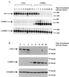
FIG. 1
FIG. 1
(A) The level of c-Myc RNA decreases following the induction of C/EBPα gene expression. U937 stable cell lines that contain either a rat C/EBPα cDNA under the control of the human metallothionein promoter (U937α#2) or the empty metallothionein expression vector alone were harvested for total RNA at the indicated time points following the addition of ZnSO4 to the culture medium. (Top) Northern hybridization with a c-Myc cDNA probe. (Middle) Northern hybridization with a C/EBPα cDNA probe. (Bottom) The Northern blot was stripped and rehybridized with a glyceraldehyde-3-phosphate dehydrogenase (GAPDH) cDNA probe to control for RNA loading and integrity. (B) The expression of c-Myc protein decreases as the level of C/EBPα protein increases. The U937α#2 stable line was harvested for cell lysates for Western analysis at the indicated time points following incubation with ZnSO4. (Top) Western blot hybridized with c-Myc antiserum. (Middle) The same Western blot hybridized with C/EBPα antiserum. (Bottom) The same Western blot hybridized with β-tubulin antibody to control for protein loading and integrity.
FIG. 2
FIG. 2
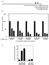
C/EBPα down-regulates c-Myc promoter activity. (A) The 6.5-kb c-Myc promoter and indicated 5′ deletions were cloned into the pXP2 luciferase reporter vector. (B) CV-1 cells were cotransfected with 200 ng of the indicated reporter gene and increasing amounts of C/EBPα expression plasmid (nanograms). Control transfection experiments indicated that C/EBPα had no effect on the pXP2 luciferase reporter vector (data not shown). (C) As a positive control for C/EBPα transactivation, CV-1 cells were cotransfected with the G-CSF receptor reporter gene containing four C/EBPα binding sites (pTK-G-CSFr). All transfection groups were normalized with a Renilla luciferase vector as an internal control. Results represent the percentages of luciferase activity with 0 ng of C/EBPα (vector alone) set to 100% activity. Results are given as the averages of at least three independent experiments, and error bars represent the standard errors of the means.
FIG. 3
FIG. 3
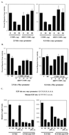
(A) Dominant-negative C/EBPα does not repress c-Myc reporter activity. CV-1 cells were cotransfected with 200 ng of the indicated c-Myc reporter construct along with wild-type C/EBPα or dominant-negative C/EBPα (4HEP C/EBPα). (B) Dominant-negative C/EBPα interferes with wild-type C/EBPα repression of c-Myc reporter activity. CV-1 cells were cotransfected with wild-type C/EBPα along with increasing amounts of dominant-negative C/EBPα and either the 2.5-kb or 0.14-kb c-Myc reporter gene. All transfection groups were cotransfected with a Renilla luciferase vector as an internal control. Results represent the percentages of luciferase activity with 0 ng of C/EBPα (vector alone) set to 100% activity. (C) Mutation of the E2F DNA binding site on c-Myc reporter constructs abolishes C/EBPα negative regulation. Wild-type and mutant sequences of the E2F DNA binding site in the c-Myc promoter located at residues −58 to −51 relative to the P2 promoter are shown at the top. CV-1 cells were cotransfected with either the 2.5-kb or 0.14-kb c-Myc reporter gene containing the wild-type or mutated E2F site along with the C/EBPα expression construct. As a control, CV-1 cells were cotransfected with a PU.1 expression construct to demonstrate that c-Myc promoter repression is specific to C/EBPα. All transfection groups were normalized with a Renilla luciferase vector as an internal control. Results are presented as the averages of at least three independent experiments, and error bars represent the standard errors of the means.
FIG. 4
FIG. 4
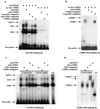
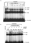
C/EBPα does not bind to the E2F DNA site in the c-Myc promoter. EMSAs using 32P-labeled, double-stranded oligonucleotides containing either an E2F site from the human c-Myc promoter (A and C) or a C/EBPα binding site from the G-CSF receptor promoter (B and D) were performed with in vitro-translated C/EBPα protein (in vitro C/EBPα) or nuclear extracts prepared from COS7 cells overexpressing C/EBPα (COS7/C/EBPα), COS7 cells overexpressing E2F1 (COS7/E2F1) as a positive control for E2F binding, or untransfected COS7 cells (UT). The migration of the free probe is indicated along with the positions of E2F1 protein complexes binding to the E2F DNA site and C/EBPα binding to the C/EBPα binding site. The asterisks indicate the positions of supershifted bands following the addition of either an E2F1 antibody or C/EBPα antisera. “competitor” refers to the 100× addition of unlabeled double-stranded E2F or C/EBPα DNA binding sites as indicated. “NSB” refers to migration of nonspecific protein complexes binding to the E2F DNA binding site. “Long” or “Short” E2F competitor refers to a double-stranded E2F oligonucleotide that contains more or less DNA sequence, respectively, flanking the E2F1 consensus site.
FIG. 5
FIG. 5
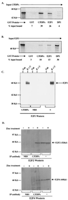
C/EBPα and E2F1 physically interact in vitro and in vivo. (A) Binding of 35S-labeled in vitro-translated C/EBPα (input C/EBPα) to GST (negative control for binding), GST-C/EBPα (positive control for binding), GST-E2F1, and GST-DP1. (B) Binding of 35S-labeled in vitro-translated E2F1 (input E2F1) to the same GST fusion proteins. Percent input bound represents the amount of in vitro-translated protein complexed with GST fusion proteins as calculated using a phosphorimager (Molecular Dynamics). (C) COS7 cells either untransfected (Unt) or transfected with E2F1 and C/EBPα expression vectors (E + C) were immunoprecipitated with C/EBPα antisera or control NRS followed by Western analysis with E2F1 antibody. As a control for E2F1 expression and migration, 1/30 of the COS7 lysate used for immunoprecipitation was resolved by SDS-PAGE (marked “−” for immunoprecipitation antibody). The position of E2F1 is indicated. (D) C/EBPα interacts with endogenous E2F proteins in myeloid cells. Uninduced (−) or induced (+) U937α#2 cells were immunoprecipitated (IP antibody) with C/EBPα antisera or control NRS followed by Western analysis with either E2F2 or E2F4 antibody. As a control for E2F2 and E2F4 expression and migration, 1/30 of the lysate used for immunoprecipitation was resolved by SDS-PAGE (marked “−” for immunoprecipitation antibody). The positions of E2F2 and E2F4 are indicated.
FIG. 6
FIG. 6
C/EBPα protein cannot disrupt the binding of E2F1 protein to DNA. (A) EMSAs using a 32P-labeled, double-stranded oligonucleotide containing the E2F site from the human c-Myc promoter were performed with nuclear extracts prepared from COS7 cells overexpressing E2F1 (COS7/E2F1). The addition of increasing amounts of in vitro-translated C/EBPα did not alter the amount of E2F1 protein binding to the E2F DNA site. The migration of the free probe is indicated along with the positions of E2F1 protein complexes. The asterisk indicates the position of a supershifted band following the addition of an E2F1 antibody. “self oligo” indicates the 100× addition of unlabeled double-stranded E2F DNA binding site. “NSB” indicates the migration of nonspecific protein complexes binding to the E2F DNA binding site. “control” indicates binding reactions performed with unprogrammed rabbit reticulocyte lysate, and “Gata-1” indicates control binding reactions performed with in vitro-translated GATA-1 protein. (B) EMSAs performed with the E2F site from the c-Myc promoter and nuclear extracts from COS7 cells cotransfected with 10 mg of E2F1 plasmid and indicated amounts of C/EBPα plasmid. For control reactions, COS7 cells were cotransfected with PU.1 or left untransfected (UT). The migration of the free probe is indicated along with the positions of the E2F1 protein complexes. “self oligo” indicates the 100× addition of unlabeled double-stranded E2F DNA binding site. “NSB” indicates the migration of nonspecific protein binding complexes.
FIG. 7
FIG. 7
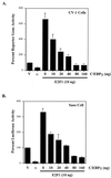
C/EBPα interferes with E2F1 transactivation of the c-Myc promoter. CV-1 cells or Saos (Rb) cells were cotransfected with 200 ng of the 0.14-kb c-Myc reporter gene (V), 20 ng of C/EBPα plasmid alone (α), 10 ng of E2F1 plasmid alone, or 10 ng of E2F1 plasmid along with the indicated amounts of C/EBPα plasmid. All transfection groups were normalized with a Renilla luciferase vector as an internal control. Results represent the percentages of reporter gene or luciferase activity with vector alone (V) set to 100% activity. Results are given as the averages of at least three independent experiments, and error bars represent the standard errors of the means.
FIG. 8
FIG. 8
Down-regulation of c-Myc is crucial to the granulocytic differentiation pathway. The 1137 cell line was derived from murine bone marrow of a transgenic line with a human c-Myc cDNA under the control of a tetracycline-responsive promoter. The addition of tetracycline to the culture medium turns off human c-Myc expression, resulting in the differentiation of these myeloblasts to neutrophils. (A) Wright-Giemsa-stained cells without (No Tet) or with (Plus Tet) treatment with tetracycline. Cells treated with tetracycline differentiated into myelocytes (M) and neutrophils (N). (B) Differential analysis of Wright-Giemsa-stained slides following treatment with tetracycline for 0, 24, and 48 h, respectively. (C) 1137 cells were harvested for cell lysates to use in Western blotting at indicated time points following treatment with tetracycline. (Top) Western blot hybridized with C/EBPα antiserum shows that the level of endogenous C/EBPα protein increased 24 h following treatment with tetracycline. This corresponds to the shift to mature cells seen in panels A and B. (Middle) The same blot hybridized with c-Myc antiserum, showing that c-Myc protein levels dramatically decreased 2 h following treatment with tetracycline. (Bottom) The same blot hybridized with a β-tubulin antibody to control for protein loading and integrity. (D) Western analysis of 1137 stable lines with (+) and without (−) treatment with zinc. “parental” indicates the 1137 parental line, “vector” indicates the 1137 stable line with metallothionein vector, and “C/EBPα ” indicates the 1137 line with metallothionein-driven C/EBPα. (E) Western analysis of cell lysates prepared from the 1137/C/EBPα stable line with the indicated treatment with tetracycline. The level of human c-Myc protein was titrated by 100-fold dilutions of tetracycline. A 20-ng/ml concentration turns off c-Myc expression, while a 2-ng/ml concentration results in a low level of c-Myc expression. Lower concentrations of tetracycline result in no decrease in c-Myc protein expression. (F) Differential analysis of Wright-Giemsa-stained slides following treatment of 1137 stable lines with tetracycline, zinc, or the combination of tetracycline and zinc for 48 h.
FIG. 8
FIG. 8
Down-regulation of c-Myc is crucial to the granulocytic differentiation pathway. The 1137 cell line was derived from murine bone marrow of a transgenic line with a human c-Myc cDNA under the control of a tetracycline-responsive promoter. The addition of tetracycline to the culture medium turns off human c-Myc expression, resulting in the differentiation of these myeloblasts to neutrophils. (A) Wright-Giemsa-stained cells without (No Tet) or with (Plus Tet) treatment with tetracycline. Cells treated with tetracycline differentiated into myelocytes (M) and neutrophils (N). (B) Differential analysis of Wright-Giemsa-stained slides following treatment with tetracycline for 0, 24, and 48 h, respectively. (C) 1137 cells were harvested for cell lysates to use in Western blotting at indicated time points following treatment with tetracycline. (Top) Western blot hybridized with C/EBPα antiserum shows that the level of endogenous C/EBPα protein increased 24 h following treatment with tetracycline. This corresponds to the shift to mature cells seen in panels A and B. (Middle) The same blot hybridized with c-Myc antiserum, showing that c-Myc protein levels dramatically decreased 2 h following treatment with tetracycline. (Bottom) The same blot hybridized with a β-tubulin antibody to control for protein loading and integrity. (D) Western analysis of 1137 stable lines with (+) and without (−) treatment with zinc. “parental” indicates the 1137 parental line, “vector” indicates the 1137 stable line with metallothionein vector, and “C/EBPα ” indicates the 1137 line with metallothionein-driven C/EBPα. (E) Western analysis of cell lysates prepared from the 1137/C/EBPα stable line with the indicated treatment with tetracycline. The level of human c-Myc protein was titrated by 100-fold dilutions of tetracycline. A 20-ng/ml concentration turns off c-Myc expression, while a 2-ng/ml concentration results in a low level of c-Myc expression. Lower concentrations of tetracycline result in no decrease in c-Myc protein expression. (F) Differential analysis of Wright-Giemsa-stained slides following treatment of 1137 stable lines with tetracycline, zinc, or the combination of tetracycline and zinc for 48 h.

Similar articles

Cited by

References

    1. Anfossi G, Gewirtz A M, Calabretta B. An oligomer complementary to c-myb-encoded mRNA inhibits proliferation of human myeloid leukemia cell lines. Proc Natl Acad Sci USA. 1989;86:3379–3383. - PMC - PubMed
    1. Antonson P, Pray M G, Jacobsson A, Xanthopoulos K G. Myc inhibits CCAAT/enhancer-binding protein alpha-gene expression in HIB-1B hibernoma cells through interactions with the core promoter region. Eur J Biochem. 1995;232:397–403. - PubMed
    1. Battey J, Moulding C, Taub R, Murphy W, Stewart T, Potter H, Lenoir G, Leder P. The human c-myc oncogene: structural consequences of translocation into the IgH locus in Burkitt lymphoma. Cell. 1983;34:779–787. - PubMed
    1. Behre G, Smith L T, Tenen D G. Use of a promoterless Renilla luciferase vector as an internal control plasmid for transient co-transfection assays of Ras-mediated transcription activation. BioTechniques. 1999;26:24–28. - PubMed
    1. Bellon T, Perrotti D, Calabretta B. Granulocytic differentiation of normal hematopoietic precursor cells induced by transcription factor PU.1 correlates with negative regulation of the c-myb promoter. Blood. 1997;90:1828–1839. - PubMed

Publication types

MeSH terms

LinkOut - more resources