. 2001 Jun;21(11):3775-88.
doi: 10.1128/MCB.21.11.3775-3788.2001.

Mutations in the novel membrane protein spinster interfere with programmed cell death and cause neural degeneration in Drosophila melanogaster

Affiliations

Mutations in the novel membrane protein spinster interfere with programmed cell death and cause neural degeneration in Drosophila melanogaster

Y Nakano et al. Mol Cell Biol. 2001 Jun.

Abstract

Mutations in the spin gene are characterized by an extraordinarily strong rejection behavior of female flies in response to male courtship. They are also accompanied by decreases in the viability, adult life span, and oviposition rate of the flies. In spin mutants, some oocytes and adult neural cells undergo degeneration, which is preceded by reductions in programmed cell death of nurse cells in ovaries and of neurons in the pupal nervous system, respectively. The central nervous system (CNS) of spin mutant flies accumulates autofluorescent lipopigments with characteristics similar to those of lipofuscin. The spin locus generates at least five different transcripts, with only two of these being able to rescue the spin behavioral phenotype; each encodes a protein with multiple membrane-spanning domains that are expressed in both the surface glial cells in the CNS and the follicle cells in the ovaries. Orthologs of the spin gene have also been identified in a number of species from nematodes to humans. Analysis of the spin mutant will give us new insights into neurodegenerative diseases and aging.


Figures

FIG. 1
FIG. 1
Rejection behavior displayed by spin mutant females. (A) Typical repelling postures taken by spinP1 female flies. The pictures were selected from continuous videotape recordings. The picture illustrating extrusion was obtained using a wild-type fertilized female, as spinP1 females do not exhibit this behavior. (B) Relative contributions of different types of repelling actions compared among wild-type, spinP1, and spinP1/spinP2 females. The wild-type females were divided into three categories, 1-day-old virgin, 4-day-old virgin, and 4-day-old fertilized females. The females of spinP1, spinP1/spinP2, and spinR4 lines were 4 days old, and the males were all wild type. The behavior of single females was recorded on a video tape for 10 min after pairing, and the numbers of females exhibiting decamping (D), fending (Fe), flicking (F1), kicking (K), curling (C), spreading (S), extrusion (E), and punching (P) actions were counted and are shown as percentages with the respect to the total number of females observed. A value of 100% means that all of the females observed exhibited that action at least once during the recorded period, while 0% means that none of the females did so. The counts obtained from at least 20 females were summed and are illustrated as frequency histograms. CS, CS strain. (C) The percentage of mating success (dark columns) was decreased in spinP1 and spinP1/spinP2 females compared to that of CS wild-type and spinR4 females. The differences in levels of mating success between the wild-type and mutant (spinP1 and spinP1/spinP2) females were found to be statistically significant at a P of <0.001 using Student's t-test. No difference was detected between the four genotypes in the females' ability to elicit male courtship songs (SAPI, paler columns). The bars on the columns represent the standard errors of the mean values for SAPI. The numbers of flies observed in order to estimate the mating success were 114 (wild type), 62 (spinP1), 44 (spinP1/spinP2), and 44 (spinR4). The number of flies observed to calculate the SAPI was 20 for each of the three genotypes. Males were from the CS wild-type strain, and all flies were aged for 3 days following eclosion. (D) Longevity of adult flies. The percentage of survival after eclosion is plotted for wild-type (CS) females spinP1 females, and spinP1 males.
FIG. 2
FIG. 2
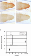
Reduced number of cells undergoing apoptosis in the spin mutant VNC. (A to D) VNCs at 6 h APF stained by the TUNEL method as viewed from the dorsal (A and C) or ventral (B and D) side. (A and B) EP822/CyO; (C and D) EP822/EP822. (E) Numbers of cells undergoing apoptosis at three different time points APF (4, 6, and 24 h) in spin heterozygotes (open squares) or homozygotes (filled squares). The mean and standard deviation of the mean are shown by a symbol and bar, respectively. The values for heterozygotes and homozygotes are significantly different at 6 h APF (P < 0.01 by Student's t test). At other time points, the difference between the values for heterozygotes and homozygotes is statistically insignificant (P > 0.05).
FIG. 3
FIG. 3
CNS abnormalities in spin mutant flies. (A) VNCs of the spinP1/spinP1 and spinP1/CyO flies 24 h after eclosion and staining with SPIF DNA stain. The spinP1 homozygous fly has a long abdominal ganglion. X, the length from the center of the third thoracic ganglion to the posterior edge of the abdominal ganglion; Y, the total length of the thoracic and abdominal ganglion. (B) Autofluorescence in the spinP1 mutant VNC. Tissues were fixed by 4% paraformaldehyde for 1 h, and the images were obtained by confocal microscopy using the blue excited light channel. The spin mutant CNS exhibits autofluorescence. (C) Expression of the spin gene and the distribution of the autofluorescent material. spin gene expression was examined in spinP1/spinP2 transheterozygotes. Only a portion of the autofluorescence overlaps with the expression of the spin gene. EM analysis of spinP1/CyO (Da) and spinP1/spinP1 (Db) cells in the abdominal ganglia 24 h after eclosion and spinP1/spinP1 cells at an early pupal stage (Dc). Nerve cells in the spinP1 homozygotes have lipofuscin-like materials inside them. Arrows indicate multilamellate bodies. Arrowheads indicate electron-dense lobulated granules. Scale bars, 2 μm.
FIG. 4
FIG. 4
Fluorescence spectra of spin mutant brain extract. Fluorescence excitation and emission spectra of the lipid extracts from the heads of spin flies (upper curves). The lower curves were obtained with the lipid extracts from the heads of wild-type flies. Ex, excitation spectrum; Em, emission spectrum.
FIG. 5
FIG. 5
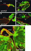
The spin gene is required for proper ovarian development. Confocal images of control (A and B) and spin (C to E) mutant egg chambers labeled with Texas Red-X phalloidin (red) and SPIF DNA stain (green). (A) Stage 12 (st 12) control egg chamber showing nurse cell nuclear accumulation at anterior and cytoplasmic actin bundles. (B) Stage 14 control egg chamber showing dorsal appendage (da) formation and no nurse cell nuclei. (C and D) Stage 14 EP822/EP822 and spinP1 mutants egg chambers. Nurse cell nuclei still remain (arrows), cytoplasmic actin bundles are now absent, but the dorsal appendages (da) are well formed. Some earlier-stage egg chambers are degraded (asterisks). (E) Mature ovaries in the spinP1 mutant. Nurse cell nuclei are accumulated at the basal stalk region (arrowheads).
FIG. 6
FIG. 6
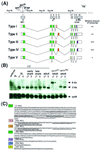
Molecular characterization of the spin gene and its products. (A) Genomic structure of the spin gene and the structures of five transcripts produced by alternative splicing. The restriction sites are shown for the EcoRI restriction enzyme. The P-element insertion site in the spinP1 mutant is located 8 bp downstream of the transcription initiation site and is indicated by a triangle. The enhancer trap vectors in the spinP2 and EP822 genes are inserted in the middle of exon 1. The relative amount of each type of transcript is represented as follows: +++, abundant; ++; modest; or +, rare. No obvious differences were found during development between the sexes or between wild-type and spinP1 mutant flies. The primers used for RT-PCR were primers A and B. (B) Developmental Northern blots probed by spin cDNA. Twenty microgram samples of poly(A)+ RNAs extracted from embryos, second-instar larvae, third-instar larvae, early pupae, late pupae, adults of the wild type, adults with the spinP1 mutation, and adults with the revertant spinR4 mutation were loaded onto the gel. The blots were then probed with the spin type I cDNA as well as the ribosomal protein gene rp49 cDNA in order to assess the amount of RNA loaded in each lane. (C) Amino acid sequences of each of the domains of Spin. C1 and C2 represent common domains. The V1, V2, and V3 domains are variable (see panel A). The predicted transmembrane domains are underlined.
FIG. 7
FIG. 7
Comparison of the spin gene products in various species. (A) Alignment of the nematode (C39E9.10, C13C4.5, and CEF09A5), fly (type I), mouse (Mspin1), and human (Hspin1) spin gene products. Sequences are aligned using MacDNAsis, and residues that are identical with fly Spin are highlighted. The predicted cyclic-AMP- and cyclic-GMP-dependent protein kinase phosphorylation sites are underlined. (B) Hydrophobicity profiles of fly (type I), nematode (C13C4.5), and human Spin proteins. Eight putative transmembrane domains are present.
FIG. 8
FIG. 8
Expression of the spin gene during development. (A) In situ hybridization of the spin mRNA probe to whole-mount wild-type embryos at stage 12 is shown in the first set of images. A dorsal view (top) and a dorsolateral view (middle) of an embryo are shown. The boxed region is illustrated at a higher magnification in the bottom image. Expression is evident in the developing VNC and brain. The three middle panels illustrate mRNA localization in the CNS of a third-instar larva (3L) (left), pupa (middle), and adult (right) of the CS wild type. The eye-antennal disks attached to the third-instar brain can also be seen. Expression in the larval ring gland is shown in the inset. The far-right images show mRNA expression in the spinP1 adult. The four CNS images show the dorsal side up. (B) The top three images show the double staining of the spinP2/CyO larval brains for the spin reporter β-Gal (green) and the Repo protein (red). Colocalization of β-Gal and the Repo protein results in a yellow signal. The bottom three images show the double staining of the spinP2/CyO pupal brains for spin reporter β-Gal (green) and the Elav protein (red). The spin gene is not expressed in the neurons. α, antibody. (C) The left two images show β-Gal expression in spinP2/CyO ovaries. The right two images show the in situ hybridization analysis of spin gene expression in ovaries at stage 10A (st10A) and st12. The spin gene is expressed in the follicle cells at st10B and st12.

Similar articles

Cited by

References

    1. Altschul S F, Gish W, Miller W, Myers E W, Lipman D J. Basic local alignment search tool. J Mol Biol. 1990;215:403–410. - PubMed
    1. Becker L E, Prior T W, Yates A J. Metabolic disease. In: Davis R L, Robertson D M, editors. Textbook of neuropathology. Baltimore, Md: The Williams & Willkins Co.; 1997. pp. 407–509.
    1. Brand A H, Perrimon N. Targeted gene expression as a means of altering cell fates and generating dominant phenotypes. Development. 1993;118:401–415. - PubMed
    1. Buchanan R L, Benzer S. Defective glia in the Drosophila brain degeneration mutant drop-dead. Neuron. 1993;10:839–850. - PubMed
    1. Buege J A, Aust S D. Microsomal lipid peroxidation. Methods Enzymol. 1978;52:302–310. - PubMed

Publication types

MeSH terms