. 2001 May;21(10):3325-35.
doi: 10.1128/MCB.21.10.3325-3335.2001.

Transcriptional repression by RB-E2F and regulation of anchorage-independent survival

Affiliations

Transcriptional repression by RB-E2F and regulation of anchorage-independent survival

J T Yu et al. Mol Cell Biol. 2001 May.

Abstract

Mutations that lead to anchorage-independent survival are a hallmark of tumor cells. Adhesion of integrin receptors to extracellular matrix activates a survival signaling pathway in epithelial cells where Akt phosphorylates and blocks the activity of proapoptotic proteins such as the BCL2 family member Bad, the forkhead transcription factor FKHRL-1, and caspase 9. Insulin-like growth factor 1 (IGF-1) is a well-established epithelial cell survival factor that also triggers activation of Akt and can maintain Akt activity after cells lose matrix contact. It is not until IGF-1 expression diminishes (~16 h after loss of matrix contact) that epithelial cells deprived of matrix contact undergo apoptosis. This suggests that IGF-1 expression is linked to cell adhesion and that it is the loss of IGF-1 which dictates the onset of apoptosis after cells lose matrix contact. Here, we examine the linkage between cell adhesion and IGF-1 expression. While IGF-1 is able to maintain Akt activity and phosphorylation of proapoptotic proteins in cells that have lost matrix contact, Akt is not able to phosphorylate and inactivate another of its substrates, glycogen synthase kinase 3beta (GSK-3beta), under these conditions. The reason for this appears to be a rapid translocation of active Akt away from GSK-3beta when cells lose matrix contact. One target of GSK-3beta is cyclin D, which is turned over in response to this phosphorylation. Therefore, cyclin D is rapidly lost when cells are deprived of matrix contact, leading to a loss of cyclin-dependent kinase 4 activity and accumulation of hypophosphorylated, active Rb. This facilitates assembly of a repressor complex containing histone deacetylase (HDAC), Rb, and E2F that blocks transcription of the gene for IGF-1, leading to loss of Akt activity, accumulation of active proapoptotic proteins, and apoptosis. This feedback loop containing GSK-3beta, cyclin D, HDAC-Rb-E2F, and IGF-1 then determines how long Akt will remain active after cells lose matrix contact, and thus it serves to regulate the onset of apoptosis in such cells.


Figures
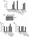
FIG. 1
FIG. 1
Regulation of apoptosis in cells deprived of matrix contact. (A) LNCaP prostate epithelial cells either adherent to or detached from the matrix (Det.) were transfected with the indicated expression vectors and/or treated with the indicated inhibitors (PD is the MEK inhibitor PD98059; LY is the PI-3K inhibitor LY294002). Cells were detached for 24 h unless otherwise indicated. Apoptosis was monitored by a TUNEL assay and by trypan blue exclusion. Apoptosis was further confirmed in cells by DNA laddering and electron microscopy. Similar results were seen in primary cultures of HUVEC, primary human tracheal epithelial cells, and MCF10A mammary epithelial cells. (B) Caspase 3 is cleaved to an active form when cells are deprived of matrix contact. A Western blot is shown for caspase 3 at different time points after LNCaP cells were detached from the matrix. The arrow indicates the cleaved, active form of the protein. (C) LNCaP prostate epithelial cells were cotransfected with the indicated expression vectors along with the pSV-luciferase reporter. Retention of luciferase activity was used as a measure of cell viability in these assays as described previously (74). CD2-p110, Myr-Akt, and V12 Ras are constitutively active constructs; CD2-p110KD is a kinase-dead mutant control. The results shown are all representative of at least three independent experiments, each done in duplicate. (D) MCF10A cells were cotransfected with the indicated expression vectors along with the pCMV-luciferase reporter. E2F-DB is a dominant-negative E2F. Results are standardized with respect to an empty-vector control for V12 Ras and CD2-p110, a myristoylation mutant form of MyrAkt, and E2F-DB, an E132 binding mutant form of E2F-DB.
FIG. 2
FIG. 2
Phosphorylation of Akt and Bad is regulated by cell adhesion and by IGF-1. (A) Western blot for phosphorylated, active Akt (pAkt) (Thr 308; similar results were seen with an antibody specific for Ser 473) in extracts from LNCaP cells either adherent, deprived of matrix contact (Det.) for 24 h with or without the addition of IGF-1 for the indicated times, or allowed to reattach for 6 h. The blot was reprobed with a pan-Akt antibody to detect total Akt. (B) Western blot analysis using antibodies specific for Bad phosphorylation on Ser 136 or Ser 112. LNCaP cells were detached for 12 h and then pretreated with LY294002 (LY) at 50 μM (where indicated) for 10 min prior to addition of IGF-1 (10−7 M). Bad (total) indicates that a pan-Bad antibody was used to detect total Bad protein. The same blot was reprobed with the three different antibodies. (C) Western blot for phosphorylated active Akt (pAkt) (Thr 308) in extracts from LNCaP cells deprived of matrix contact for the indicated times. The blot was reprobed with a pan-Akt antibody to detect total Akt. (D) Western blot for phosphorylated Akt (pAkt) in extracts from MCF10A cells deprived of matrix contact for the indicated times. The blot was reprobed with a pan-Akt antibody to detect total Akt. (E) Western blot for PTEN in adherent LNCaP cell: HUVEC, and MCF10A cell extracts.
FIG. 3
FIG. 3
IGF-1 regulates the onset of apoptosis in cells deprived of matrix contact. (A) MCF10A mammary epithelial cells were detached from the matrix for 48 h; where indicated, the cells were detached in the presence of 10−7 M IGF-1 or insulin with or without 50 μM LY294002 (LY). Apoptosis was analyzed by trypan blue exclusion. The results shown are representative of at least three independent experiments. (B) RT-PCR analysis for IGF-1 and glyceraldehyde-3-phosphate dehydrogenase (GAPDH) control mRNA. Wild-type (WT) LNCaP cells or a clone stably expressing cyclin D1 (clone 3 in Fig. 8A; similar results were seen with clone 5) were detached from the matrix for the indicated periods of time, and cellular RNA was analyzed by RT-PCR. (C) HUVEC were treated with the insulin receptor inhibitor HNMPA(AM)3, and the effect of the inhibitor on apoptosis of cells deprived of matrix contact was analyzed by trypan blue exclusion. Similar results were seen with LNCaP cells. (D) Western blot of phosphorylated active Akt (pAkt) (Thr 308) in LNCaP cells treated with the insulin receptor inhibitor. The blot was reprobed with a pan-Akt antibody to detect total Akt.
FIG. 4
FIG. 4
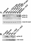
Cell adhesion regulates the subcellular localization of Akt and its ability to phosphorylate GSK-3β. (A) LNCaP cells were treated as described in the legend to Fig. 2A and Western blot assayed for phosphorylated GSK-3β (GSK-3α is also recognized by this phosphorylation-specific antibody against GSK-3β). The blot was reprobed with a pan-GSK-3β antibody to show total GSK-3β. (B) Immunoprecipitation kinase assay showing phosphorylation of GSK-3β. Total Akt was immunoprecipitated from LNCaP cells treated as indicated and used to phosphorylate GSK-3β purified from bacteria. (C to E) Immunofluorescent staining for active Akt (Thr 308) (red) and GSK-3β (green). Nuclear staining with 4′,6′-diamidino-2-phenylindole (DAPI) is shown in blue-purple. The merge is shown at the top of each panel. Panel C shows adherent cells, and panel D shows cells detached (Det.) from the matrix for 16 h and then treated for 15 min with IGF-1. Panel E is the same as panel D, but the cells were not stimulated with IGF-1 (the cells are also shown at a higher magnification). Similar results were seen when using an antibody specific for Akt phosphorylated on Ser 473. LY, LY294002.
FIG. 4
FIG. 4
Cell adhesion regulates the subcellular localization of Akt and its ability to phosphorylate GSK-3β. (A) LNCaP cells were treated as described in the legend to Fig. 2A and Western blot assayed for phosphorylated GSK-3β (GSK-3α is also recognized by this phosphorylation-specific antibody against GSK-3β). The blot was reprobed with a pan-GSK-3β antibody to show total GSK-3β. (B) Immunoprecipitation kinase assay showing phosphorylation of GSK-3β. Total Akt was immunoprecipitated from LNCaP cells treated as indicated and used to phosphorylate GSK-3β purified from bacteria. (C to E) Immunofluorescent staining for active Akt (Thr 308) (red) and GSK-3β (green). Nuclear staining with 4′,6′-diamidino-2-phenylindole (DAPI) is shown in blue-purple. The merge is shown at the top of each panel. Panel C shows adherent cells, and panel D shows cells detached (Det.) from the matrix for 16 h and then treated for 15 min with IGF-1. Panel E is the same as panel D, but the cells were not stimulated with IGF-1 (the cells are also shown at a higher magnification). Similar results were seen when using an antibody specific for Akt phosphorylated on Ser 473. LY, LY294002.
FIG. 5
FIG. 5
Loss of G1 cyclin expression, induction of the cdk2 inhibitor p27, and loss of cdk4 and cdk2 activity in cells deprived of matrix contact. (A and B) Western analysis for cyclin D1 and p27 in MCF10A cells deprived of matrix contact (det.) for increasing lengths of time or allowed to reattach. (C) Western analysis for cyclin D, cyclin E, and p27 in LNCaP cells deprived of matrix contact or treated with LY294002 (LY) or PD98059 (PD). (D) Cyclin D1 mRNA levels decay more slowly than cyclin D1 protein when cells are deprived of matrix contact or treated with LY294002. Cyclin D1 and control glyceraldehyde-3-phosphate dehydrogenase (GAPDH) mRNA from MCF10A cells were analyzed by RT-PCR. (E) Anchorage is required for IGF-1 to maintain cyclin D1 expression. A Western blot for cyclin D1 is shown examining MCF10A cells detached for 48 h or detached in the presence of 10−7 M IGF-1, similar to Fig. 3A. (F and G) cdk4 and cdk2 activities are lost in cells deprived of matrix contact, but stable expression of cyclin D1 overcomes the loss of cdk4 activity. cdk4 and cdk2 were immunoprecipitated from wild-type (WT) or cyclin D1-expressing (clone 3 in Fig. 8A; similar results were seen with clone 5) adherent LNCaP cells or cells placed in suspension for 6 h. Phosphorylation of the Rb C-terminal region (Rb-C; amino acids 792 to 928) or histone H1 was used to assess kinase activity. Western blots for total cdk4 or cdk2 are shown.
FIG. 6
FIG. 6
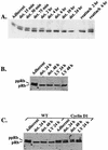
Hypophosphorylated (active) Rb accumulates in cells deprived of matrix contact. (A to C) Western blot showing that hypophosphorylated Rb (pRb indicates hypophosphorylated Rb; ppRb indicates hyperphosphorylated Rb) accumulates in MCF10A cells (A) and LNCaP cells (B and C) deprived of matrix contact (det.) or treated with LY294002 (LY; similar results were seen in HUVEC and human tracheal epithelial cells) and that stable expression of cyclin D1 prevents this accumulation. WT, wild-type cells. Cyclin D1, clone 3 in Fig. 8A (similar results were also seen with clone 5).
FIG. 7
FIG. 7
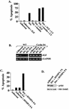
Transcriptional repression by HDAC-Rb-E2F. (A) Maintenance of cyclin D1 or expression of E2F-DB prevents apoptosis in cells deprived of matrix contact. Percent apoptosis was determined by trypan blue exclusion and confirmed by TUNEL reaction. WT, wild-type cells. Cyclin D1, clone 3 in Fig. 8A (similar results were seen with clone 5). The data shown are representative of at least three independent experiments. (B) Western blot of FL5.12 clone stably expressing E2F-DB. (C) Withdrawal of IL-3 results in apoptosis at the indicated times in both wild-type (open bars) and E2F-DB-expressing (closed bars) FL5.12 cells. Percent apoptosis was determined by trypan blue exclusion. The data shown are representative of at least three independent experiments. (D) Adherent and detached HUVEC were treated with the HDAC inhibitor TSA (48), and apoptosis was evaluated by trypan blue exclusion. (E) Adherent HUVEC were treated with TNF-α with or without TSA for 48 h. Apoptosis was evaluated by trypan blue exclusion. (F) The indicated reporter constructs (2 μg) were transfected into 60-mm plates of U2OS cells. Where indicated, TSA was added 18 h after transfection. (G) IGF-1 CAT (2 μg) was cotransfected along with the indicated expression vector (2 μg for Rb, 0.5 μg for E2F-1) into 60-mm plates of U2OS cells. An empty vector was transfected into plates that did not receive Rb or the E2F-1 expression vector. Det., detached; LY, LY294002.
FIG. 8
FIG. 8
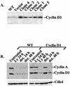
Stable expression of cyclin D1. (A) Western blot of LNCaP clones stably expressing cyclin D1. Clones 3 and 5 were used in the studies here with similar results. (B) Western blot analysis of cyclin D1, cyclin A, and cdk4 in wild-type (WT) cells or clone 3 (Cyclin D1). det., detached; LY, LY294002.
FIG. 9
FIG. 9
HDAC-Rb-E2F and the regulation of apoptosis in cells deprived of matrix contact. Adherent cells have active PI-3K and Akt, which inhibit proapoptotic effectors of Akt and prevent the formation of an active Rb repressor complex. When epithelial cells are deprived of matrix contact, Akt activity is lost; however, apoptosis does not ensue immediately. The onset of apoptosis does not occur until the Rb complex represses expression of IGF-1. This then dictates the timing of apoptosis in anchorage-deprived cells. See the text for details.

Similar articles

Cited by

References

    1. Alexander C M, Howard E W, Bissell M J, Werb Z. Rescue of mammary epithelial cell apoptosis and entactin degradation by a tissue inhibitor of metalloproteinases-1 transgene. J Cell Biol. 1996;135:1669–1677. - PMC - PubMed
    1. Andjelkovic M, Alessi D R, Meier R, Fernandez A, Lamb N J, Frech M, Cron P, Cohen P, Lucocq J M, Hemmings B A. Role of translocation in the activation and function of protein kinase B. J Biol Chem. 1997;272:31515–31524. - PubMed
    1. Aveyard J S, Skilleter A, Habuchi T, Knowles M A. Somatic mutation of PTEN in bladder carcinoma. Br J Cancer. 1999;80:904–908. - PMC - PubMed
    1. Baserga R, Sell C, Porcu P, Rubini M. The role of the IGF-1 receptor in the growth and transformation of mammalian cells. Cell Prolif. 1994;27:63–71. - PubMed
    1. Bellacosa A, de Feo D, Godwin A K, Bell D W, Cheng J Q, Altomare D A, Wan M, Dubeau L, Scambia G, Masciullo V, et al. Molecular alterations of the AKT2 oncogene in ovarian and breast carcinomas. Int J Cancer. 1995;64:280–285. - PubMed

Publication types