. 2001 Apr 16;153(2):367-80.
doi: 10.1083/jcb.153.2.367.

Regulation and localization of the Bloom syndrome protein in response to DNA damage

Affiliations

Regulation and localization of the Bloom syndrome protein in response to DNA damage

O Bischof et al. J Cell Biol. .

Abstract

Bloom syndrome (BS) is an autosomal recessive disorder characterized by a high incidence of cancer and genomic instability. BLM, the protein defective in BS, is a RecQ-like helicase, presumed to function in DNA replication, recombination, or repair. BLM localizes to promyelocytic leukemia protein (PML) nuclear bodies and is expressed during late S and G2. We show, in normal human cells, that the recombination/repair proteins hRAD51 and replication protein (RP)-A assembled with BLM into a fraction of PML bodies during late S/G2. Biochemical experiments suggested that BLM resides in a nuclear matrix-bound complex in which association with hRAD51 may be direct. DNA-damaging agents that cause double strand breaks and a G2 delay induced BLM by a p53- and ataxia-telangiectasia mutated independent mechanism. This induction depended on the G2 delay, because it failed to occur when G2 was prevented or bypassed. It coincided with the appearance of foci containing BLM, PML, hRAD51 and RP-A, which resembled ionizing radiation-induced foci. After radiation, foci containing BLM and PML formed at sites of single-stranded DNA and presumptive repair in normal cells, but not in cells with defective PML. Our findings suggest that BLM is part of a dynamic nuclear matrix-based complex that requires PML and functions during G2 in undamaged cells and recombinational repair after DNA damage.


Figures
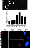
Figure 1
Figure 1
Cell cycle–dependent localization of BLM. Cells were synchronized, immunostained for BLM or PML, stained for nuclear DNA (DAPI), and pulsed (1 h) with [3H]thymidine to determine the percentage of cells in S phase (% LN), as described in Materials and Methods. (a and b) BLM antibody specificity. Proliferating BS fibroblasts (HG2654, shown; GM11492F, not shown) were stained with DAPI (a) to visualize nuclei and the anti-BLM antibody (b). (c) BLM foci during the cell cycle. WI-38 cells were arrested in G0 (Q) and then stimulated with serum for 8 h (Q,8) to enrich for cells in mid-G1. Alternatively, cells were arrested at the G1/S boundary (HU) and released for varying intervals to enrich for cells in mid-S (HU,4), late S/G2 (HU,8), G2/M/early G1 (HU,10), or G1/early S (HU,12). The percent of LN was determined in parallel cultures. Nuclei (≥200 per data point) were scored for the presence of >10 BLM foci. (d–k) BLM and PML were identified by immunostaining using fluorescein isothiocyanate (green) or Texas red secondary antibodies. Red and green fluorescent images were superimposed (MERGE). Nuclei were identified by DAPI staining. (d) PML localization in quiescent cells. (e) BLM localization in quiescent cells. (f) Merged image of PML and BLM localization in quiescent cells. (g) DAPI staining of nuclei in d–f. (h) PML localization in cells in late S/G2. (i) BLM localization in cells in late S/G2. (j) Merged image of PML and BLM localization in cells in late S/G2. (k) DAPI staining of nuclei in h–k. Bars, ∼10 μm.
Figure 2
Figure 2
BLM, PML, hRAD51, and RP-A localization in cells in G0 or late S/G2. WI-38 cells were made quiescent (Quiescent), or released from a G1/S arrest for 8 h (Late S/G2). Cells were stained for BLM, PML, hRAD51, or RP-A, nuclei were visualized (DAPI), and fluorescent images were superimposed (MERGE) as described in the legend to Fig. 1. (a–d) hRAD51 and PML localization in quiescent cells. (e–h) hRAD51 and BLM localization in cells in late S/G2. (i–l) RP-A and BLM localization in cells in late S/G2. Bars, ∼10 μm.
Figure 3
Figure 3
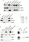
BLM associates with the nuclear matrix and hRAD51. Nuclear matrices were prepared from proliferating WI-38 cells by high salt extraction (NaCl), or low salt extraction and amine modification (NH2SO4). After extraction, 30 μg of protein was analyzed from whole cell (Total), nuclear (Nuclear), and cytoplasmic (Cytosol) lysates, and the supernatants (S) and nuclear matrix pellets (P). Proteins were analyzed for BLM, α-tubulin (Tubulin; cytosolic marker), lamin B (nuclear matrix marker), PARP, and Ku70 (DNA-associated) by Western blotting. (a–b) Results of two independent fractionations. (c) Recombinant GST-BLM. GST-BLM was produced by baculovirus in insect cells. Nuclear proteins from infected cells were bound to glutathione-Sepharose, the resin was transferred to a column, and bound proteins (200 ng) were eluted and analyzed by silver-stained SDS-PAGE. (d) hRAD51 associates with BLM. GST or GST-BLM, bound to glutathione-Sepharose beads, were incubated with SAOS-2 nuclear lysates (Input) and transferred to a column. After washing, proteins were eluted from the columns (GST, Eluted; GST-BLM, Eluted), and proteins resistant to elution were released by boiling in SDS-PAGE sample buffer (GST, Boiled; GST-BLM, Boiled). Input, eluted, and released proteins were analyzed for BLM, hRAD51, PARP, and Ku70 by Western blotting. (e) hRAD51 coimmunoprecipitates with BLM from nuclear lysates. Nuclear lysates from SOAS-2 cells (Input) were precleared and immunoprecipitated with nonspecific (IgG) or anti-hRAD51 (α-RAD51) antibody, and the immunoprecipitates were analyzed for BLM and hRAD51 by SDS-PAGE and Western blotting, as described in Materials and Methods.
Figure 4
Figure 4
BLM responds to IR. Proliferating WI-38 cells were X-irradiated (IR) with 5 Gy (a, b, and d) or 0–10 Gy (c). RNA and protein were isolated, or cells were harvested for flow cytometry, at the indicated intervals (h) thereafter. BLM mRNA was measured by quantitative PCR using QM as a control; BLM protein was assessed by Western blotting using α-tubulin (Tubulin) as a control. A value of one was assigned to the normalized levels of BLM mRNA and protein in unirradiated cells (0 h). Autoradiograms of the Western analyses are shown above the histograms. (a) BLM mRNA after IR. (b) BLM protein after IR. (c) IR dose response. Cells were analyzed for BLM protein 4 h (autoradiogram) or 8 h (autoradiogram, histogram) after irradiation. (d) Cell cycle arrest after IR. Cells were analyzed for DNA content by flow cytometry. The G1 (2N) and G2 (4N) peaks are indicated and the fraction of cells in G1, S, and G2/M is given in the text.
Figure 5
Figure 5
BLM response to DNA damage depends on the G2 delay. WI-38 (a–e), AT-2SF, or WI38-E6 (f) cells were treated with the indicated agents while quiescent (a) or proliferating (b–f). Protein lysates were prepared from untreated cells (0 h) or at the indicated times after treatment (h), and analyzed for BLM and α-tubulin (Tubulin; control) by Western blotting. (a) Quiescent cells were X-irradiated (5 Gy) and immediately stimulated with serum. Parallel cultures were pulsed for 24 h with [3H]thymidine to determine the percentage of cells that synthesized DNA (% LN). (b) Proliferating cells were X-irradiated (5 Gy) and immediately given medium containing 5 mM caffeine. Parallel cultures were analyzed for mitotic figures (Mitotic index; 1,000 nuclei/point). The mitotic index of the unirradiated culture was given a value of 1. (c) Cells were given bleomycin (10 μg/ml) for 1 h in serum-containing medium. (d) Cells were given etoposide (10 μM) for 1 h in serum-containing medium. (e) Cells were irradiated with UVC (1.6 J/m2/s) in PBS and returned to serum-containing medium. (f) Proliferating AT-2SF or WI38-E6 cells were X-irradiated (5 Gy), protein lysates were prepared, and BLM protein level was normalized to α-tubulin.
Figure 6
Figure 6
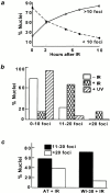
BLM focus formation after DNA damage. Proliferating WI-38 (a–c) and AT-2SF (c) cells were X-irradiated (5 Gy) or UV-irradiated (1.6 J/m2/s) and immunostained for BLM at the indicated intervals thereafter. BLM foci were counted in 200 nuclei per point. (a) BLM foci increase after IR. Nuclei were scored for the presence of >10 or <10 BLM foci. (b) Effect of IR versus UV. Cells were unirradiated (−IR) or irradiated with X-rays (+IR) or UV (+UV). 10 h later, nuclei were scored for the presence of 0–10, 11–20, or >20 BLM foci. (c) BLM foci formation in irradiated AT cells. Proliferating AT-2SF or WI-38 cells were X-irradiated. 10 h later, nuclei were scored for the presence of 11–20 or >20 BLM foci.
Figure 7
Figure 7
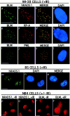
BLM localizes with hRAD51, RP-A, and PML after IR. Proliferating WI-38, BS HG2654, and NB4 cells were X-irradiated (5 Gy) where indicated. 10 h after irradiation, the cells were immunostained for BLM, PML, hRAD51, or RP-A; nuclei were visualized (DAPI); and fluorescent images were superimposed (MERGE) as described in the legend to Fig. 1. (a–d) BLM and hRAD51 localization in irradiated WI-38 cells. (e–h) BLM and RP-A localization in irradiated WI-38 cells. (i–l) BLM and PML localization in irradiated WI-38 cells. (m–o) hRAD51 localization in irradiated BS cells. (p–s) hRAD51 and BLM localization in unirradiated (−IR) and irradiated (+IR) NB4 cells. Bars, ∼10 μm.
Figure 8
Figure 8
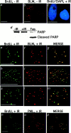
BLM and PML localize to sites of putative DNA repair after IR. Proliferating WI-38 were labeled for two doublings with BrdU and X-irradiated (5 Gy) where indicated. 10–12 h after irradiation, the cells were fixed under nondenaturing conditions and immunostained for BrdU, BLM, and PML. Nuclei were visualized (DAPI) and fluorescent images were superimposed (MERGE) as described in the legend to Fig. 1. (a) BrdU staining in unirradiated cells. (b) BLM foci in unirradiated, BrdU-labeled cells. (c) BrdU and DAPI staining in irradiated cells. The BrdU and DAPI images were merged. (d) PARP integrity after irradiation. BrdU-labeled cells were left untreated (−IR), X-irradiated (+IR), or treated with Fas ligand (Fas; positive control for apoptosis). 10 h later, protein lysates were prepared and analyzed for PARP by Western blotting. (e–m) BLM and BrdU localization in irradiated cells. e–g show a cell with 80–90% colocalization of BLM and BrdU foci; 30–50% of cells with BrdU foci showed this staining pattern. h–m show cells in which BLM and BrdU colocalized to varying degrees. (n–p) PML and BrdU localization in irradiated cells. Bar, ∼10 μm.

Similar articles

Cited by

References

    1. Aurias A., Antoine J.L., Assathiany R., Odievre M., Dutrillaux B. Radiation sensitivity of Bloom's syndrome lymphocytes during S and G2 phases. Cancer Genet. Cytogenet. 1985;16:131–136. - PubMed
    1. Banin S., Moyal L., Shieh S., Taya Y., Anderson C.W., Chessa L., Smorodinsky N.I., Prives C., Reiss Y., Shiloh Y., Ziv Y. Enhanced phosphorylation of p53 by ATM in response to DNA damage. Science. 1998;281:1674–1677. - PubMed
    1. Baumann P., West S.C. The human Rad51 proteinpolarity of strand transfer and stimulation by hRP-A. EMBO (Eur. Mol. Biol. Organ.) J. 1997;16:5198–5206. - PMC - PubMed
    1. Beamish H., Khanna K.K., Lavin M.F. Ionizing radiation and cell cycle progression in ataxia telangiectasia. Radiat. Res. 1994;138:130–133. - PubMed
    1. Brosh R.M., Li J.L., Kenny M.K., Karow J.K., Cooper M.P., Kureekattil R.P., Hickson I.D., Bohr V.A. Replication protein A physically interacts with the Bloom's syndrome protein and stimulates its helicase activity. J. Biol. Chem. 2000;275:23500–23508. - PubMed

Publication types

MeSH terms