. 2001 May;21(9):3220-33.
doi: 10.1128/MCB.21.9.3220-3233.2001.

Identification of TFII-I as the endoplasmic reticulum stress response element binding factor ERSF: its autoregulation by stress and interaction with ATF6

Affiliations

Identification of TFII-I as the endoplasmic reticulum stress response element binding factor ERSF: its autoregulation by stress and interaction with ATF6

R Parker et al. Mol Cell Biol. 2001 May.

Abstract

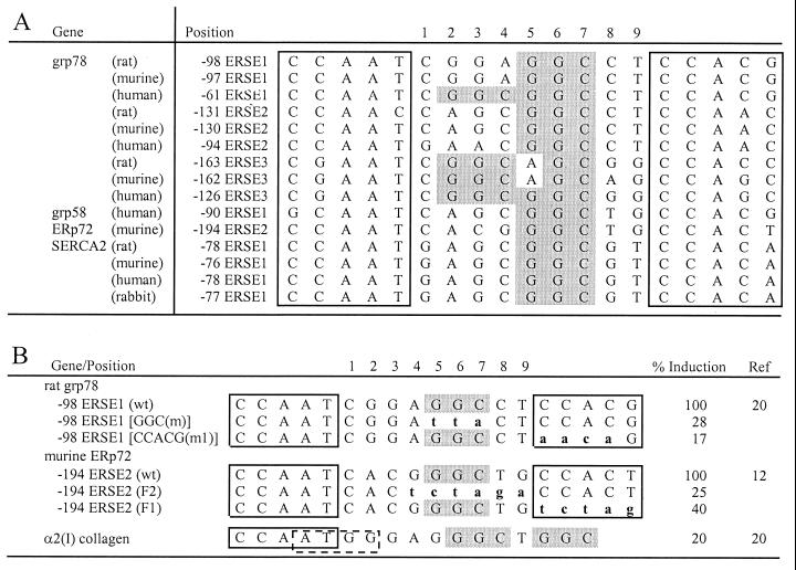
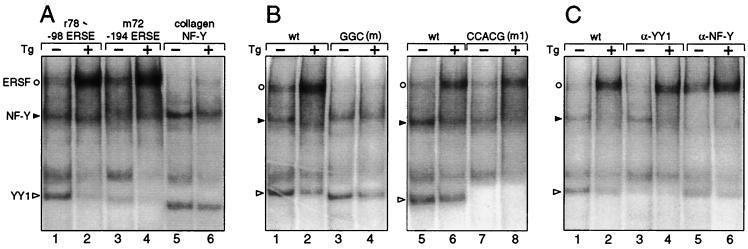
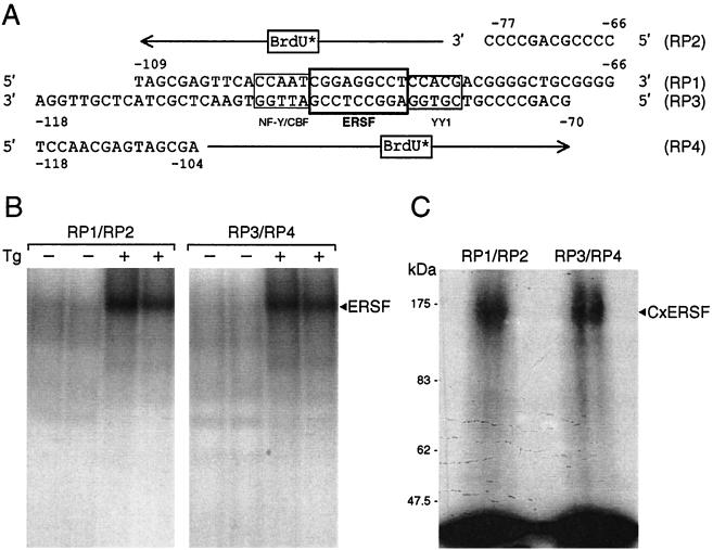
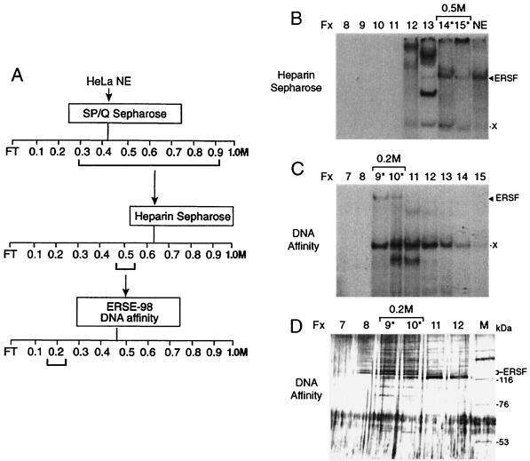
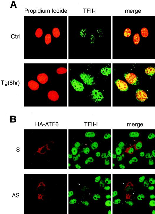
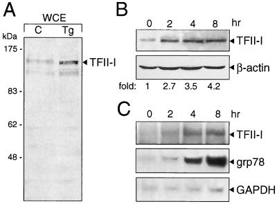
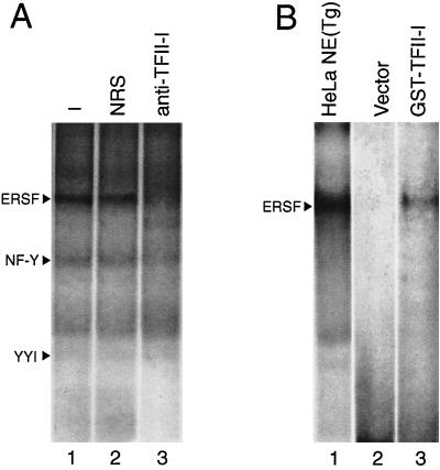
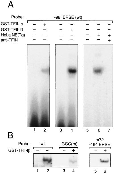
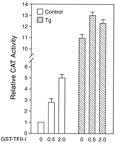
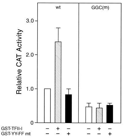
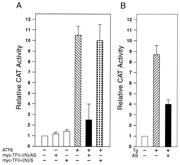
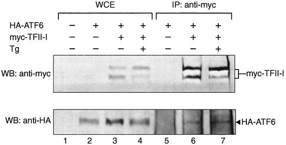
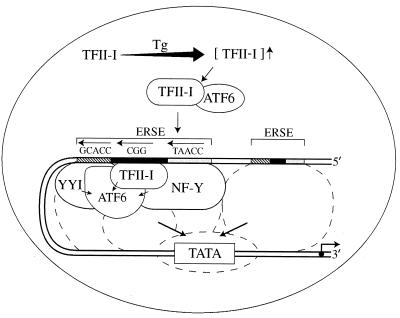
When mammalian cells are subjected to stress targeted to the endoplasmic reticulum (ER), such as depletion of the ER Ca(2+) store, the transcription of a family of glucose-regulated protein (GRP) genes encoding ER chaperones is induced. The GRP promoters contain multiple copies of the ER stress response element (ERSE), consisting of a unique tripartite structure, CCAAT(N(9))CCACG. Within a subset of mammalian ERSEs, N(9) represents a GC-rich sequence of 9 bp that is conserved across species. A novel complex (termed ERSF) exhibits enhanced binding to the ERSE of the grp78 and ERp72 promoters using HeLa nuclear extracts prepared from ER-stressed cells. Optimal binding of ERSF to ERSE and maximal ERSE-mediated stress inducibility require the conserved GGC motif within the 9-bp region. Through chromatographic purification and subsequent microsequencing, we have identified ERSF as TFII-I. Whereas TFII-I remains predominantly nuclear in both nontreated NIH 3T3 cells and cells treated with thapsigargin (Tg), a potent inducer of the GRP stress response through depletion of the ER Ca(2+) store, the level of TFII-I transcript was elevated in Tg-stressed cells, correlating with an increase in TFII-I protein level in the nuclei of Tg-stressed cells. Purified recombinant TFII-I isoforms bind directly to the ERSEs of grp78 and ERp72 promoters. The stimulation of ERSE-mediated transcription by TFII-I requires the consensus tyrosine phosphorylation site of TFII-I and the GGC sequence motif of the ERSE. We further discovered that TFII-I is an interactive protein partner of ATF6 and that optimal stimulation of ERSE by ATF6 requires TFII-I.


Figures

FIG. 1
FIG. 1
Sequence and spatial conservation of the GC-rich sequence motif within the N9 region of ERSE. (A) Sequence alignment of ERSEs from the promoter regions of the ER protein genes grp78, grp58, ERp72, and SERCA2 (1, 12, 20, 23, 26). The CCACT and CCACG motifs are boxed. For each ERSE, the negative number indicates the distance in base count of the first C of the CCAAT element with the transcription initiation site (set at +1). For each gene, the ERSEs are numbered (1 to 3) in the order of increasing distance upstream of the transcription initiation site. The GGC triplets within the 9-bp region of the ERSEs are shaded in gray. (B) The GGC sequence is required for optimal stress inducibility of ERSE-mediated transcription. The sequences of −98 ERSE1 of grp78 and −194 ERSE of ERp72 are shown with the CCAAT and CCACG/CCACT motifs boxed. The mutated bases of each ERSE are in lowercase and aligned against the wild-type (wt) sequences. For the grp78 reporter gene, ER stress was induced by Tg (20). For the ERp72 reporter gene, stress was induced by accumulation of μ heavy chain in the ER (12). The α2(I) collagen promoter contained an inverted YY1 binding site, CCAT (indicated by the dashed box), overlapping with the CCAAT sequence. Its induction by Tg was directly compared to that of the wild-type −98 ERSE of grp78 (20).
FIG. 2
FIG. 2
Requirement for stress-induced enhanced binding of ERSF to ERSE. (A) The EMSAs were performed with HeLa NEs prepared from either control (−) or Tg-treated (+) cells with the different probes as indicated at the top. (B) ERSF binding to ERSE requires the GGC motif. The EMSAs were performed with either the wild-type (wt) −98 ERSE of grp78 or the GGC(m) mutant as a probe. (C) The probe used was wt −98 ERSE. No antibody was added to the first two lanes. Anti-YY1 or anti-NF-Y antibody was added to the reaction mixture as indicated at the top. The ERSF, NF-Y, and YY1 complexes formed are indicated by an open circle, a closed arrowhead, and an open arrowhead, respectively.
FIG. 3
FIG. 3
UV cross-linking of the ERSF complex. (A) Sequence of −98 ERSE with the locations of the NF-Y, YY1, and ERSF binding sites boxed. The primers for the BrdU substitution and the orientation of the extended products are indicated. (B) Preparative EMSA gel for isolating ERSF complexes using either RP1-RP2 or RP3-RP4 as template-primer. The EMSA binding reactions included HeLa NE from either control or Tg-treated cells and were performed in duplicate. The autoradiograms are shown. The position of the ERSF complex is indicated. (C) Estimation of the molecular size of the ERSF UV-cross-linked complex. The preparative gel shown in panel B was subjected to UV cross-linking. The bands corresponding to ERSF obtained with RP1-RP2 or RP3-RP4 were excised and subjected to SDS-PAGE. The autoradiogram is shown. The positions of the ERSF cross-linked complex (CxERSF) and the molecular size markers are indicated.
FIG. 4
FIG. 4
Protein purification of ERSF binding activity. (A) Scheme for ERSF purification. Salt concentrations at which ERSF binding activity eluted from the columns are bracketed. FT, flowthrough. (B) Heparin Sepharose column chromatography of pooled SP/Q Sepharose fractions. The EMSAs for eluted fractions (Fx) 8 through 15 (corresponding to 0.1 to 0.5 M KCl) are shown, with HeLa NE providing a positive control for the electrophoretic mobility of the ERSF complex. Fractions 14 and 15 were pooled for their ability to form the ERSF complex. The positions of the ERSF complex and an unknown copurifying complex (X) are indicated. The column fractions containing major ERSF binding activity are indicated with an asterisk. (C) DNA affinity column fractionation of ERSF binding activity. The EMSAs for fractions 7 through 15 (corresponding to 0.1 to 0.5 M KCl) are shown. (D) SDS-PAGE analysis of fractions from the DNA affinity column. Fifteen-microliter portions of fractions 7 through 12 were electrophoresed alongside high-molecular-weight protein markers (lane M) on an SDS–8% polyacrylamide gel. The gel was silver stained. The sizes of markers are indicated. Fractions 9 and 10 contain proteins which correlate with the major ERSF binding activity as defined by EMSAs.
FIG. 5
FIG. 5
Alignment of peptide sequence derived from ERSF with human TFII-I. The amino acid sequence of human TFII-I is shown. The matching peptides are underlined.
FIG. 6
FIG. 6
Nuclear localization of TFII-I. (A) NIH 3T3 cells were grown to 80% confluence in chamber slides. The cells were either not treated (Ctrl) or treated with 300 nM Tg for 8 h. The cells were stained with anti-TFII-I antibody (Ab2240) and viewed with a 40× oil immersion lens yielding a magnification of ×400, using a Zeiss confocal microscope with LSM 510 imaging software. The left panels show propidium iodide-stained nuclei, the middle panels show the TFII-I staining, and the right panels show the merge of the two immunofluorescence images. (B) COS cells were grown to 80% confluence in chamber slides. At 36 h following transient transfection, the cells were fixed and stained with an anti-HA epitope antibody and anti-TFII-I antibody (Ab2240). The upper set of images shows cells transfected with the sense (S) vector [myc-TFII-I(N)/S], and the lower set shows cells transfected with the antisense (AS) vector [myc-TFII-I(N)/AS]. Both sets were cotransfected with HA-ATF6. The left panel shows HA staining, the middle panel shows endogenous TFII-I staining, and the right panel shows the merge of the two images. The transfected cells, as indicated by high-level expression of the HA epitope, are indicated with arrowheads.
FIG. 7
FIG. 7
Tg stress induction of endogenous TFII-I. (A) NIH 3T3 cells were either grown under normal culture conditions (lane C) or treated with Tg for 6 h. The whole-cell extract (WCE) was prepared by lysing the cells directly in radioimmunoprecipitation assay buffer. The levels of TFII-I were detected by Western blotting using an anti-TFII-I antibody (Ab2240). (B) The cells were subjected to Tg treatment for the time indicated. TFII-I and β-actin protein levels were detected by Western blotting. The fold induction of TFII-I after normalization to the β-actin level is indicated. (C) Total RNA was prepared from NIH 3T3 cells treated with Tg for the time indicated. The levels of TFII-I, grp78, and glyceraldehyde-3-phosphate dehydrogenase (GAPDH) transcripts were detected by Northern blotting.
FIG. 8
FIG. 8
TFII-I is an ERSE binding protein. (A) Antibody inhibition of ERSF complex formation. EMSAs were performed using HeLa NE from Tg-treated cells and 32P-labeled −98 ERSE as a probe. Lane 1, standard EMSA reaction; lane 2, preincubation of the extract with 2 μl of normal rabbit serum (NRS); lane 3, binding reaction with 2 μl of the anti-TFII-I antibody. The positions of the ERSF, NF-Y, and YY1 complexes are indicated. (B) Binding of GST–TFII-I to −98 ERSE. An EMSA is presented for reactions of 32P-labeled −98 ERSE as a probe and GST–TFII-I purified from extracts of COS cells transfected with either the empty vector (lane 2) or the expression vector for GST-TFII-I (lane 3). The band detected in lane 3 comigrated with the ERSF complex formed between −98 ERSE and HeLa NE prepared from Tg-treated cells (lane 1).
FIG. 9
FIG. 9
Binding of purified GST–TFII-I isoforms to −98 ERSE. (A) GST–TFII-I was purified from COS cells transfected with expression vector for the Δ form of GST–TFII-I (120 kDa) or the β form of GST–TFII-I (128 kDa). These were used in the EMSAs with 32P-labeled −98 ERSE as a probe performed under conditions that optimized TFII-I isoform binding (see Materials and Methods). Lanes 1 and 3, probe alone; lanes 2 and 4, isoform complexes being formed. HeLa NE from Tg-treated cells was mixed with the −98 ERSE probe under the same EMSA conditions used for the TFII-I isoforms in the absence (lane 6) or presence (lane 7) of anti-TFII-I antibody. Lane 5, probe alone. wt, wild type. (B) The probes used were the −98 ERSE wt (lanes 1 and 2), the −98 ERSE GGC mutant (lanes 3 and 4), and the −194 ERSE from ERp72 (lanes 5 and 6) as indicated. GST–TFII-Iβ was used in the EMSA reactions.
FIG. 10
FIG. 10
Effect of overexpression of TFII-I on the −98 ERSE-mediated CAT activity. The construct (−109/−74)MCAT was used as the reporter gene and was cotransfected with empty vector alone or increasing amounts (in micrograms) of the expression vector for GST–TFII-I (pEBGTFII-I) into COS cells. An expression vector for β-galactosidase activity was included to normalize for transfection efficiency. The amount of total DNA in each transfection was adjusted to be the same by addition of the empty vector in the reaction mixture. The transfected cells were either grown under normal culture conditions (open bars) or treated with Tg (hatched bars). The CAT activity in nonstressed cells transfected with the empty vector was set at 1. The relative promoter activities are shown with standard deviations.
FIG. 11
FIG. 11
Requirements for TFII-I stimulation of −98 ERSE. COS cells were transfected either with (−109/−74)MCAT containing the wild-type (wt) rat grp78 −98 ERSE as the reporter gene (left panel) or with the GGC mutant [GGC(m)] of −98 ERSE as the reporter gene (right panel). The cells were transfected with either the empty vector, the wild-type GST–TFII-I, or the tyrosine phosphorylation site mutant (mt) of GST-TFII-I (YY-FF) as indicated. Error bars indicate standard deviations.
FIG. 12
FIG. 12
Suppression of ATF6 and Tg stimulation of −98 ERSE by antisense TFII-I. (A) COS cells were transfected with (−109/−74)MCAT as the reporter gene. The cells were cotransfected with either the empty CMV vector, pCGN-ATF6, or TFII-I(N)/AS or TFII-I(N)/S, alone and in combination, as indicated. The CAT activity in cells transfected with the empty CMV vector was set at 1. The relative promoter activities are shown with standard deviations. (B) COS cells were transfected with (−109/−74)MCAT as the reporter gene. The cells were either not treated or treated with Tg in the presence of the empty vector or the antisense TFII-I (AS) vector as indicated.
FIG. 13
FIG. 13
In vivo interaction of TFII-I with ATF6. COS cells were transfected with either the empty vector, HA-tagged-ATF6, or myc-tagged TFII-I, alone and in combination, as indicated. The cells were either not treated or treated with Tg for 16 h prior to preparation of the whole-cell extract (WCE) in NP-40 buffer. Fifty micrograms of the WCE was directly applied to SDS–8% PAGE for Western blots (WB) (lanes 1 to 4). To detect ATF6 and TFII-I interaction, 500 μg of the WCE was immunoprecipitated (IP) with 2 μg of anti-myc antibody (lanes 5 to 7). The immunoprecipitates were applied to denaturing SDS–8% PAGE and Western blotted with either anti-myc (upper panel) or anti-HA (lower panel) antibody. The positions of myc–TFII-I and HA-ATF6 are indicated.
FIG. 14
FIG. 14
Model for TFII-I action on ERSE following Tg stress. TFII-I is a nuclear protein expressed at a basal level in nonstressed cells. Upon Tg treatment, the level of TFII-I increases. TFII-I can associate with ATF6. These two factors become part of a multiprotein complex binding onto the ERSE of the grp78 promoter. TFII-I, NF-Y, and YY1 serve as coactivators for ATF6. Other Tg-induced modifications of the transcription factors may also occur. Components of this multiprotein complex have the potential to serve as bridging proteins between the upstream ERSE regulatory elements and the basal transcription machinery. Acting synergistically, they promote activation of the grp78 promoter in response to Tg stress.

Similar articles

Cited by

References

    1. Caspersen C, Pedersen P S, Treiman M. The sarco/endoplasmic reticulum calcium-ATPase 2b is an endoplasmic reticulum stress-inducible protein. J Biol Chem. 2000;275:22363–22372. - PubMed
    1. Cheriyath V, Novina C D, Roy A L. TFII-I regulates Vβ promoter activity through an initiator element. Mol Cell Biol. 1998;18:4444–4454. - PMC - PubMed
    1. Cheriyath V, Roy A L. Alternatively spliced isoforms of TFII-I. Complex formation, nuclear translocation, and differential gene regulation. J Biol Chem. 2000;275:26300–26308. - PubMed
    1. Foti D M, Welihinda A, Kaufman R J, Lee A S. Conservation and divergence of the yeast and mammalian unfolded protein response. Activation of specific mammalian endoplasmic reticulum stress element of the grp78/BiP promoter by yeast Hac1. J Biol Chem. 1999;274:30402–30409. - PubMed
    1. Grueneberg D A, Henry R W, Brauer A, Novina C D, Cheriyath V, Roy A L, Gilman M. A multifunctional DNA-binding protein that promotes the formation of serum response factor/homeodomain complexes: identity to TFII-I. Genes Dev. 1997;11:2482–2493. - PMC - PubMed

Publication types

MeSH terms

LinkOut - more resources