

Role of tumor-host interactions in interstitial diffusion of macromolecules: cranial vs. subcutaneous tumors
- PMID: 11274375
- PMCID: PMC31885
- DOI: 10.1073/pnas.081626898
Role of tumor-host interactions in interstitial diffusion of macromolecules: cranial vs. subcutaneous tumors
Abstract
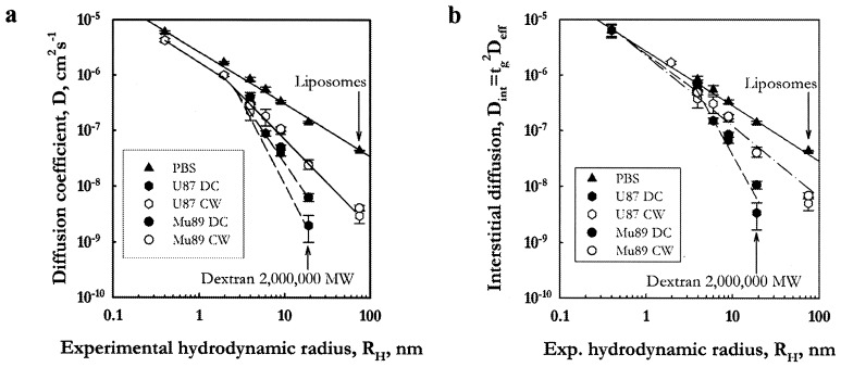
The large size of many novel therapeutics impairs their transport through the tumor extracellular matrix and thus limits their therapeutic effectiveness. We propose that extracellular matrix composition, structure, and distribution determine the transport properties in tumors. Furthermore, because the characteristics of the extracellular matrix largely depend on the tumor-host interactions, we postulate that diffusion of macromolecules will vary with tumor type as well as anatomical location. Diffusion coefficients of macromolecules and liposomes in tumors growing in cranial windows (CWs) and dorsal chambers (DCs) were measured by fluorescence recovery after photobleaching. For the same tumor types, diffusion of large molecules was significantly faster in CW than in DC tumors. The greater diffusional hindrance in DC tumors was correlated with higher levels of collagen type I and its organization into fibrils. For molecules with diameters comparable to the interfibrillar space the diffusion was 5- to 10-fold slower in DC than in CW tumors. The slower diffusion in DC tumors was associated with a higher density of host stromal cells that synthesize and organize collagen type I. Our results point to the necessity of developing site-specific drug carriers to improve the delivery of molecular medicine to solid tumors.
Figures
Similar articles
-
Comparison of IgG diffusion and extracellular matrix composition in rhabdomyosarcomas grown in mice versus in vitro as spheroids reveals the role of host stromal cells.Br J Cancer. 2002 May 20;86(10):1639-44. doi: 10.1038/sj.bjc.6600270. Br J Cancer. 2002. PMID: 12085216 Free PMC article.
-
Measurement of macromolecular diffusion coefficients in human tumors.Microvasc Res. 2004 May;67(3):231-6. doi: 10.1016/j.mvr.2004.02.001. Microvasc Res. 2004. PMID: 15121448
-
Enhanced macromolecule diffusion deep in tumors after enzymatic digestion of extracellular matrix collagen and its associated proteoglycan decorin.FASEB J. 2008 Jan;22(1):276-84. doi: 10.1096/fj.07-9150com. Epub 2007 Aug 29. FASEB J. 2008. PMID: 17761521
-
The tumor microenvironment: a critical determinant of neoplastic evolution.Eur J Cell Biol. 2003 Nov;82(11):539-48. doi: 10.1078/0171-9335-00346. Eur J Cell Biol. 2003. PMID: 14703010 Review.
-
Stromal reaction in cutaneous melanoma.Crit Rev Oncol Hematol. 2004 Mar;49(3):269-75. doi: 10.1016/j.critrevonc.2003.10.007. Crit Rev Oncol Hematol. 2004. PMID: 15036266 Review.
Cited by
-
Intratumoral nanofluidic system enhanced tumor biodistribution of PD-L1 antibody in triple-negative breast cancer.Bioeng Transl Med. 2023 Sep 15;8(6):e10594. doi: 10.1002/btm2.10594. eCollection 2023 Nov. Bioeng Transl Med. 2023. PMID: 38023719 Free PMC article.
-
Development and Application of a Novel Model System to Study "Active" and "Passive" Tumor Targeting.Mol Cancer Ther. 2016 Oct;15(10):2541-2550. doi: 10.1158/1535-7163.MCT-16-0051. Epub 2016 Aug 2. Mol Cancer Ther. 2016. PMID: 27486224 Free PMC article.
-
Modulation of invasive phenotype by interstitial pressure-driven convection in aggregates of human breast cancer cells.PLoS One. 2012;7(9):e45191. doi: 10.1371/journal.pone.0045191. Epub 2012 Sep 18. PLoS One. 2012. PMID: 23028839 Free PMC article.
-
Investigating the optimum size of nanoparticles for their delivery into the brain assisted by focused ultrasound-induced blood-brain barrier opening.Sci Rep. 2020 Oct 26;10(1):18220. doi: 10.1038/s41598-020-75253-9. Sci Rep. 2020. PMID: 33106562 Free PMC article.
-
Tumor Priming by SMO Inhibition Enhances Antibody Delivery and Efficacy in a Pancreatic Ductal Adenocarcinoma Model.Mol Cancer Ther. 2019 Nov;18(11):2074-2084. doi: 10.1158/1535-7163.MCT-18-0354. Epub 2019 Jul 30. Mol Cancer Ther. 2019. PMID: 31363010 Free PMC article.
References
Publication types
MeSH terms
Substances
Grants and funding
LinkOut - more resources
Full Text Sources
Other Literature Sources
Medical
