Egress of alphaherpesviruses: comparative ultrastructural study
- PMID: 11264357
- PMCID: PMC114859
- DOI: 10.1128/JVI.75.8.3675-3684.2001
Egress of alphaherpesviruses: comparative ultrastructural study
Abstract
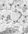
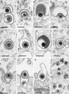
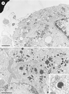
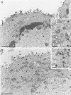
Egress of four important alphaherpesviruses, equine herpesvirus 1 (EHV-1), herpes simplex virus type 1 (HSV-1), infectious laryngotracheitis virus (ILTV), and pseudorabies virus (PrV), was investigated by electron microscopy of infected cell lines of different origins. In all virus-cell systems analyzed, similar observations were made concerning the different stages of virion morphogenesis. After intranuclear assembly, nucleocapsids bud at the inner leaflet of the nuclear membrane, resulting in enveloped particles in the perinuclear space that contain a sharply bordered rim of tegument and a smooth envelope surface. Egress from the perinuclear cisterna primarily occurs by fusion of the primary envelope with the outer leaflet of the nuclear membrane, which has been visualized for HSV-1 and EHV-1 for the first time. The resulting intracytoplasmic naked nucleocapsids are enveloped at membranes of the trans-Golgi network (TGN), as shown by immunogold labeling with a TGN-specific antiserum. Virions containing their final envelope differ in morphology from particles within the perinuclear cisterna by visible surface projections and a diffuse tegument. Particularly striking was the addition of a large amount of tegument material to ILTV capsids in the cytoplasm. Extracellular virions were morphologically identical to virions within Golgi-derived vesicles, but distinct from virions in the perinuclear space. Studies with gB- and gH-deleted PrV mutants indicated that these two glycoproteins, which are essential for virus entry and direct cell-to-cell spread, are dispensable for egress. Taken together, our studies indicate that the deenvelopment-reenvelopment process of herpesvirus maturation also occurs in EHV-1, HSV-1, and ILTV and that membrane fusion processes occurring during egress are substantially different from those during entry and direct viral cell-to-cell spread.
Figures


Similar articles
-
The UL48 tegument protein of pseudorabies virus is critical for intracytoplasmic assembly of infectious virions.J Virol. 2002 Jul;76(13):6729-42. doi: 10.1128/jvi.76.13.6729-6742.2002. J Virol. 2002. PMID: 12050386 Free PMC article.
-
Ultrastructural analysis of the replication cycle of pseudorabies virus in cell culture: a reassessment.J Virol. 1997 Mar;71(3):2072-82. doi: 10.1128/JVI.71.3.2072-2082.1997. J Virol. 1997. PMID: 9032339 Free PMC article.
-
Herpes simplex virus glycoproteins gB and gH function in fusion between the virion envelope and the outer nuclear membrane.Proc Natl Acad Sci U S A. 2007 Jun 12;104(24):10187-92. doi: 10.1073/pnas.0703790104. Epub 2007 Jun 4. Proc Natl Acad Sci U S A. 2007. PMID: 17548810 Free PMC article.
-
Herpesvirus Nuclear Egress across the Outer Nuclear Membrane.Viruses. 2021 Nov 24;13(12):2356. doi: 10.3390/v13122356. Viruses. 2021. PMID: 34960625 Free PMC article. Review.
-
Intriguing interplay between viral proteins during herpesvirus assembly or: the herpesvirus assembly puzzle.Vet Microbiol. 2006 Mar 31;113(3-4):163-9. doi: 10.1016/j.vetmic.2005.11.040. Epub 2005 Dec 5. Vet Microbiol. 2006. PMID: 16330166 Review.
Cited by
-
Why Cells and Viruses Cannot Survive without an ESCRT.Cells. 2021 Feb 24;10(3):483. doi: 10.3390/cells10030483. Cells. 2021. PMID: 33668191 Free PMC article. Review.
-
Nuclear Exodus: Herpesviruses Lead the Way.Annu Rev Virol. 2016 Sep 29;3(1):387-409. doi: 10.1146/annurev-virology-110615-042215. Epub 2016 Jul 22. Annu Rev Virol. 2016. PMID: 27482898 Free PMC article.
-
Advances in the immunoescape mechanisms exploited by alphaherpesviruses.Front Microbiol. 2024 Jun 19;15:1392814. doi: 10.3389/fmicb.2024.1392814. eCollection 2024. Front Microbiol. 2024. PMID: 38962133 Free PMC article. Review.
-
The UL48 tegument protein of pseudorabies virus is critical for intracytoplasmic assembly of infectious virions.J Virol. 2002 Jul;76(13):6729-42. doi: 10.1128/jvi.76.13.6729-6742.2002. J Virol. 2002. PMID: 12050386 Free PMC article.
-
Directional transneuronal spread of α-herpesvirus infection.Future Virol. 2009 Nov 1;4(6):591. doi: 10.2217/fvl.09.62. Future Virol. 2009. PMID: 20161665 Free PMC article.
References
-
- Babic N, Klupp B G, Makoschey B, Karger A, Flamand A, Mettenleiter T C. Glycoprotein gH of pseudorabies virus is essential for penetration and propagation in cell culture and in the nervous system of mice. J Gen Virol. 1996;77:2277–2285. - PubMed
Publication types
MeSH terms
Substances
LinkOut - more resources
Full Text Sources
Miscellaneous

