. 2001 Apr;21(7):2423-34.
doi: 10.1128/MCB.21.7.2423-2434.2001.

S338 phosphorylation of Raf-1 is independent of phosphatidylinositol 3-kinase and Pak3

Affiliations

S338 phosphorylation of Raf-1 is independent of phosphatidylinositol 3-kinase and Pak3

A Chiloeches et al. Mol Cell Biol. 2001 Apr.

Abstract

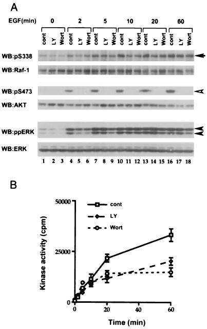
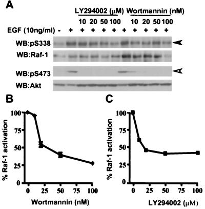
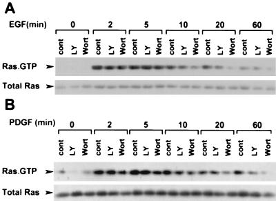
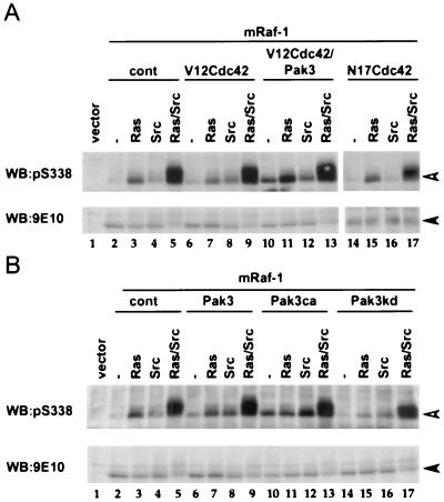
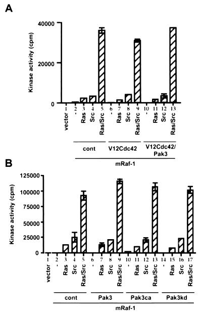
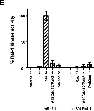
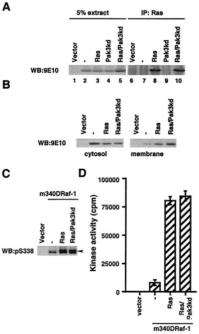
The Raf-1 serine/threonine protein kinase requires phosphorylation of the serine at position 338 (S338) for activation. Ras is required to recruit Raf-1 to the plasma membrane, which is where S338 phosphorylation occurs. The recent suggestion that Pak3 could stimulate Raf-1 activity by directly phosphorylating S338 through a Ras/phosphatidylinositol 3-kinase (Pl3-K)/-Cdc42-dependent pathway has attracted much attention. Using a phospho-specific antibody to S338, we have reexamined this model. Using LY294002 and wortmannin, inhibitors of Pl3-K, we find that growth factor-mediated S338 phosphorylation still occurs, even when Pl3-K activity is completely blocked. Although high concentrations of LY294002 and wortmannin did suppress S338 phosphorylation, they also suppressed Ras activation. Additionally, we show that Pak3 is not activated under conditions where S338 is phosphorylated, but when Pak3 is strongly activated, by coexpression with V12Cdc42 or by mutations that make it independent of Cdc42, it did stimulate S338 phosphorylation. However, this occurred in the cytosol and did not stimulate Raf-1 kinase activity. The inability of Pak3 to activate Raf-1 was not due to an inability to stimulate phosphorylation of the tyrosine at position 341 but may be due to its inability to recruit Raf-1 to the plasma membrane. Taken together, our data show that growth factor-stimulated Raf-1 activity is independent of Pl3-K activity and argue against Pak3 being a physiological mediator of S338 phosphorylation in growth factor-stimulated cells.


Figures

FIG. 1
FIG. 1
PI3-kinase inhibitors do not block S338 phosphorylation on Raf-1 in COS cells. (A) Effects of PI3-kinase inhibitors on the phosphorylation of S338 on Raf-1 and of S473 on Akt and double phosphorylation of ERK1 and ERK2. COS cells were pretreated with dimethyl sulfoxide (cont), 20 μM LY294002 (LY), or 100 nM wortmannin (Wort) for 20 min and then stimulated with EGF (10 ng/ml) for the indicated times. For S338 phosphorylation (upper rows, arrow), endogenous Raf-1 was immunoprecipitated with a Raf-1 monoclonal antibody and S338 phosphorylation (pS338) was detected by Western blotting (WB) using our specific antibody as described in Materials and Methods. Phosphorylations of S473 on Akt (middle rows, open arrowhead) and ERK (lower rows, closed arrowheads) were detected in the same extracts, using appropriate phospho-specific antibodies. For each pair of rows, an image of the phospho-specific blot is shown with its appropriate reprobed image with antibodies against Raf-1, Akt, and ERK. Similar results were obtained in three independent experiments. (B) Effects of PI3-kinase inhibitors on Raf-1 kinase activity. COS cells were treated as described above. The activity of endogenous Raf-1 was measured as described in Materials and Methods. The results presented are for one experiment assayed in triplicate, with error bars to represent standard deviations from the mean. Similar results were obtained in three independent experiments.
FIG. 2
FIG. 2
PI3-kinase inhibitors do not block S338 phosphorylation on Raf-1 in NIH 3T3 cells. (A) Effects of PI3-kinase inhibitors on Raf-1, Akt, and ERK phosphorylation in NIH 3T3 cells. NIH 3T3 cells were treated as described for Fig. 1A except that they were stimulated with PDGF (50 ng/ml) for the indicated times. Phosphorylation of Raf-1 on S338 (upper row, arrow), Akt on S473 (middle row, open arrowhead), and doubly phosphorylated ERK (lower row, closed arrowheads) was tested as described for Fig. 1. To ensure equivalent protein loading, the blots were reprobed for Raf-1, Akt, and ERK after the membranes had been stripped (data not shown). Similar results were obtained in two independent experiments. (B) Effects of PI3-kinase inhibitors on Raf-1 kinase activity. NIH 3T3 cells were treated as described above, and Raf-1 kinase activity was determined as for Fig. 1. The results presented are for one experiment assayed in triplicate, with error bars to represent standard deviations from the mean. Similar results were obtained in two independent experiments.
FIG. 3
FIG. 3
LY294002 and wortmannin suppress S338 phosphorylation at high concentrations. (A) Effect of the concentration of PI3-K inhibitors on the phosphorylation of Raf-1 on S338 and AKT on S473. COS cells were pretreated for 20 min with dimethyl sulfoxide DMSO as vehicle control or increasing concentration of LY294002 or wortmannin for 20 min. Then cells were left untreated (−) or stimulated with EGF (10 ng/ml; +) for 20 min, and S338 phosphorylation on Raf-1 (upper rows, closed arrowhead) and S473 on Akt (lower rows open arrowhead) were detected as for Fig 1. For each pair of rows, the phospho-specific blot is shown with its appropriate reprobed image with antibodies against Raf-1 and Akt, respectively. (B and C) Effects of increasing concentrations of LY294002 or wortmannin on Raf-1 kinase activity. Raf-1 kinase activity was measured as described for Fig 1, using the same extracts as used for panel A. The results presented are for one experiment assayed in triplicate, with error bars to represent standard deviations from the mean.
FIG. 4
FIG. 4
Effect of PI3-kinase on Ras activity. COS (A) or NIH 3T3 (B) cells were serum starved and either preincubated with dimethyl sulfoxide (cont), 20 μM LY294002 (LY), or 100 nM wortmannin (Wort) for 20 min and then stimulated with EGF (10 ng/ml) (COS cells) or PDGF (50 ng/ml) (NIH 3T3 cells) for the indicated times. Active GTP-bound Ras (upper rows) was extracted from the lysates and detected as described in Materials and Methods. An immunoblot of 5% total Ras in the extracts is shown as a loading control (lower rows). Blots are for one representative experiment performed three times with similar results.
FIG. 5
FIG. 5
Pak3 is not activated by EGF, V12Ras, or F527Src in COS cells. COS cells were transfected with wild-type HA-Pak3 (Pak3) and then treated with EGF (10 ng/ml) for the indicated times. Equivalent amounts of HA-Pak3 were immunoprecipitated with antibody 12CA5, and kinase activity was determined in the immunoprecipitate using MBP as substrate. For each assay, the tops of the gels were transferred and blotted with antibody 12CA5 as a control of the immunoprecipitation (data not shown). As a positive control, COS cells transfected with HA-Pak3ca (Pak3ca) were included in the assays (lane 8). Gels presented are for one representative experiment of three performed with identical results. (A) EGF stimulation of HA-Pak3. Following transfection, cells were stimulated with EGF (10 ng/ml) for the indicated times, and the activity of HA-Pak3 was determined. (B) Activation of HA-Pak3 by small G proteins and F527Src. HA-Pak3 (Pak3) was coexpressed with V12Cdc42 (V12Cdc42), V12Rac (V12Rac), V12Ras (Ras), or F527Src (Src) as indicated, and the activity of HA-Pak3 was determined.
FIG. 6
FIG. 6
Activated Pak3 stimulates S338 phosphorylation on Raf-1. COS cells were transfected with mRaf-1 alone (−) or with V12Ras (Ras) or F527Src (Src) in the absence or presence of V12Cdc42 (V12Cdc42), HA-Pak3 (Pak3), or N17Cdc42 (N17Cdc42) (A) or with HA-Pak3ca (Pak3ca) or HA-Pak3kd (Pak3kd) (B) as indicated. Equivalent amounts of mRaf-1 were immunoprecipitated with antibody 9E10 for Western blot (WB) analysis, using the pS338 phospho-specific antibody (open arrowhead) or for mRaf-1 using antibody 9E10 (closed arrowhead). Data shown are from one representative experiment performed three times with similar results.
FIG. 7
FIG. 7
Effects of Cdc42 and Pak3 on Raf-1 kinase activity. COS cells were transfected with the indicated vectors as described for Fig 6. Equivalent amounts of mRaf-1 were immunoprecipitated with antibody 9E10 for kinase assays as described in Materials and Methods. The results presented are from one experiment assayed in triplicate, with error bars to represent the standard deviation from the mean. Similar result were obtained in three independent experiments.
FIG. 8
FIG. 8
Pak3 phosphorylates S338 of Raf-1 in the cytosol and does not translocate Raf-1 to the plasma membrane. mRaf-1 or m89LRaf was transiently expressed in COS cells alone (−) or with V12Ras (Ras), N17Ras (N17Ras), F527Src (Src), V12Cdc42 (V12Cdc42), HA-Pak3 (Pak3), or HA-Pak3ca (Pak3ca) as indicated. (A) Phospho-S338 blot. The samples were processed as for Fig. 6. The phospho-specific Western blot (WB) image (upper row, open arrowhead) is shown together with its appropriate 9E10 expression blot (lower row, closed arrowhead). (B) EGF-stimulated S338 phosphorylation on Raf-1. The transfected cells were unstimulated (−) or stimulated with EGF for 20 min (+) as indicated. The samples were processed as in Fig. 6, and the phospho-specific image (upper row, open arrowhead) is shown together with the appropriate 9E10 expression blot (lower row, closed arrowhead). (C) Akt partitions into the cytosolic fraction. COS cells were fractionated into cytosol (upper row) and membranes (lower row) preparations. The samples were probed for endogenous Akt, the position of migration of which is shown by the arrows. (D) Pak3ca does not recruit Raf-1 to the membrane. COS cells extracts (upper row), as separated in panel C, were probed for Raf-1 in the cytosol (middle row) and in the membrane (lower row) by blotting with antibody 9E10. Raf-1 is indicated by the arrowheads. (E) Raf-1 kinase assay, performed as for Fig. 7. Results are the means ± standard errors for one representative experiment performed in triplicate. Similar results were obtained in three independent experiments.
FIG. 8
FIG. 8
Pak3 phosphorylates S338 of Raf-1 in the cytosol and does not translocate Raf-1 to the plasma membrane. mRaf-1 or m89LRaf was transiently expressed in COS cells alone (−) or with V12Ras (Ras), N17Ras (N17Ras), F527Src (Src), V12Cdc42 (V12Cdc42), HA-Pak3 (Pak3), or HA-Pak3ca (Pak3ca) as indicated. (A) Phospho-S338 blot. The samples were processed as for Fig. 6. The phospho-specific Western blot (WB) image (upper row, open arrowhead) is shown together with its appropriate 9E10 expression blot (lower row, closed arrowhead). (B) EGF-stimulated S338 phosphorylation on Raf-1. The transfected cells were unstimulated (−) or stimulated with EGF for 20 min (+) as indicated. The samples were processed as in Fig. 6, and the phospho-specific image (upper row, open arrowhead) is shown together with the appropriate 9E10 expression blot (lower row, closed arrowhead). (C) Akt partitions into the cytosolic fraction. COS cells were fractionated into cytosol (upper row) and membranes (lower row) preparations. The samples were probed for endogenous Akt, the position of migration of which is shown by the arrows. (D) Pak3ca does not recruit Raf-1 to the membrane. COS cells extracts (upper row), as separated in panel C, were probed for Raf-1 in the cytosol (middle row) and in the membrane (lower row) by blotting with antibody 9E10. Raf-1 is indicated by the arrowheads. (E) Raf-1 kinase assay, performed as for Fig. 7. Results are the means ± standard errors for one representative experiment performed in triplicate. Similar results were obtained in three independent experiments.
FIG. 9
FIG. 9
Pak3ca does not activate m340DRaf-1. COS cells were transfected with m340DRaf-1 alone (−), with V12Ras (Ras), or with HA-Pak3ca (Pak3ca). (A) m340DRaf-1 kinase activity was determined as for Fig. 7. Data presented are the means ± standard errors for one representative experiment performed in triplicate. Similar results were observed in three independent experiments. (B) Phosphorylation of S338 of m340DRaf-1 was determined as for Fig. 6. The Western blot (WB) shown is from one representative experiment. Similar results were obtained in three independent experiments.
FIG. 10
FIG. 10
Pak3kd does not affect association with Ras or membrane translocation of mRaf-1 and does not suppress activation of m340DRaf-1. (A) Association of mRaf with V12Ras. COS cells were transfected with mRaf-1 alone (−) or with V12Ras (Ras) or with HA-Pak3kd (Pak3kd). The levels of mRaf-1 in the 5% of the total cell extracts (lanes 1 to 5) and the levels that coimmunoprecipitated (IP) with V12 Ras (lanes 6 to 10) were determined. Data shown are from one experiment; similar results were obtained in two independent experiments. WB, Western blot. (B) Pak3kd does not affect Ras-stimulated membrane recruitment of mRaf-1. COS cell were transfected with mRaf-1 alone (−) or with V12Ras (Ras) in the absence or presence of HA-Pak3kd (Pak3kd). Membrane or cytosol fractions were prepared, and the levels of mRaf-1 in the different fractions were detected by immunoblotting. Data shown are from one representative experiment of two performed with similar results. (C) Pak3kd does not suppress S338 phosphorylation on m340DRaf-1. COS cells were transfected with m340DRaf-1 in the absence or presence of V12Ras (Ras) with or without HA-Pak3kd (Pak3kd). S338 phosphorylation of m340DRaf-1 was determined as in Fig. 6. (D) Pak3kd does not suppress m340DRaf-1 kinase activity. Using the same cell extracts as for panel C, m340DRaf-1 kinase activity was determined as described for Fig. 7. Presented data are for one experiment assayed in triplicate, with error bars to represent standard deviations from the mean. Similar results were observed in three experiments.

Similar articles

Cited by

References

    1. Arcaro A, Wymann M P. Wortmannin is a potent phosphatidylinositol 3-kinase inhibitor: the role of phosphatidylinositol 3,4,5-trisphosphate in neutrophil responses. Biochem J. 1993;296:297–301. - PMC - PubMed
    1. Bagrodia S, Cerione R A. PAK to the future. Trends Cell Biol. 1999;9:350–355. - PubMed
    1. Barnard D, Diaz B, Clawson D, Marshall M. Oncogenes, growth factors and phorbol esters regulate Raf-1 through common mechanisms. Oncogene. 1998;17:1539–1547. - PubMed
    1. Bos J. All in the family? New insights and questions regarding interconnectivity of Ras, Rap1 and Ral. EMBO J. 1998;17:6776–6782. - PMC - PubMed
    1. Bradford M M. A rapid and sensitive method for quantitation of microgram quantities of protein utilizing the principle of protein-dye binding. Anal Biochem. 1976;72:248–254. - PubMed

Publication types

Substances