. 2001 Mar 19;152(6):1135-43.
doi: 10.1083/jcb.152.6.1135.

Diacylglycerol kinase zeta regulates Ras activation by a novel mechanism

Affiliations

Diacylglycerol kinase zeta regulates Ras activation by a novel mechanism

M K Topham et al. J Cell Biol. .

Abstract

Guanine nucleotide exchange factors (GEFs) activate Ras by facilitating its GTP binding. Ras guanyl nucleotide-releasing protein (GRP) was recently identified as a Ras GEF that has a diacylglycerol (DAG)-binding C1 domain. Its exchange factor activity is regulated by local availability of signaling DAG. DAG kinases (DGKs) metabolize DAG by converting it to phosphatidic acid. Because they can attenuate local accumulation of signaling DAG, DGKs may regulate RasGRP activity and, consequently, activation of Ras. DGK zeta, but not other DGKs, completely eliminated Ras activation induced by RasGRP, and DGK activity was required for this mechanism. DGK zeta also coimmunoprecipitated and colocalized with RasGRP, indicating that these proteins associate in a signaling complex. Coimmunoprecipitation of DGK zeta and RasGRP was enhanced in the presence of phorbol esters, which are DAG analogues that cannot be metabolized by DGKs, suggesting that DAG signaling can induce their interaction. Finally, overexpression of kinase-dead DGK zeta in Jurkat cells prolonged Ras activation after ligation of the T cell receptor. Thus, we have identified a novel way to regulate Ras activation: through DGK zeta, which controls local accumulation of DAG that would otherwise activate RasGRP.


Figures
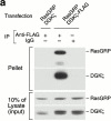
Figure 1
Figure 1
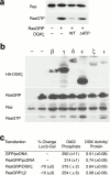
RasGRP coimmunoprecipitates with DGKζ. (a) DGKζ with or without a FLAG epitope tag was cotransfected into HEK293 cells along with HA-RasGRP. 48 h later, anti-FLAG antibodies were used to immunoprecipitate DGKζ. Normal mouse IgG was used as a control. After SDS-PAGE of the pellets or 10% of the lysates, RasGRP was detected by immunoblotting with anti-HA. The blot was then stripped and reprobed for DGKζ. (b) HA-RasGRP was transfected along with a control vector or progressive COOH-terminal truncations of DGKζ containing FLAG epitope tags. The DGK proteins were immunoprecipated with anti-FLAG and then coimmunoprecipitation of HA-RasGRP was detected by immunoblotting with polyclonal anti-HA. The blot was stripped and then the DGKs were detected using anti-DGKζ. The three DGK constructs shown encode amino acids 1–748, 1–605, or 1–467 with FLAG epitope tags at their COOH termini. (c) A172 cells, either control or treated with PMA (90 ng/ml, 30 min), were lysed and then RasGRP was immunoprecipitated using an affinity purified antibody. To verify the specificity of the immunoprecipitation, the antibody was preincubated with its affinity peptide or a control peptide before the immunoprecipitation. The precipitates were then used either for Western blotting to detect RasGRP (top) or for DGK activity assays (bottom). (d) RasGRP was immunoprecipitated from control or PMA-treated A172 cells as described in the legend to c and then the precipitates were used for Western blotting to detect coimmunoprecipitation of DGKζ.
Figure 1
Figure 1
RasGRP coimmunoprecipitates with DGKζ. (a) DGKζ with or without a FLAG epitope tag was cotransfected into HEK293 cells along with HA-RasGRP. 48 h later, anti-FLAG antibodies were used to immunoprecipitate DGKζ. Normal mouse IgG was used as a control. After SDS-PAGE of the pellets or 10% of the lysates, RasGRP was detected by immunoblotting with anti-HA. The blot was then stripped and reprobed for DGKζ. (b) HA-RasGRP was transfected along with a control vector or progressive COOH-terminal truncations of DGKζ containing FLAG epitope tags. The DGK proteins were immunoprecipated with anti-FLAG and then coimmunoprecipitation of HA-RasGRP was detected by immunoblotting with polyclonal anti-HA. The blot was stripped and then the DGKs were detected using anti-DGKζ. The three DGK constructs shown encode amino acids 1–748, 1–605, or 1–467 with FLAG epitope tags at their COOH termini. (c) A172 cells, either control or treated with PMA (90 ng/ml, 30 min), were lysed and then RasGRP was immunoprecipitated using an affinity purified antibody. To verify the specificity of the immunoprecipitation, the antibody was preincubated with its affinity peptide or a control peptide before the immunoprecipitation. The precipitates were then used either for Western blotting to detect RasGRP (top) or for DGK activity assays (bottom). (d) RasGRP was immunoprecipitated from control or PMA-treated A172 cells as described in the legend to c and then the precipitates were used for Western blotting to detect coimmunoprecipitation of DGKζ.
Figure 2
Figure 2
RasGRP and DGK colocalize. (a) A172 cells were immunostained for DGKζ (top) or RasGRP (bottom). Phalloidin was used to identify actin filaments. To assure that the immunostaining was specific, the antibodies were preincubated with their affinity peptide before staining the cells. Several areas of intense staining common to actin and DGKζ or RasGRP are indicated by the arrows. (b) Cos-7 cells were cotransfected with GFP-RasGRP and DGKζ. 24 h later, they were suspended and then allowed to spread for 30 min on glass slides coated with fibronectin. The cells were then immunostained to detect DGKζ (red), nuclei were counterstained (blue), and immunofluorescence images were obtained. (c) To view migrating cells, A172 cell monolayers were wounded with a pipet tip 12 h before immunostaining. Using antibodies directly conjugated with separate fluorophores, the cells were immunostained to detect RasGRP (green) and DGKζ (red) and then viewed by confocal microscopy (Bio-Rad Laboratories), and digital images were obtained. One representative cell is shown migrating into the wounded area, with arrows indicating overlapping localization that was also apparent in most cells. The boxed area is magnified in the lower panels to demonstrate colocalization at the leading edge. Bars, 10 μm.
Figure 3
Figure 3
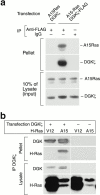
DGKζ and A15-Ras coimmunoprecipitate. (a) DGKζ with or without a FLAG epitope tag was cotransfected into HEK293 cells along with A15-Ras. 48 h later, anti-FLAG antibodies were used to immunoprecipitate DGKζ. Normal mouse IgG was used as a control. After SDS-PAGE of the pellets or 10% of the lysates, A15-Ras was detected by immunoblotting with anti–H-Ras. The blot was then stripped and reprobed for DGKζ. (b) DGKζ-FLAG or a control vector was transfected into Cos-7 cells with H-Ras mutants: either V12-Ras or A15-Ras. After 48 h, anti-FLAG was used to immunoprecipitate DGKζ. After SDS-PAGE of both the lysates and pellets, H-Ras or DGKζ was detected with specific antibodies.
Figure 4
Figure 4
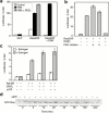
DGKζ attenuates RasGRP activity. (a) HEK293 cells were transfected with myc-Ras and either GFP or GFP-RasGRP along with DGKζ or a kinase-dead, mutant DGKζ (ΔATP). After 48 h, GTP-Ras was affinity-precipitated (Taylor and Shalloway 1996) and then detected by immunoblotting. GFP-RasGRP protein expression was not altered by the DGKs (not shown). (b) Myc-Ras was transfected into HEK293 cells along with GFP or GFP-RasGRP and a control vector or a DGK isotype (β, γ, δ, ε, ζ, or ι) with an NH2-terminal HA epitope tag. After 48 h, GTP-Ras was affinity-precipitated and then detected by immunoblotting. RasGRP, DGK, and H-Ras were detected in the cell lysates by immunoblotting with anti-GFP, anti-HA, or anti-H-Ras. (c) DGKζ or an alternatively spliced variant (ζ2) was transfected into HEK293 cells along with GFP or GFP-RasGRP and the luciferase reporter vectors. 48 h later, the cells were harvested and luciferase activity, DGK activity, and cellular DAG were determined in triplicate. These values were normalized to either β-galactosidase activity (luciferase), lipid phosphate (DAG), or total protein (DGK activity). Expression levels of the two DGKs were similar (not shown).
Figure 5
Figure 5
DGKζ inhibits RasGRP at the level of Ras activation. Elk-1 activity was detected by a luciferase reporter (Stratagene). In all cases, luciferase activity in the cell lysates was determined in triplicate and normalized to β-galactosidase activity, which was included in the transfection. (a) HEK293 cells were transfected with GFP or GFP-RasGRP. 16 h later, a PKC inhibitor (PKCi; 100 nM Ro-32-0432) or control vehicle was added. After 20 min, PMA (0.5 ng/ml) was added for 24 h and then luciferase and β-galactosidase activities in the cell lysates were determined. Shown are the mean and standard deviation. (b) HEK293 cells were transfected with GFP or GFP-RasGRP along with a control vector or DGKζ. 16 h later, one of two PKC inhibitors (a, 200 nM Ro-31-7549; b, 100 nM Ro-32-0432) or control vehicle was added. After 24 h, the cells were harvested and luciferase and β-galactosidase activities in the cell lysates were determined. (c) Raf:ER or a control vector was transfected into HEK293 cells along with DGKζ, ΔATP, or a control vector. 24 h later, estrogen (1 μM) or control vehicle was added for 10 h. The cells were harvested and luciferase and β-galactosidase activities in the cell lysates were determined. (d) Jurkat cells were transfected by electroporation with myc-Ras and either GFP or kinase-dead DGKζ (ΔATP). 48 h later, TCR signaling was activated with anti-CD3 (5 μg/ml, CRIS-7) for up to 4 h. GTP-Ras was affinity-precipitated and then detected by immunoblotting.
Figure 6
Figure 6
Proposed mechanism for precise regulation of RasGRP activity. After TCR stimulation, PLCγ1 is activated and initiates DAG signaling. One protein activated by the DAG is RasGRP. DAG accumulation also recruits DGKζ to the RasGRP signaling complex, where it attenuates RasGRP activity by converting the DAG to PA.

Similar articles

Cited by

References

    1. Bishop W.R., Bell R.M. Attenuation of sn-1,2-diacylglycerol second messengers. J. Biol. Chem. 1986;261:12513–12519. - PubMed
    1. Bunting M., Tang W., Zimmerman G.A., McIntyre T.M., Prescott S.M. Molecular cloning and characterization of a novel human diacylglycerol kinase ζ. J. Biol. Chem. 1996;271:10230–10236. - PubMed
    1. Chang J.-S., Noh D.Y., Park I.A., Kim M.J., Song H., Ryu S.H., Suh P.-G. Overexpression of phospholipase C-γ1 in rat 3Y1 fibroblast cells leads to malignant transformation. Cancer Res. 1997;57:5465–5468. - PubMed
    1. Chin L., Tam A., Pomerantz M.W., Wong M., Holash J., Bardeesy N., Shen Q., O'Hagan R., Pantginis J., Zhou H. Essential role for oncogenic Ras in tumor maintenance. Nature. 1999;400:468–472. - PubMed
    1. Ding L., Bunting M., Topham M.K., McIntyre T.M., Zimmerman G.A., Prescott S.M. Alternative splicing of the human diacylglycerol kinase ζ gene in muscle. Proc. Natl. Acad. Sci. USA. 1997;94:5519–5524. - PMC - PubMed

Publication types

MeSH terms