

Antimycobacterial agent based on mRNA encoding human beta-defensin 2 enables primary macrophages to restrict growth of Mycobacterium tuberculosis
- PMID: 11254636
- PMCID: PMC98208
- DOI: 10.1128/IAI.69.4.2692-2699.2001
Antimycobacterial agent based on mRNA encoding human beta-defensin 2 enables primary macrophages to restrict growth of Mycobacterium tuberculosis
Abstract
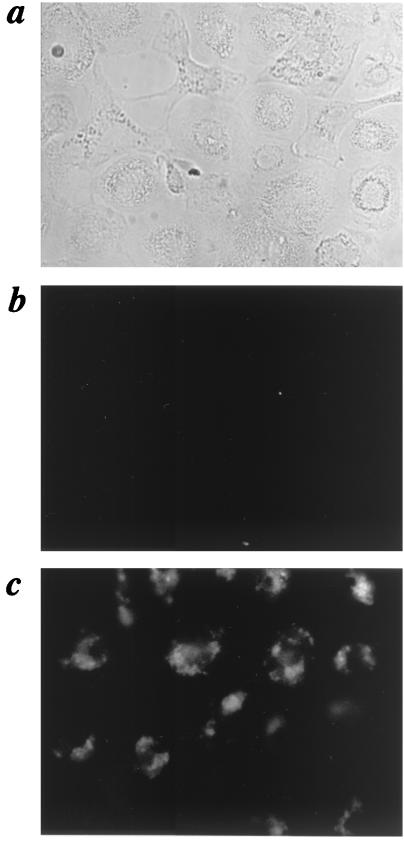
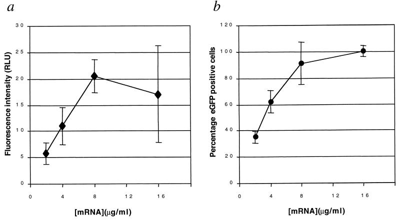
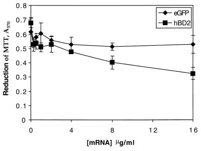
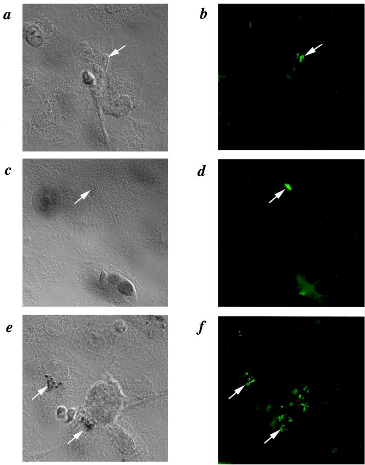
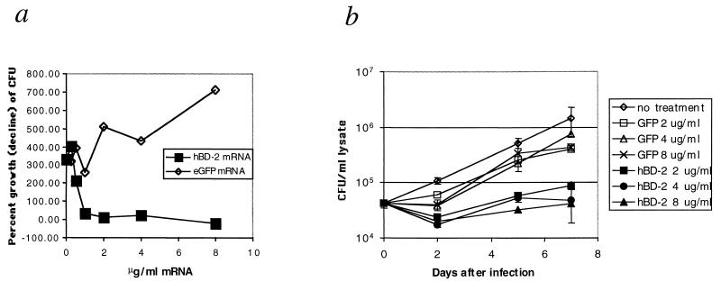
Human macrophages are hosts for Mycobacterium tuberculosis, the causative agent of tuberculosis, which killed approximately 1.87 million people in 1997. Human alveolar macrophages do not express alpha- or beta-defensins, broad-spectrum antimicrobial peptides which are expressed in macrophages from other species more resistant to infection with M. tuberculosis. It has been previously reported that M. tuberculosis is susceptible to killing by defensins, which may explain the difference in resistance. Defensin peptides have been suggested as a possible therapeutic strategy for a variety of infectious diseases, but development has been hampered by difficulties in their large-scale production. Here we report the cellular synthesis of human beta-defensin 2 via highly efficient mRNA transfection of human macrophages. This enabled mycobactericidal and mycobacteristatic activity by the macrophages. Although human macrophages are difficult to transfect with plasmid vectors, these studies illustrate that primary macrophages are permissive for mRNA transfection, which enabled expression of a potentially therapeutic protein.
Figures
Similar articles
-
Metformin promotes Mycobacterium tuberculosis killing and increases the production of human β-defensins in lung epithelial cells and macrophages.Microbes Infect. 2020 Apr;22(3):111-118. doi: 10.1016/j.micinf.2019.10.002. Epub 2019 Nov 2. Microbes Infect. 2020. PMID: 31689532
-
Hypoxia triggers the expression of human β defensin 2 and antimicrobial activity against Mycobacterium tuberculosis in human macrophages.J Immunol. 2012 Apr 15;188(8):4001-7. doi: 10.4049/jimmunol.1100976. Epub 2012 Mar 16. J Immunol. 2012. PMID: 22427634
-
Steroid hormone modulates the production of cathelicidin and human β-defensins in lung epithelial cells and macrophages promoting Mycobacterium tuberculosis killing.Tuberculosis (Edinb). 2021 May;128:102080. doi: 10.1016/j.tube.2021.102080. Epub 2021 Mar 24. Tuberculosis (Edinb). 2021. PMID: 33799143
-
Killing intracellular mycobacteria in in vitro macrophage systems: what may be the role of known host microbicidal mechanisms?Res Microbiol. 1990 Feb;141(2):217-30. doi: 10.1016/0923-2508(90)90034-n. Res Microbiol. 1990. PMID: 2111923 Review. No abstract available.
-
Tuberculosis Resistance and Nanoparticles: Combating the Dual Role of Reactive Oxygen Species in Macrophages for Tuberculosis Management.Crit Rev Ther Drug Carrier Syst. 2020;37(2):161-182. doi: 10.1615/CritRevTherDrugCarrierSyst.2020029870. Crit Rev Ther Drug Carrier Syst. 2020. PMID: 32865904 Review.
Cited by
-
Lung epithelial cells interact with immune cells and bacteria to shape the microenvironment in tuberculosis.Thorax. 2022 Apr;77(4):408-416. doi: 10.1136/thoraxjnl-2021-217997. Epub 2022 Jan 11. Thorax. 2022. PMID: 35017314 Free PMC article. Review.
-
Design, synthesis and structure-activity relationship study of wollamide B; a new potential anti TB agent.PLoS One. 2017 Apr 19;12(4):e0176088. doi: 10.1371/journal.pone.0176088. eCollection 2017. PLoS One. 2017. PMID: 28423019 Free PMC article.
-
Antimicrobial Peptides in Innate Immunity against Mycobacteria.Immune Netw. 2011 Oct;11(5):245-52. doi: 10.4110/in.2011.11.5.245. Epub 2011 Oct 31. Immune Netw. 2011. PMID: 22194707 Free PMC article.
-
Human {beta}-defensin 2 is expressed and associated with Mycobacterium tuberculosis during infection of human alveolar epithelial cells.Infect Immun. 2005 Aug;73(8):4505-11. doi: 10.1128/IAI.73.8.4505-4511.2005. Infect Immun. 2005. PMID: 16040961 Free PMC article.
-
Mycobacterium tuberculosis cell-wall and antimicrobial peptides: a mission impossible?Front Immunol. 2023 May 17;14:1194923. doi: 10.3389/fimmu.2023.1194923. eCollection 2023. Front Immunol. 2023. PMID: 37266428 Free PMC article. Review.
References
-
- Deretic V, Fratti R A. Mycobacterium tuberculosis phagosome. Mol Microbiol. 1999;31:1603–1609. - PubMed
Publication types
MeSH terms
Substances
Grants and funding
LinkOut - more resources
Full Text Sources
Other Literature Sources
Medical
