Testicular FasL is expressed by sperm cells
- PMID: 11248076
- PMCID: PMC30651
- DOI: 10.1073/pnas.051566098
Testicular FasL is expressed by sperm cells
Abstract
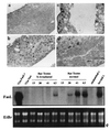
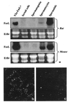
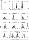
The testis is the main source of Fas ligand (FasL) mRNA in rodents; it is generally believed that this molecule, expressed on bordering somatic Sertoli cells, bestows an immune-privileged status in the testis by eliminating infiltrating inflammatory Fas-bearing leukocytes. Our results demonstrate that the attribution of testicular expression of FasL to Sertoli cells is erroneous and that FasL transcription instead occurs in meiotic and postmeiotic germ cells, whereas the protein is only displayed on mature spermatozoa. These findings point to a significant role of the Fas system in the biology of mammalian reproduction.
Figures


Similar articles
-
The Fas system is a key regulator of germ cell apoptosis in the testis.Endocrinology. 1997 May;138(5):2081-8. doi: 10.1210/endo.138.5.5110. Endocrinology. 1997. PMID: 9112408
-
The Fas system in the seminiferous epithelium and its possible extra-testicular role.Andrologia. 2003 Feb;35(1):64-70. doi: 10.1046/j.1439-0272.2003.00538.x. Andrologia. 2003. PMID: 12558530 Review.
-
Ontogenesis and cell specific localization of Fas ligand expression in the rat testis.Int J Androl. 2004 Oct;27(5):304-10. doi: 10.1111/j.1365-2605.2004.00483.x. Int J Androl. 2004. PMID: 15379972
-
Participation of the Fas-signaling system in the initiation of germ cell apoptosis in young rat testes after exposure to mono-(2-ethylhexyl) phthalate.Toxicol Appl Pharmacol. 1999 Nov 1;160(3):271-8. doi: 10.1006/taap.1999.8786. Toxicol Appl Pharmacol. 1999. PMID: 10544061
-
The role of the Fas/FasL signaling pathway in environmental toxicant-induced testicular cell apoptosis: An update.Syst Biol Reprod Med. 2018 Apr;64(2):93-102. doi: 10.1080/19396368.2017.1422046. Epub 2018 Jan 4. Syst Biol Reprod Med. 2018. PMID: 29299971 Review.
Cited by
-
Immunologic Environment of the Testis.Adv Exp Med Biol. 2021;1288:49-67. doi: 10.1007/978-3-030-77779-1_3. Adv Exp Med Biol. 2021. PMID: 34453731
-
Expression of the Fas-ligand gene in ejaculated sperm from adolescents with and without varicocele.J Assist Reprod Genet. 2010 Feb;27(2-3):103-9. doi: 10.1007/s10815-010-9384-9. Epub 2010 Feb 18. J Assist Reprod Genet. 2010. PMID: 20165911 Free PMC article.
-
Gene expression study in the juvenile mouse testis: identification of stage-specific molecular pathways during spermatogenesis.Mamm Genome. 2006 Sep;17(9):956-75. doi: 10.1007/s00335-006-0029-3. Epub 2006 Sep 8. Mamm Genome. 2006. PMID: 16964443
-
Estrogens up-regulate the Fas/FasL apoptotic pathway in lactotropes.Endocrinology. 2005 Nov;146(11):4737-44. doi: 10.1210/en.2005-0279. Epub 2005 Aug 11. Endocrinology. 2005. PMID: 16099864 Free PMC article.
-
Nonlymphoid Fas ligand in peptide-induced peripheral lymphocyte deletion.Proc Natl Acad Sci U S A. 2002 Dec 10;99(25):16174-9. doi: 10.1073/pnas.262660999. Epub 2002 Nov 26. Proc Natl Acad Sci U S A. 2002. PMID: 12454289 Free PMC article.
References
-
- Nagata S. Cell. 1997;88:355–365. - PubMed
-
- Takahashi T, Tanaka M, Brannan C I, Jenkins N A, Copeland N G, Suda T, Nagata S. Cell. 1994;76:969–976. - PubMed
-
- Li J H, Rosen D, Ronen D, Behrens C K, Krammer P H, Clark W R, Berke G. J Immunol. 1998;161:3943–3949. - PubMed
-
- Abbas A K. Cell. 1996;84:655–657. - PubMed
-
- Head J R, Neaves W B, Billingham R E. Transplantation. 1983;36:423–431. - PubMed
MeSH terms
Substances
LinkOut - more resources
Full Text Sources
Research Materials
Miscellaneous

